
Method for introducing changes into a eukaryotic genome in vivo and a kit

a technology of eukaryotic genome and kit, applied in the field of molecular biology, can solve the problems of over-frequent loading, and achieve the effect of good viability of transfected cells and good model for in vivo experimentation

Inactive Publication Date: 2010-11-04
UNIV OF TARTU
View PDF0 Cites 0 Cited by
  • Summary
  • Abstract
  • Description
  • Claims
  • Application Information

AI Technical Summary

Benefits of technology

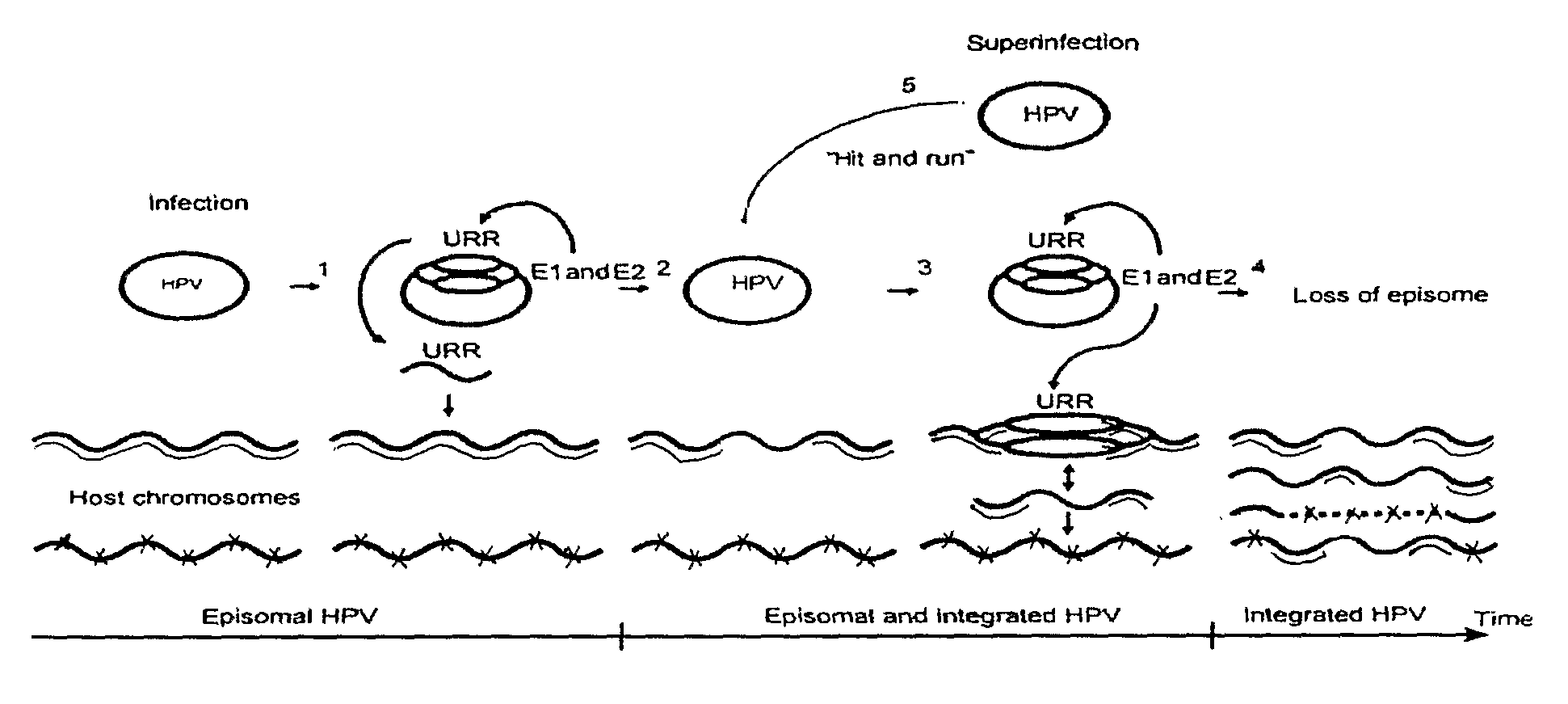
[0012]The presented data suggest that papillomavirus DNA replication machinery can actively induce irreversible changes in the genomic make-up of the cell at sites of HPV origin integration. The results show that papillomavirus replication proteins are capable of mobilizing integrated HPV origin and that simultaneous DNA replication of episomal and integrated HPV origins may occur in HeLa cells. These kinds of changes provide a useful tool for research, if one wishes to amplify, excise or translocate a genomic sequence, either adjacent to the integration site of the HPV sequence or wishes to introduce foreign sequences to the cell with respectively constructed HPV vector. The kit including HPV replication origin and overexpression of genes encoding HPV E1 and E2 for introducing duplications, multiplications, insertions, deletions, inversions and / or translocations of a DNA sequence into a eukaryotic cell is particularly important as it exhibits unexpectedly good viability of the transfected cells thus making the system a good model for in vivo experimentation. Moreover, amplification of a DNA sequence encoding functional units of heredity allows this combination to serve as basis for gene expression and overexpression experiments. The beforementioned kit comprises a vector carrying HPV genome or a part of HPV genome including HPV replication origin sequence, and expression vector or vectors encoding HPV early proteins, e.g. E1 and E2.

Problems solved by technology

Increase of E1 concentration above a certain level results in overly frequent loading of the viral hexameric helicase and cellular replication complexes at the viral origin and induction of ‘onion skin’-type replication intermediates on the viral genome (Männik et al., 2002).

Method used


Image

  • Method for introducing changes into a eukaryotic genome in vivo and a kit
  • Method for introducing changes into a eukaryotic genome in vivo and a kit
  • Method for introducing changes into a eukaryotic genome in vivo and a kit

Examples


example 1

E1 Protein Induces DNA Replication in Cells with Integrated HPV

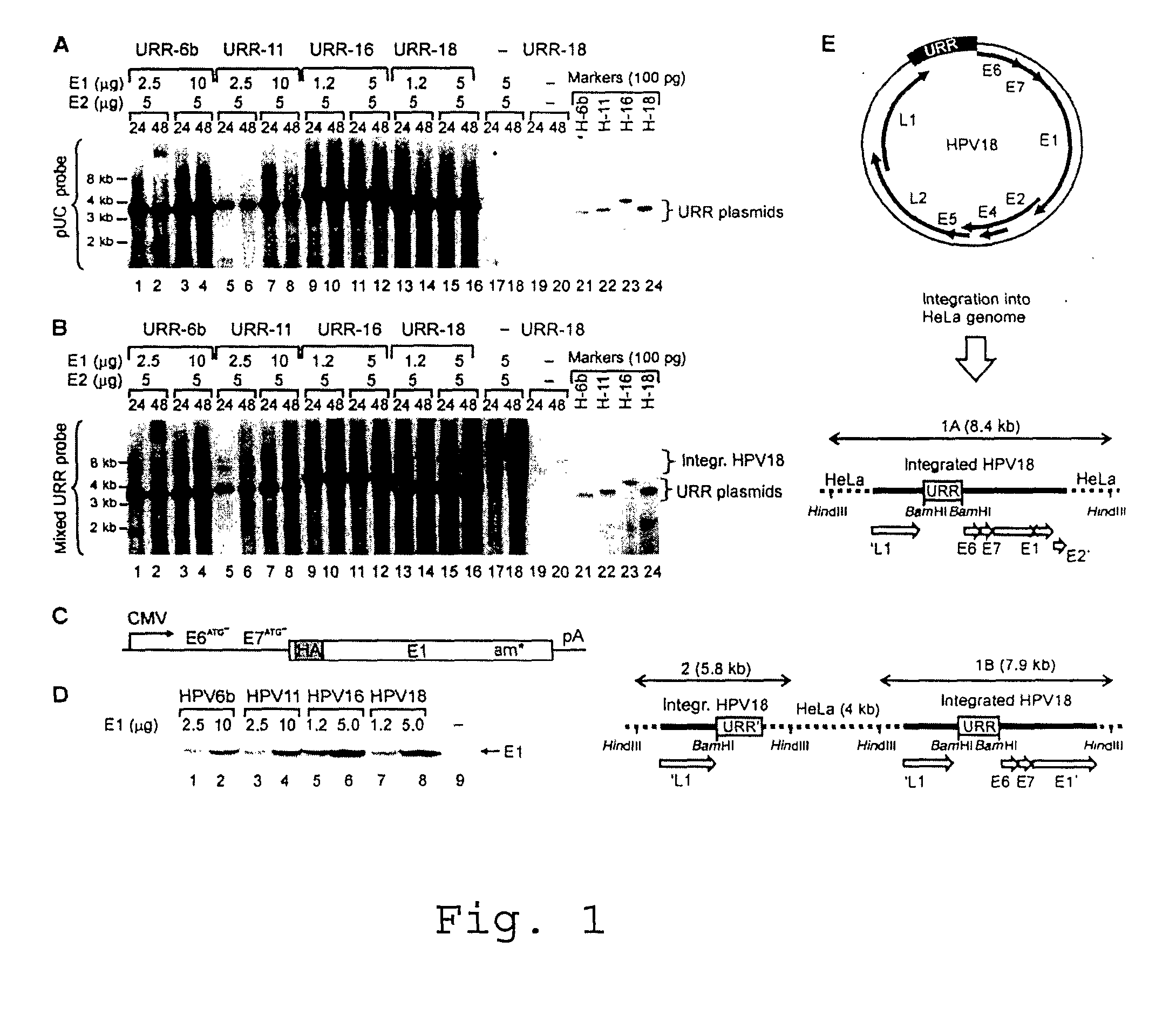
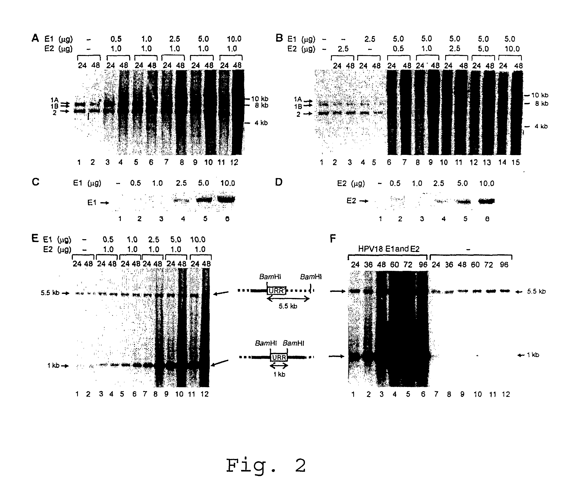
[0027]Titration of the HPV18 E1 and E2 proteins in the transient assays showed that the efficiency of DNA replication initiation depends on the E1 protein concentration (FIG. 2A), while modulation of E2 concentration in a quite wide range had little effect on the efficiency of initiation of the DNA replication of the integrated HPV (FIG. 2B). The replication reached a plateau at 0.5 pg of transfected E2 expression plasmid (FIG. 2B, lanes 6 and 7) and changed little at higher vector concentrations. The E2 expression level in our replication assays did not induce senescence or apoptosis of the transfected cells. High E1 levels caused smearing of the integrated HPV18 origin replication signals characteristic to the ‘onion skin’ type of replication mode. Cleavage of the integrated HPV18 sequences in HeLa cells (FIGS. 1A, B and E) with BamHI generates a 1 kb URR fragment, which includes the complete functional HPV replication...

example 2

Expression of E1 and E2 Proteins Induces Amplification of Episomal and Integrated HPV18 Origins in HeLa Cells

[0028]To test E1 expression constructs and to identify the conditions conducive to viral origin replication, the origin plasmids (pUC / URRs) of all HPVs, together with the homologous expression vectors for E1 and E2, were cotransfected into HeLa cells. The DpnI resistant replication signal from the transient assays was examined by Southern blot analysis of total DNA with common plasmid probe (FIG. 1A). Replication of all origin plasmids was clearly detected in HeLa cells. Western blot analysis to monitor E1 protein levels in transfected cells (FIG. 1D) confirms its effective and comparable expression in HeLa cells. E2 levels were kept constant and considerably low (5 pg of transfected E2 plasmid) in order to support replication without suppression of E6 promoter. HPV18 and HPV18 E1 proteins seem to be the most efficient in initiation of DNA replication within the used expressi...

example 3

HPV E1 and E2 Proteins Efficiently Initiate Replication from Integrated HPV16 Origin in SiHa Cells

[0029]SiHa cell line derived from the cervical carcinoma of a 55-year-old Japanese female is aneusomic and has been found to contain 66-72 chromosomes. However, this cell line has been shown to be disomic with respect to chromosome 13, which contains one copy of the HPV16 genome (Meissner, 1999; Szuhai et al., 2000). Integration of the HPV16 genome has occurred, with disruption in the E2 and E4 ORFs at nucleotides 3132 and 3384 of HPV16 genome (FIG. 3A). Expression plasmids for the E1 and E2 proteins were transfected into SiHa cells and replication of integrated HPV16 origin DNA was analyzed using HPV16 URR probe. The E2 expression plasmid concentration was kept constant at the level where replication of integrated HPV16 had reached a plateau (data not shown). Increasing amounts of E1 expression plasmids were used in replication assays. In order to reach comparable levels of E1 expressi...


PUM

PropertyMeasurementUnit
molecular-weightaaaaaaaaaa
timeaaaaaaaaaa
voltageaaaaaaaaaa

Abstract

The invention relates to a method for introducing changes into a eukaryotic genome in vivo wherein the HPV genome, which comprises HPV replication origin sequence, is used together with HPV early proteins in order to achieve DNA replication in vivo. There is also disclosed a kit for in vivo amplification, excision, translocation and / or inversion of a DNA sequence, which comprises a vector carrying HPV genome or a part of HPV genome including HPV replication origin sequence, and expression vector or vectors encoding HPV early proteins.

Description

CROSS-REFERENCE TO RELATED APPLICATIONS[0001]This application is a national phase application pursuant to 35 U.S.C. §371 of International Application No. PCT / EE2008 / 000004, filed Mar. 27, 2008, which claims priority to Estonia Application No. P200700012, filed Mar. 28, 2007.TECHNICAL FIELD OF THE INVENTION[0002]The current invention is most widely in the field of molecular biology, more particularly in the field of virology and provides a means for introducing changes into a genome of a eukaryotic cell, which may be duplications, multiplications, insertions, deletions, inversions and / or translocations of a DNA sequence.BACKGROUND OF THE INVENTION[0003]Papillomaviruses are small species-specific DNA tumor viruses that establish latent infection in the basal cells of the differentiating epithelium with the help of viral oncoproteins and maintain their small, about 8 kb, circular doublestranded (ds) genomes as episomal multicopy nuclear plasmids in proliferating transformed cells (Howl...

Claims


Application Information

Patent Timeline
Patent Type & Authority Applications(United States)
IPC IPC(8): C12N15/86C12N15/63
CPCC12N15/86C12N2800/80C12N2710/20043C12N2510/00
Inventor USTAV, MARTUSTAV, ENEKADAJA, MEELISSUMERINA, ALINA
Owner UNIV OF TARTU
Learn More