Fused perfusion and functional 3D rotational angiography rendering

a technology of perfusion and rotation angiography, applied in the field of system and method for rendering images/views of anatomical structures, can solve the problems of unsatisfactory presentation mode and the inability of a clinician to interpret the 3d morphology of a vessel

Active Publication Date: 2010-11-18
KONINKLIJKE PHILIPS ELECTRONICS NV
View PDF5 Cites 1 Cited by
  • Summary
  • Abstract
  • Description
  • Claims
  • Application Information

AI Technical Summary

Benefits of technology

[0007]It is an object of the present invention to provide an improved fused visua

Problems solved by technology

This presentation mode is not ideal, because it is impossible for a clin

Method used


Image

  • Fused perfusion and functional 3D rotational angiography rendering
  • Fused perfusion and functional 3D rotational angiography rendering
  • Fused perfusion and functional 3D rotational angiography rendering

Examples


Embodiment Construction

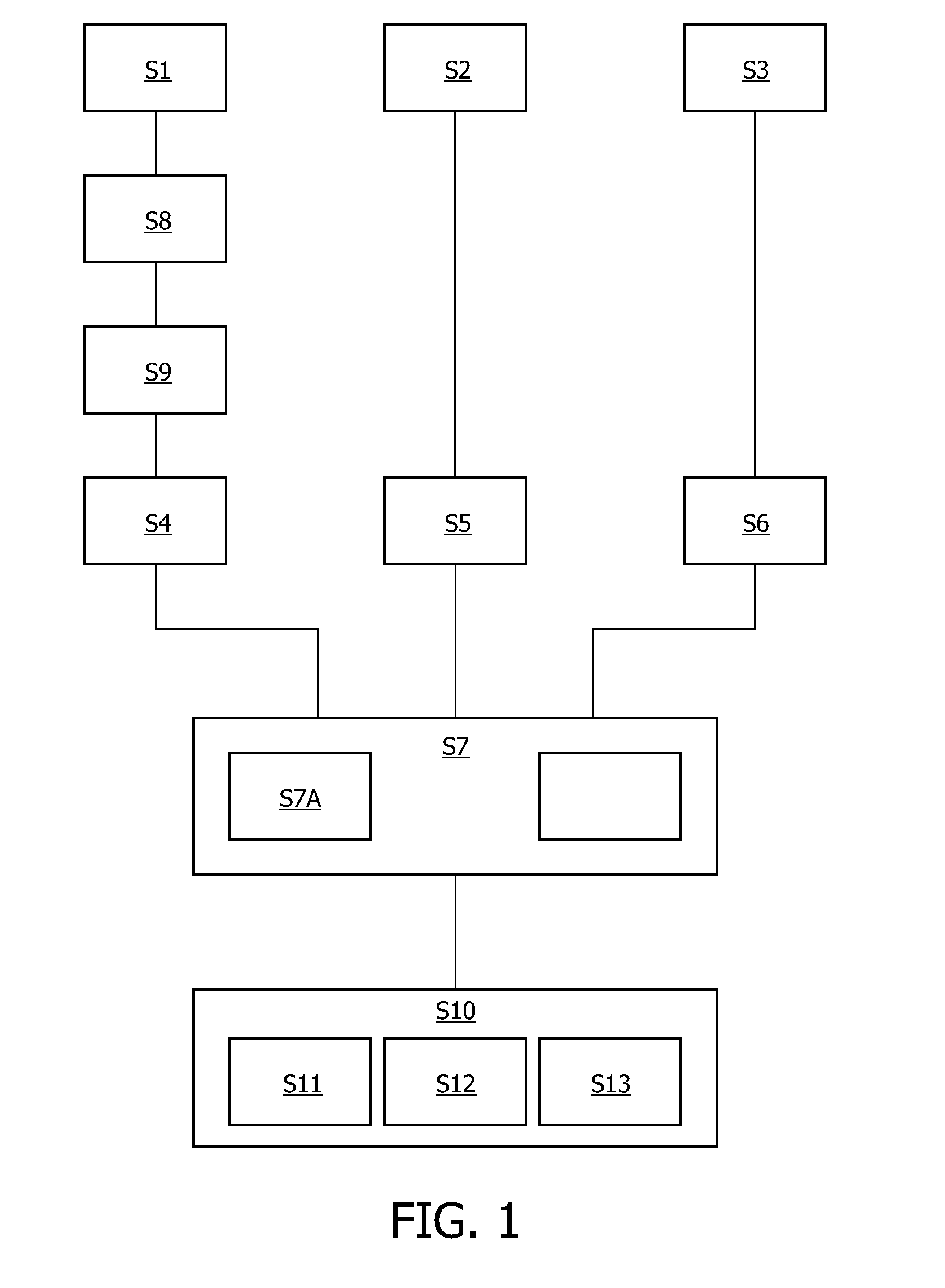
[0048]FIG. 1 shows a flow chart of a method according to an exemplary embodiment of the present invention. The method comprises obtaining a 3D rotational angiography data set of an anatomical structure S1, obtaining a physiologic data set relating to a physiologic parameter with respect to the anatomic structure S2, generating a first image that includes the anatomical structure and is derived from the 3D rotational angiography data set S4, generating a second image that includes the physiologic parameter and is derived from the physiologic data set S5, and registering the first image and the second image S7. Further, the method may comprise obtaining a diagnostic data set relating to an anatomical region S3 and generating a third image that includes the anatomical region and is derived from the diagnostic data set S6.

[0049]The generated images of the anatomical structure and the physiologic parameter, and optionally of the anatomical region are registered S7, S7a. Registering means...


PUM


Abstract

A Method and system for visualising information by combining 3DRA with diagnostic data like regular CT or MR, and colourised physiologic data like perfusion or functional data to obtain a plurality of volumes obtained from the same patient. These volumes may be a 3DRA volume, a regular greyscale CT or MR volume and a colourised physiologic parameter like a perfusion CT, a perfusion MR or a functional MR volume. Then, an anatomic structure like a vessel is segmented from the 3DRA volume, a slab out of the regular CT or MR data is rendered through the segmented vessel, and a slice out of the colourised volume of the perfusion or functional data is rendered on top of the slab.

Description

TECHNICAL FIELD[0001]The present invention relates to a system and method for rendering images / views of an anatomical structure, e.g. vessel structures. In particular, the present invention relates to a system and method for registering an image of a 3D X-ray rotational angiography (3DRA) with a scan of physiologic data, e.g. from a computer tomography (CT) or magneto resonance (MR) scan, to yield a combined image or view thereof.BACKGROUND ART[0002]Interventional X-ray angiography procedures are generally based on real time, two-dimensional (2D) minimally invasive image guidance of endovascular material through the vasculature or the human vessels. The imaging modality of choice for the interactive tracking of guide wires and catheters is an X-ray angio machine. Typically, interactive tracking of such endovascular material, e.g. a guide wire and / or catheter, have been achieved with an X-ray angiography instrument. Recently introduced three-dimensional rotational angiography (3DRA) ...

Claims


Application Information

Patent Timeline
IPC IPC(8): G06K9/00
CPCG06T2207/30101G06T7/0024G06T7/30
Inventor RUIJTERS, DANIEL SIMON ANNA
Owner KONINKLIJKE PHILIPS ELECTRONICS NV
Why Eureka
Try Eureka