

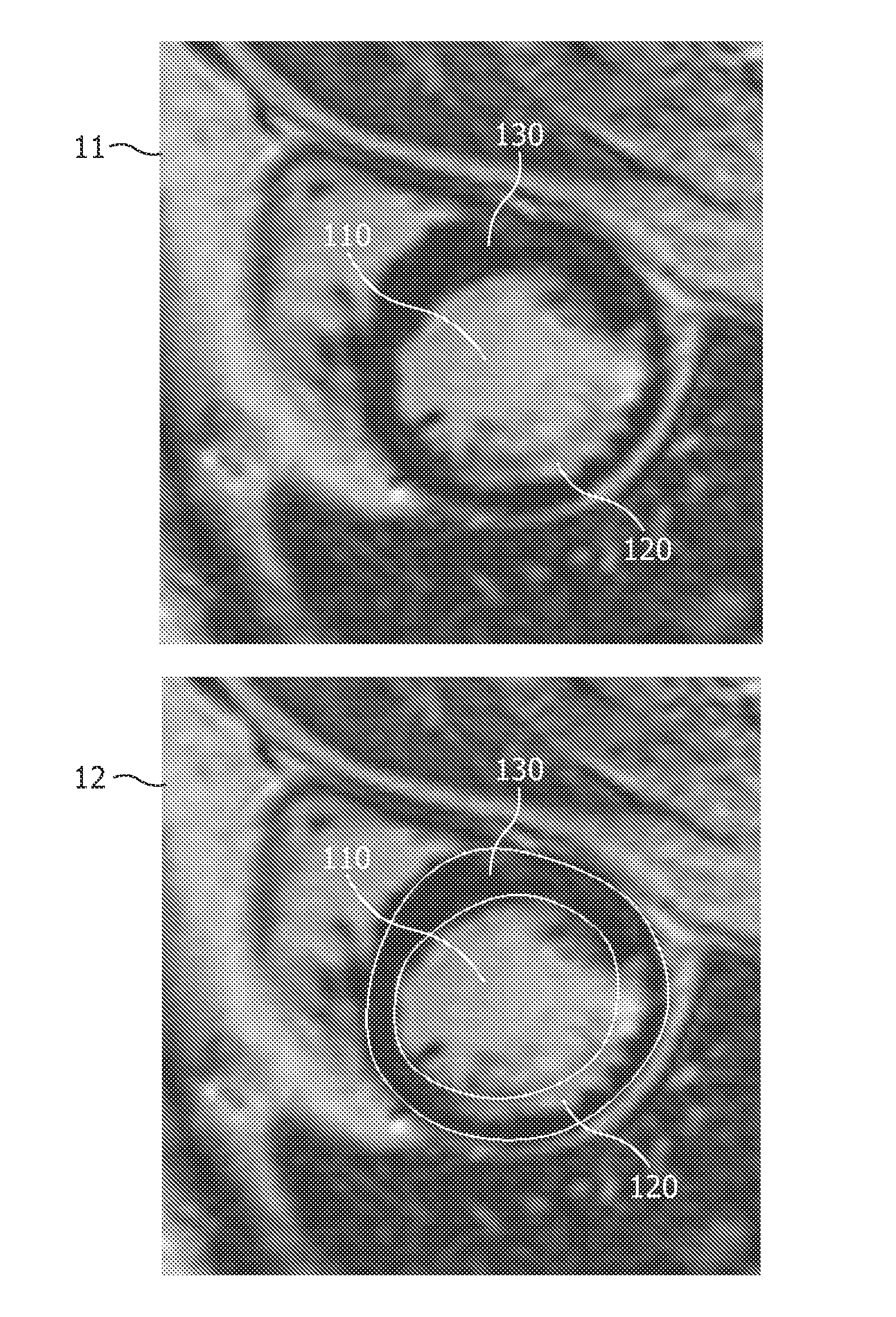
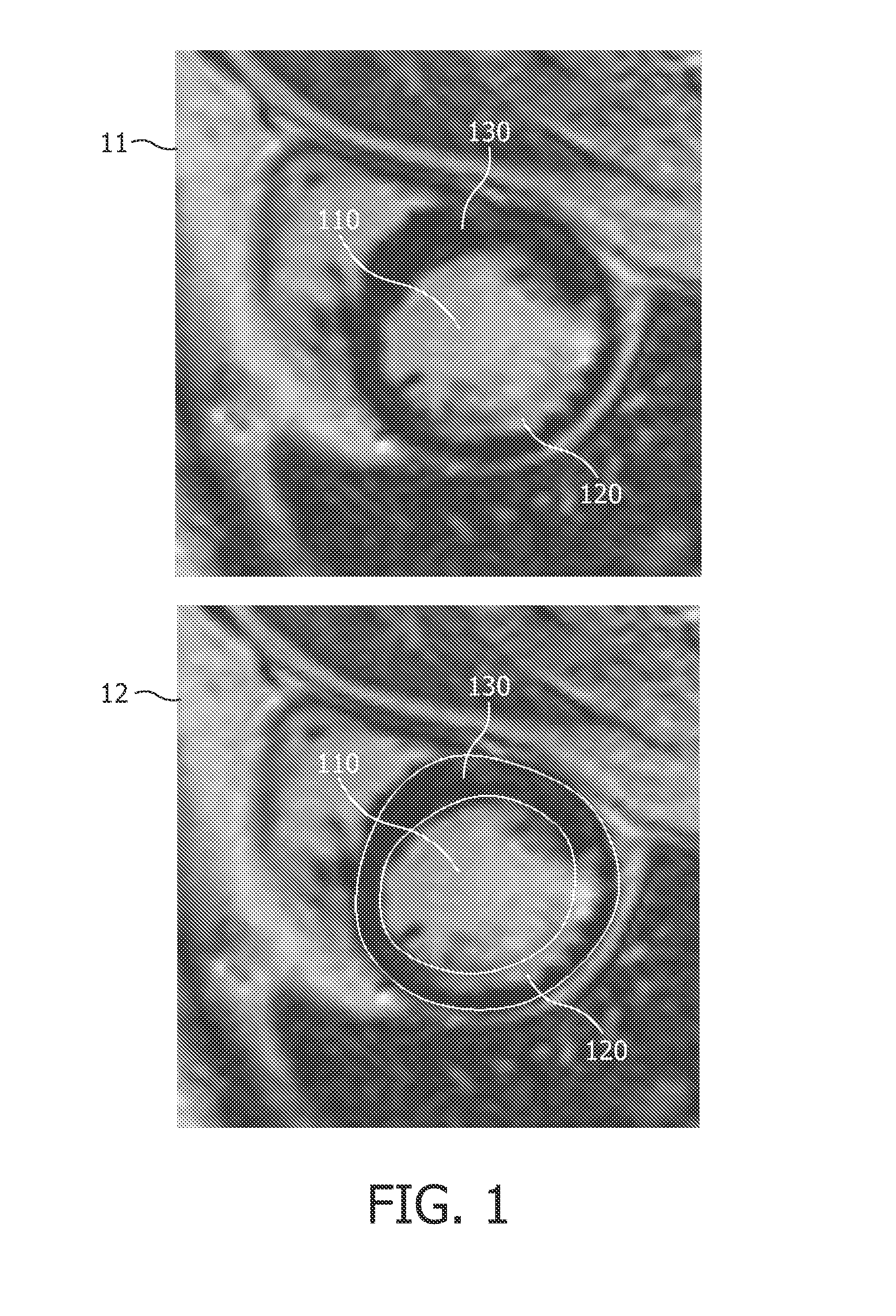
Automatic 3-d segmentation of the short-axis late-enhancement cardiac MRI
a cardiac mri and short-axis technology, applied in the field of image segmentation, can solve the problems of difficult and time-consuming manual segmentation, difficult automatic myocardium segmentation, and rarely implemented in current commercial products, and achieve the difficulty of designing an automatic method to delineate the endo- and epicardial contours
- Summary
- Abstract
- Description
- Claims
- Application Information
AI Technical Summary
Benefits of technology
Problems solved by technology
Method used
Image

Examples
Embodiment Construction
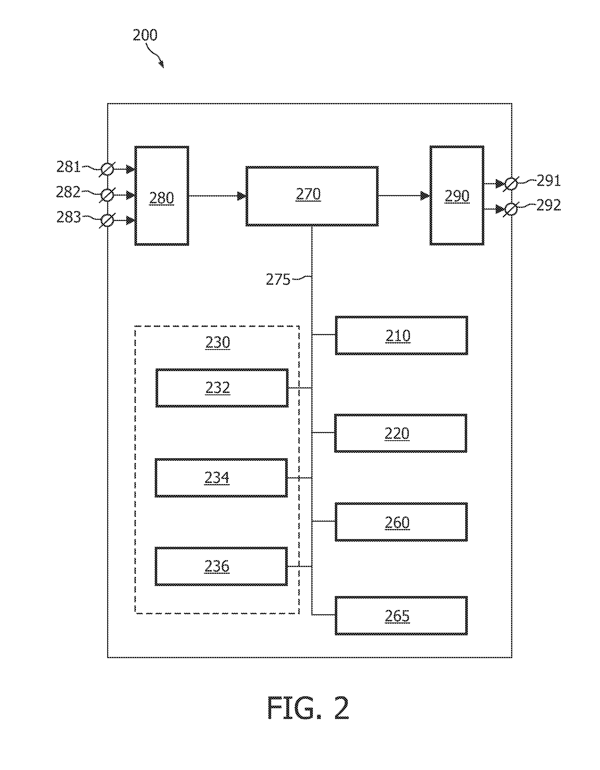
[0045]FIG. 2 schematically shows a block diagram of an exemplary embodiment of the system 200 for delineating an anatomical structure in an image computed from a slice of image data, the system 200 comprising:
[0046]a partition unit 210 for partitioning the image into a plurality of image portions, each image portion depicting a portion of the anatomical structure; and
[0047]an adaptation unit 220 for adapting a template to the image, based on a criterion function, the criterion function being a function of template parameters and of image values and their relative positions in the image, and on a criterion to be satisfied by a computed value of the criterion function,
[0048]wherein the criterion function is defined on the basis of the plurality of image portions.
[0049]The exemplary embodiment of the system 200 further comprises the following units:
[0050]a registration unit 230 for registering a surface model with a plurality of templates, wherein each template is adapted to an image c...
PUM
 Abstract
Description
Claims
Application Information