

Primed cell therapy
a technology of prilosec and cell therapy, applied in the direction of skeletal/connective tissue cells, drug compositions, peptide/protein ingredients, etc., can solve the problems of exacerbated the tendency of anti-arthritis drugs to produce serious side effects, inefficient drug delivery routes, and frequent repeated injections, so as to facilitate cell growth of primary chondrocytes and maximize cartilage tissue regeneration
- Summary
- Abstract
- Description
- Claims
- Application Information
AI Technical Summary
Benefits of technology
Problems solved by technology
Method used
Image

Examples
example 1
Preparation of Primed Chondrocytes
Example 1.1
Cell Culture and Treatments
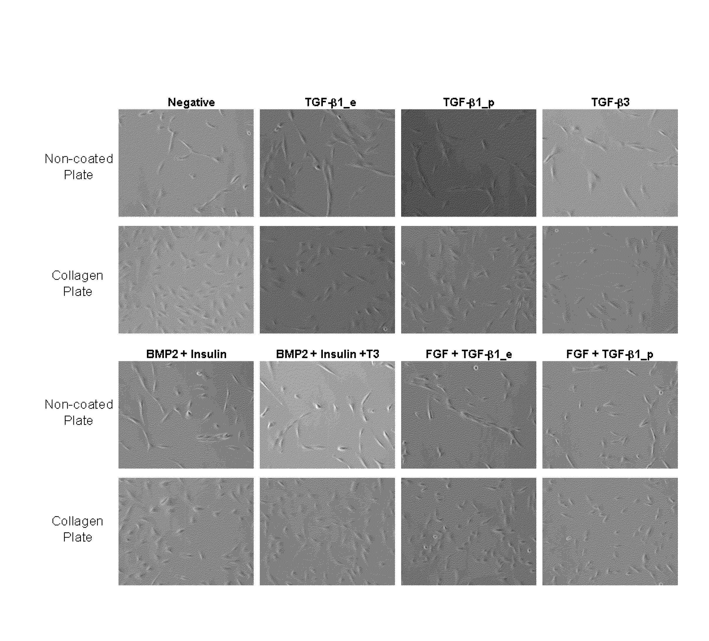
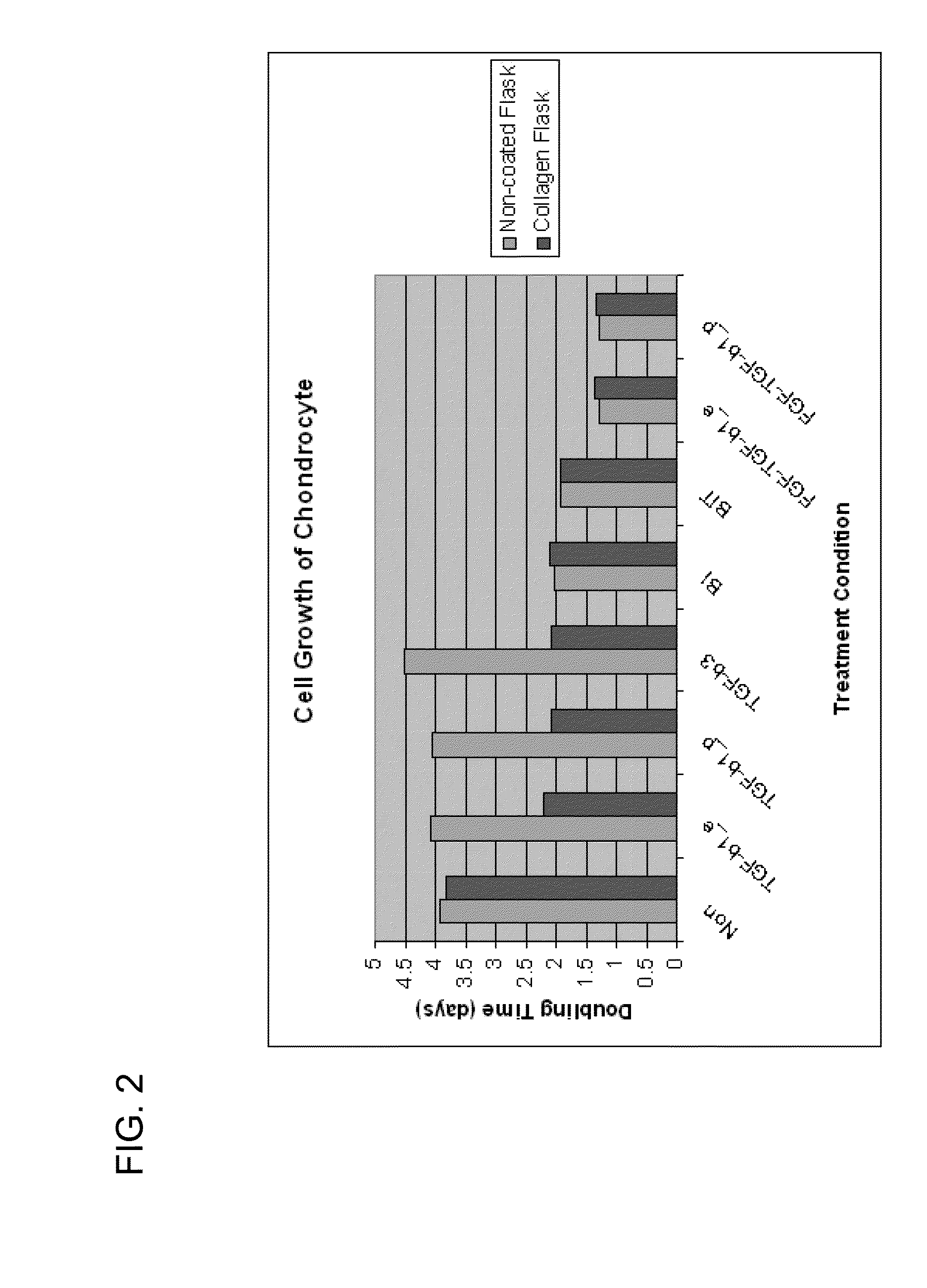
[0092]Cells used in this study originated from primary human chondrocytes that had been cultured for an extended period in vitro. These cells have assumed a morphology and phenotype that is characteristic of fibroblasts despite their origin. Experiments were performed with cultures approximately at passage seven. Cells were seeded in two different culture formats: monolayer, and micromass. Culture medium consisted of DMEM (Lonza) with 4.5 g / L glucose, supplemented with 10% fetal bovine serum (Lonza) and 1% L-Glutamine (Lonza). Cells were incubated in 37° C., 5% CO2 environment for the duration of treatment. Cells were exposed to multiple concentrations of TGF-β1 (R&D Systems) at different time points of the culture process for several lengths of incubation time.
example 1.2
Monolayer Culture
[0093]Chondrocytes were seeded at 5×10⁴ cells / well into a 6-well collagen coated plate (BioCoat, BD Biosciences). Cells were either primed with TGF-β1 at a concentration of 100 ng / mL for a period of 6 hours prior to seeding or incubated with TGF-β1 at 1 ng / mL concentration for the duration of the study. In addition to these two treatments, a co-culture of TGF-β1 producing cells and human chondrocytes were prepared at a 1:3 ratio, seeded at 5×10⁴ cells / well and similarly examined for the duration of the three week study. Cells would be harvested weekly for RNA preparation and staining.
[0094]A shorter one week study was also performed. Chondrocytes were seeded at 3×10³ cells / cm2 to each well of the collagen coated 6-well plate. Four treatment groups represented the different time points at which TGF-β1 supplementation occurs: 24 hour, 48 hour, 72 hour, and two 36 hour intervals prior to cell harvest at the end of the week long study.
example 1.3
Micromass Culture
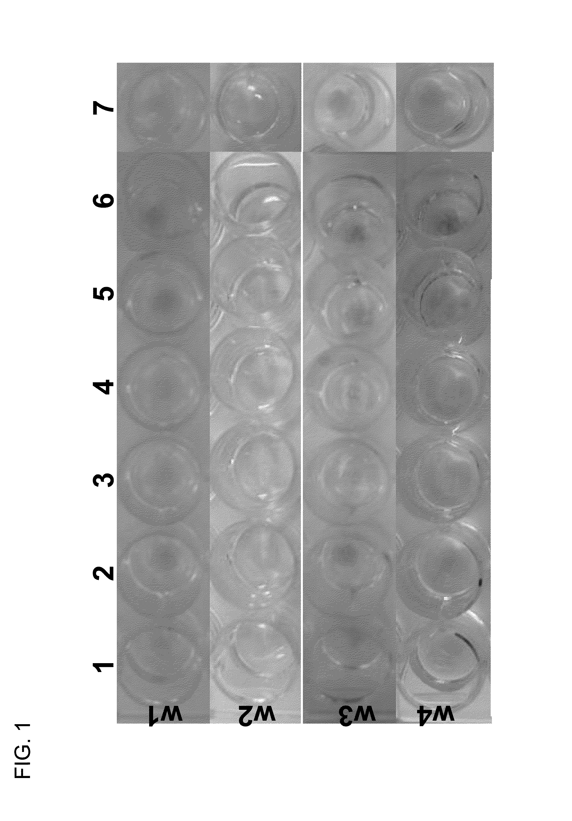
[0095]Cell suspensions were prepared for a seeding density of 3×10⁵ cells / 15 μL droplet. Cell droplets were placed in the center of the well of a 24-well collagen coated plate (BioCoat). Cell masses were incubated for 1.5 hours at 37° C. were replenished with 1 mL of complete medium once masses were set. Cells were primed through treatment with 10 ng / mL or 50 ng / mL of TGF-β1 for duration of 6 hours or 18 hours prior to the seeding of micromasses. In addition to these four treatment groups, two groups of chondrocytes were exposed to TGF-β1 at concentrations of 1 ng / mL or 10 ng / mL for the duration of the study. The final experimental group consisted of the 3:1 ratio of TGF-β1 producing cells to untreated chondrocytes. Masses were cultured for up to four weeks.
PUM
| Property | Measurement | Unit |
|---|---|---|
| temperature | aaaaa | aaaaa |
| time | aaaaa | aaaaa |
| concentration | aaaaa | aaaaa |
Abstract
Description
Claims
Application Information