
Methods for diagnosing and treating astrocytomas

a technology for astrocytoma and astrocytoma, applied in the field of methods for diagnosing and treating astrocytoma, can solve the problems of distress, no effective treatment, general non-curable resection, etc., and achieve the effect of reducing the activity or function of the polypeptid

Inactive Publication Date: 2010-12-23
LUDWIG INST FOR CANCER RES
View PDF6 Cites 4 Cited by
  • Summary
  • Abstract
  • Description
  • Claims
  • Application Information

AI Technical Summary

Benefits of technology

[0030]According to yet another aspect of the invention, methods of treating astrocytoma are provided. The methods include administering to a subject who has or is suspected or having astrocytoma a compound or composition, which reduces the level or biological activity of a KIAA0101, LOX, Melk, ASPM, HOXA5, PLP2, DKFZp762E1312, CD99, HOXC9 and / or E2F2 gene product, in an amount effective to treat the astrocytoma.
[0034]In a further aspect of the invention, methods of treating astrocytoma are provided. The methods include administering to a subject who has or is suspected or having astrocytoma a compound or composition, which binds a LOX, KIAA0101, HOXA5, DKFZp762E1312, HOXC9, MELK, ASPM, E2F2, CD99 or PLP2 polypeptide, in an amount effective to treat the astrocytoma. In some embodiments, the astrocytoma is a pilocytic astrocytoma (PA; grade I), low grade astrocytoma (grade II), anaplastic astrocytoma (grade III) and glioblastoma multiforme (GBM; grade IV). In other embodiments, the compound or composition is an antibody or an antigen-binding fragment thereof. Molecules that induce. RNA interferrence, such as siRNA molecules also can be used. In preferred embodiments, the compound or composition reduces the activity or function of the polypeptide.

Problems solved by technology

These are generally not curable by resection.
This scenario is particularly distressing for patients with glioblastoma multiforme (GBM), in which median survival stands below 16 months even with the combined use of resection and radiotherapy43.
Currently, there is no effective treatment for this highly aggressive and infiltrative tumor, and relatively little is known about the molecular processes leading to its formation.
Such development, however, can not be achieved without understanding the genetic and molecular events leading to oncogenic activation.

Method used


Image

  • Methods for diagnosing and treating astrocytomas
  • Methods for diagnosing and treating astrocytomas
  • Methods for diagnosing and treating astrocytomas

Examples


example 1

Identification of Astrocytoma Markers

Materials and Methods

Tissue Samples

[0227]Tumor specimens were obtained from patients with CNS tumors treated by the Neurosurgery Group of the Department of Neurology at Hospital das Clinicas, School of Medicine, University of São Paulo, São Paulo, Brazil, between 2000 and 2005. The tissues were categorized according to the WHO grading system1, 2 by neuropathologists from the Division of Pathological Anatomy of the same institution. Informed consent was obtained from each patient. Fresh surgical samples of different grades and non-neoplastic tissues of the CNS (temporal lobectomy from epilepsy surgeries) were macrodissected and immediately snap-frozen in liquid nitrogen upon surgical removal. All tumor tissues were microdissected prior to RNA extraction as described previously11.

Tissue Samples for IHC

[0228]Tumor specimens were obtained during therapeutic surgical approach of patients with CNS tumors by the Neurosurgery Group of the Department of N...

example 2

Validation of Additional Astrocytoma Marker Gene Expression

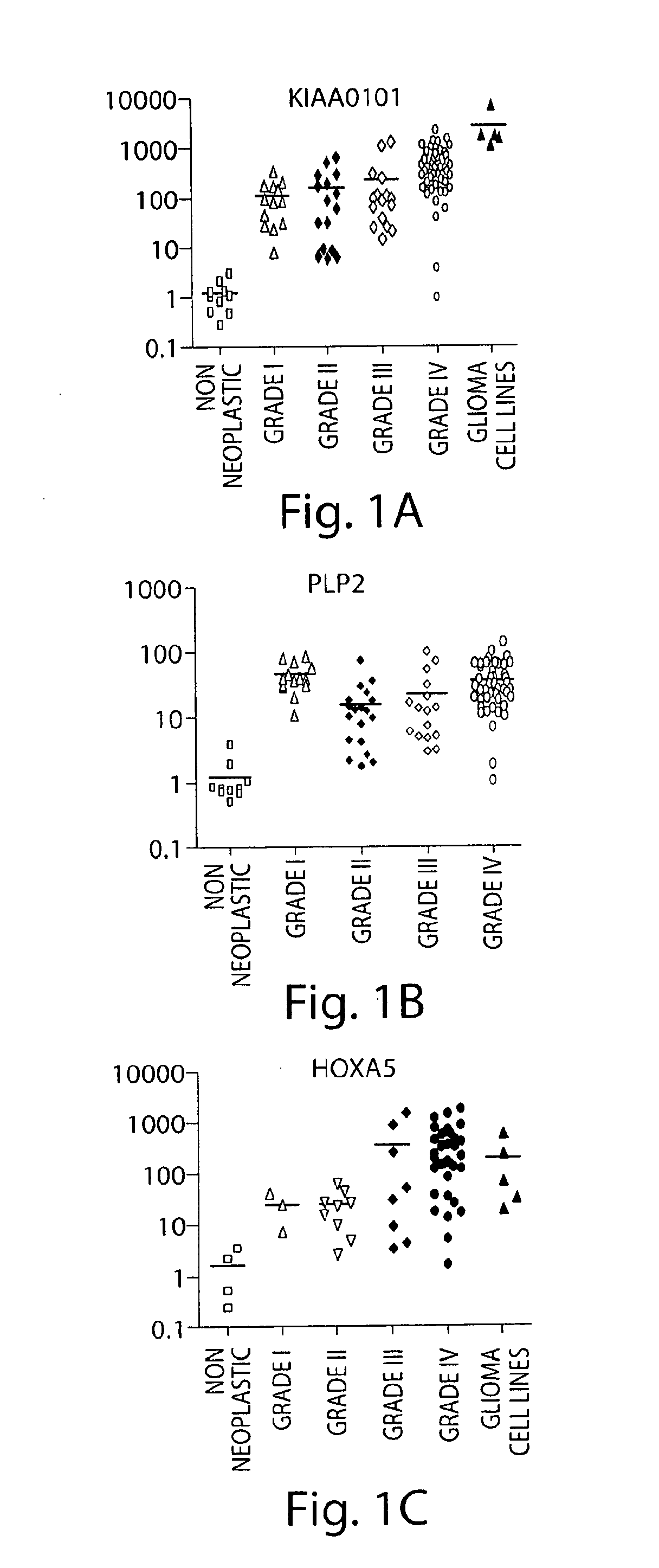
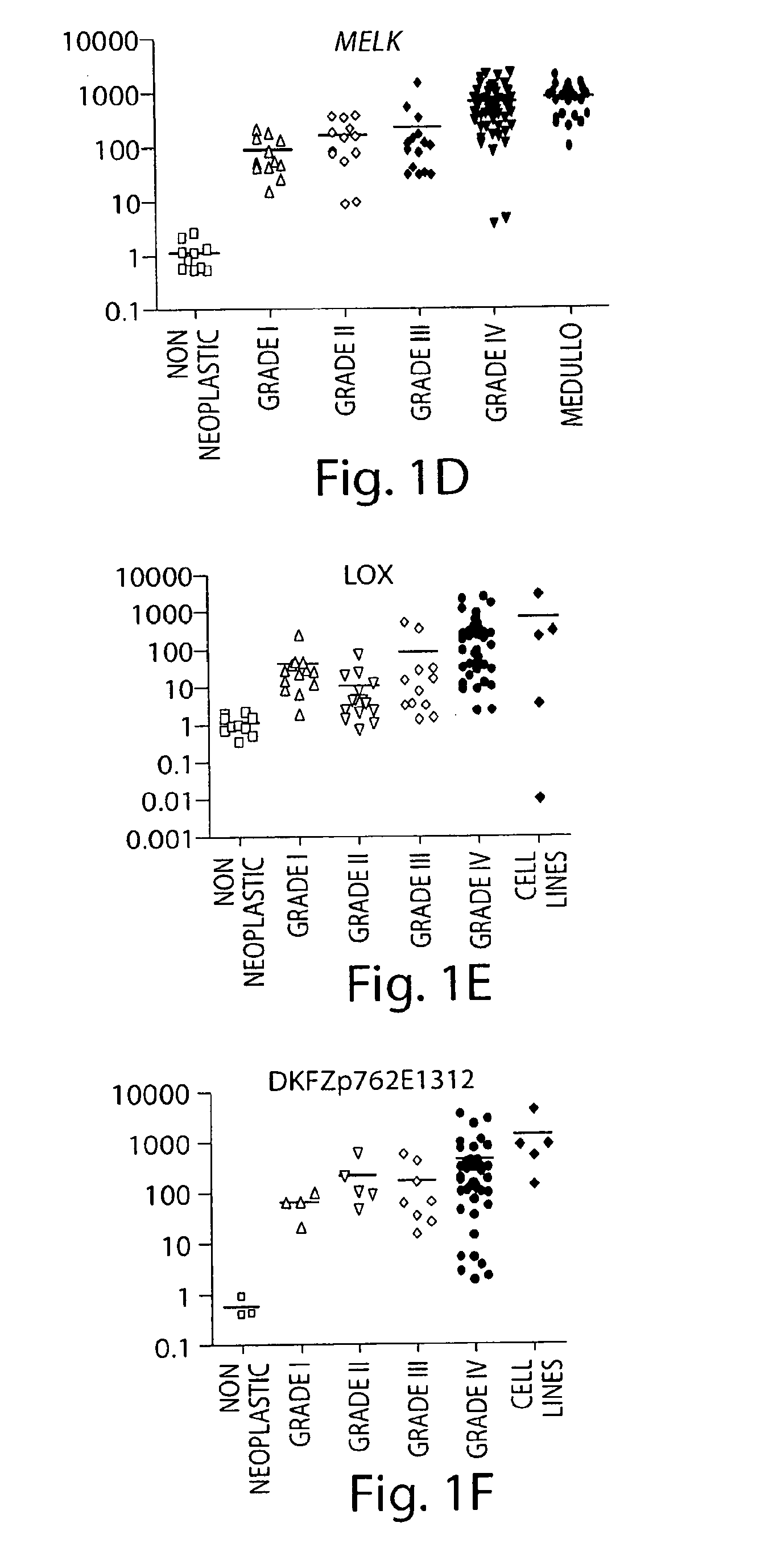
[0319]Gliomas of astrocytic origin are the most common type of primary brain tumors in the nervous system. The use of oligonucleotide microarrays to compare the expression pattern of genes between grade IV (glioblastoma) and grade I (pilocytic astrocytoma) astrocytic tumors resulted in the identification of genes involved in the progression of the disease and possible novel therapeutic targets. In further experiments, the expression of additional genes in astrocytomas, glioma cell lines and non-neoplastic tissues was analyzed.

Quantitative Real Time RT-PCR (QT-PCR)

[0320]The relative expression levels of the listed genes were analyzed by QT-PCR to validate the microarray data. To this end, we used ten non-neoplastic brain tissues, and ninety astrocytic tumor samples: twelve GI (PA) (median age 22 years), thirteen low grade astrocytomas (median age 37 years), fifteen anaplastic astrocytomas (median age 31 years), and fifty thre...

example 3

Both MELK and PLP2 are Markers for the Recurrence of Glioblastoma

[0324]FIG. 11 shows that MELK and PLP2 are markers for invasiveness. Gene expression level in recurrence was measured by QT-PCR. The relative expression of PLP2 (A) and MELK (B) in sequential samples of nine patients who were submitted to two surgeries are shown. The first surgery was diagnostic and therapeutical (white) and the second intervention was because of recurrence of tumor (black). The expression level of each gene was compared to the gene expression in non-neoplastic brain samples. As shown in FIG. 11 MELK and PLP2 can be detected highly expressed in the second operation compared to the first, which implies that they are good invasiveness markers. This suggests that a physician operating for the first time could take samples to perform a QT-PCR to detect MELK and PLP2 levels from the tissue and compare it with non-neoplastic tissue. In the case that the levels are high, the physician could then decide to sub...


PUM

PropertyMeasurementUnit
molecular weightaaaaaaaaaa
thickaaaaaaaaaa
mean survival timeaaaaaaaaaa

Abstract

The invention relates to the identification of astrocytoma markers, astrocytoma stem cells and markers of such stem cells, and diagnostic, prognostic and therapeutic methods based on an understanding of the markers and cells.

Description

RELATED APPLICATIONS[0001]This application claims the benefit under 35 U.S.C. §119(e) of U.S. provisional patent application Ser. No. 60 / 926,263, filed Apr. 26, 2007, the content of which is incorporated herein by reference in its entirety.FIELD OF THE INVENTION[0002]The invention relates to the identification of genes that are upregulated in astrocytomas, which can be used in diagnosis, staging of disease, treatment and monitoring of treatment. The invention also relates to the identification of genes that are found to be upregulated or downregulated in astrocytoma stem cells, which also can be used in diagnosis, staging of disease, treatment and monitoring of treatment.BACKGROUND OF THE INVENTION[0003]Astrocytomas are gliomas of astrocytic origin and constitute the most common type of primary brain tumor. They are classified, according to the World Health Organization (WHO), into pilocytic astrocytomas (PA) (grade I, GI), low grade astrocytomas (grade II), anaplastic astrocytomas ...

Claims


Application Information

Patent Timeline
Patent Type & Authority Applications(United States)
IPC IPC(8): A61K39/395C12Q1/00C12Q1/68C12Q1/02A61K31/713A61K31/7088A61K38/02A61P35/00G01N33/53
CPCC12Q1/6886C12Q2600/118C12Q2600/112A61P35/00
Inventor CABALLERO, OTAVIA L.MARIE, SUELY KAZUE NAGAHASHISHINJO, SUELI MIEKO OBAOKAMOTO, OSWALDO KEITH
Owner LUDWIG INST FOR CANCER RES
Learn More