Biopsy guidance by image-based x-ray guidance system and photonic needle

a biopsy and image-based technology, applied in the direction of instruments, catheters, applications, etc., to achieve the effect of reliable tissue characterization

Inactive Publication Date: 2010-12-30
KONINKLIJKE PHILIPS ELECTRONICS NV
View PDF18 Cites 44 Cited by
  • Summary
  • Abstract
  • Description
  • Claims
  • Application Information

AI Technical Summary

Benefits of technology

[0020]Another aspect of the invention is that in translating the measured optical data into a tissue type can be difficult when no information about the surrounding morphology is known. Hence the decision making of the tissue characterization improves having the morphology information coming from the non-invasive imaging system as input. Hence preferably first the optical data is registered to the non-invasive imaging data, then th

Problems solved by technology

It has been found that taking a biopsy in accordance with the above methods may have a number of drawbacks, such asdifficulties in guiding the biopsy needle to a centre of the tissue to be invest

Method used


Image

  • Biopsy guidance by image-based x-ray guidance system and photonic needle
  • Biopsy guidance by image-based x-ray guidance system and photonic needle
  • Biopsy guidance by image-based x-ray guidance system and photonic needle

Examples


first embodiment

[0037]Presented below is a short summary of the characteristics of the invention:

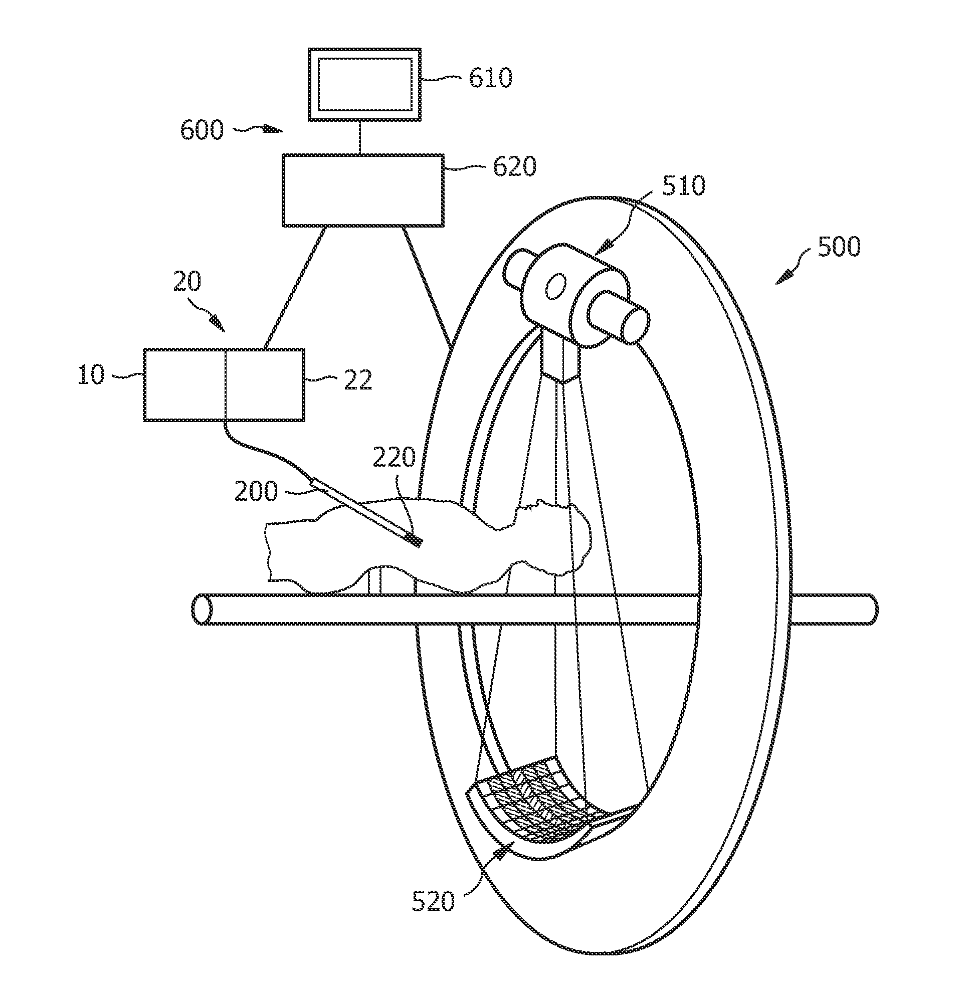
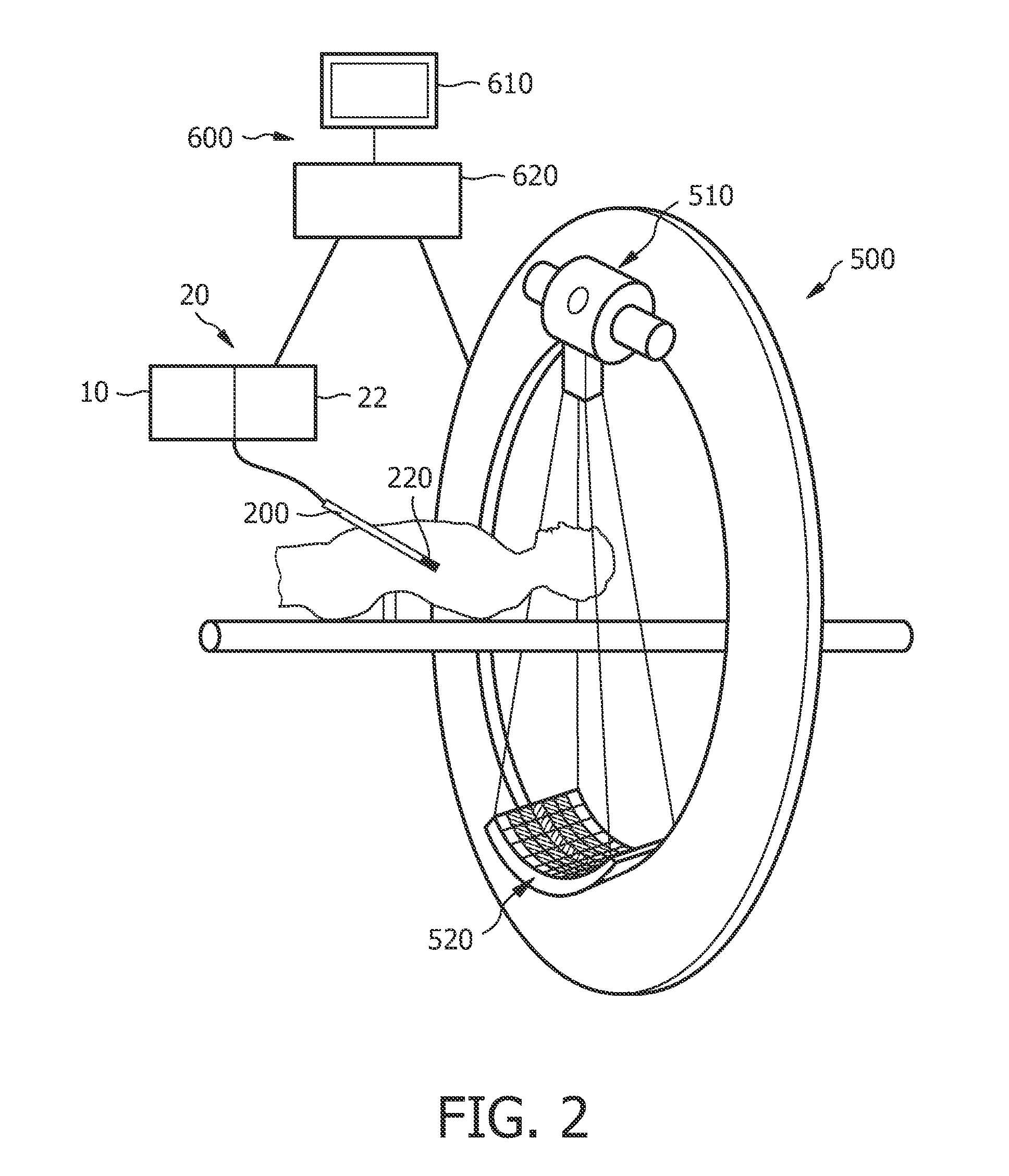
[0038]the system is able to interactively follow the biopsy needle from the incision to the target point by superimposing 2D fluro-image on 3D tissue reconstruction and provide molecular tissue information at every point along the needle trajectory that is registered to the position inside the body of the patient

[0039]the region along the needle trajectory can be scanned (scan forward and scan aside) in order to provide indications on lesion existence at the molecular level

[0040]preferably in reconstructing what tissue is in front of the needle the X-ray data and the position information of the needle is actively used in the optical reconstruction of what tissue is in front of the needle

third embodiment

[0041]tumor boundaries deduced from needle scanning and from the X-ray are compared. The X-ray information gives an estimate of the shape of the tumor, but the exact boundary cannot be determined. The needle gives detailed information of the tumor boundary but this information is only obtained along the needle trajectory. By combining the X-ray shape of the tumor with the one dimensional information of the needle, a new estimate of the 3D tumor size can be calculated (see third embodiment). The newly deduced enlarged boundary will be a better estimate for the tumor boundary

[0042]X-ray and needle information is further coupled to MRI images of the same area (MR data sets can be registered with the data sets produced by the X-ray machine)

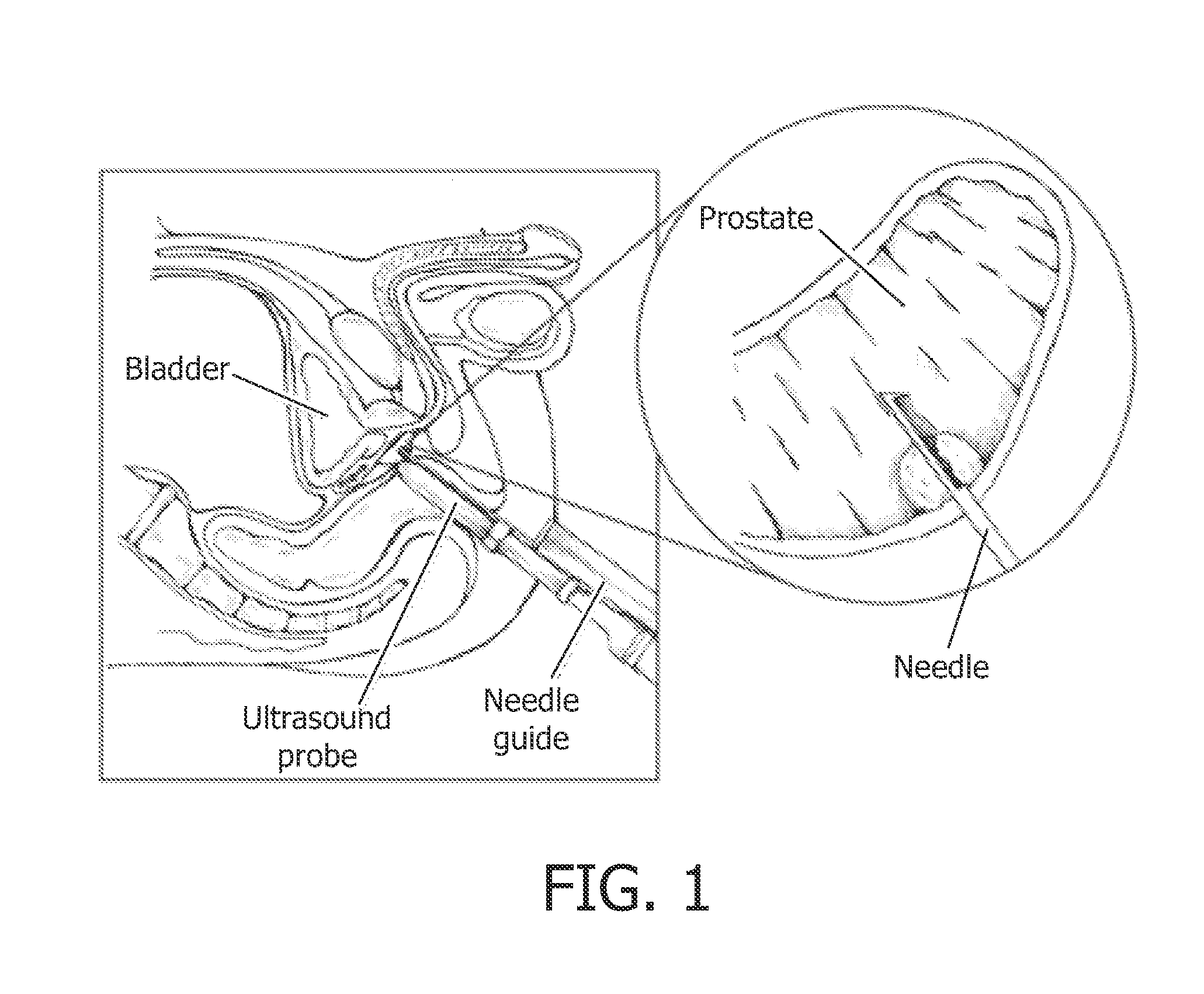
[0043]biopsy needle being equipped with an optical fiber is used to position the localization wire. The localization wire containing fixation means and may be equipped with a fiber.

[0044]To demonstrate the invention a needle intervention will be descr...


PUM


Abstract

A system for providing integrated guidance for positioning a needle in a body has two levels of guidance: a coarse guidance and a fine guidance. The system comprises a non-invasive imaging system for obtaining an image of the biopsy device in the body, for providing the coarse guidance. Furthermore, the system comprises an optical element mounted on the needle for obtaining optical information discriminating tissue in the body, for providing the fine guidance.

Description

FIELD OF THE INVENTION[0001]The present invention relates to a system for integrated guidance for positioning a biopsy device in a body, a biopsy device and a method of positioning the same. Particularly, the invention relates to a system for and a method of providing integrated guidance for positioning the biopsy device in a body.BACKGROUND OF THE INVENTION[0002]For correct diagnosis of various cancer diseases biopsies are taken. This can either be done via a lumen of an endoscope or via needle biopsies. An example of a needle biopsy is shown in FIG. 1, where a biopsy is taken from the prostate via the rectum. In order to find the correct position to take the biopsy, various imaging modalities are used such as X-ray, MRI or ultrasound. In case of prostate cancer in most cases the biopsy is guided by ultrasound (see FIG. 1). Although helpful, these methods of guidance are far from optimal.[0003]There are two major problems directly related to the biopsy:[0004]The resolution is limit...

Claims


Application Information

Patent Timeline
IPC IPC(8): A61M5/178G06K9/00
CPCA61B5/0066A61B5/6848A61B5/6852A61B6/12A61B6/5247A61B2019/5217A61B8/0841A61B8/5238A61B10/0275A61B17/3417A61B8/0833A61B2090/3614A61B6/032A61B8/5261A61B5/0035A61B5/055A61B5/4381
Inventor HENDRIKS, BERNARDUS HENDRIKUS WILHELMUSBABIC, DRAZENKO
Owner KONINKLIJKE PHILIPS ELECTRONICS NV
Why Eureka
Try Eureka