Method for detecting radiation, device thereof, and positron emission tomography scanner

a tomography and radiation detection technology, applied in tomography, x/gamma/cosmic radiation measurement, instruments, etc., can solve the problem of deviation of time determined by a difference in whether a signal rises abruptly or slowly, and no improvement in the signal-to-noise ratio

Inactive Publication Date: 2011-01-06
NAT INST FOR QUANTUM & RADIOLOGICAL SCI & TECH
View PDF7 Cites 17 Cited by
  • Summary
  • Abstract
  • Description
  • Claims
  • Application Information

AI Technical Summary

Benefits of technology

[0035]Still further, a radiation detector to be made a pair may be installed so as to be opposite behind the radiation source, thus making it possible to determine coincidence and time of flight.
[0036]In addition, radiation detectors may be arrayed in a ring shape so as to ...

Problems solved by technology

However, at the technical level at that time, a scintillator and a radiation detector used as a radiation detecting element as well as a circuit for processing electric signals from the radiation detector were insufficient in performance and others, and therefore no improvement was made in a signal-to-noise ratio.
Further, since some of the photons are absorbed by a boundary surface of the scintillator, a reflective material and others, all the photons do not necessarily arrive at a light receiver.
A third error parameter of the de...

Method used


Image

  • Method for detecting radiation, device thereof, and positron emission tomography scanner
  • Method for detecting radiation, device thereof, and positron emission tomography scanner
  • Method for detecting radiation, device thereof, and positron emission tomography scanner

Examples


embodiment 1

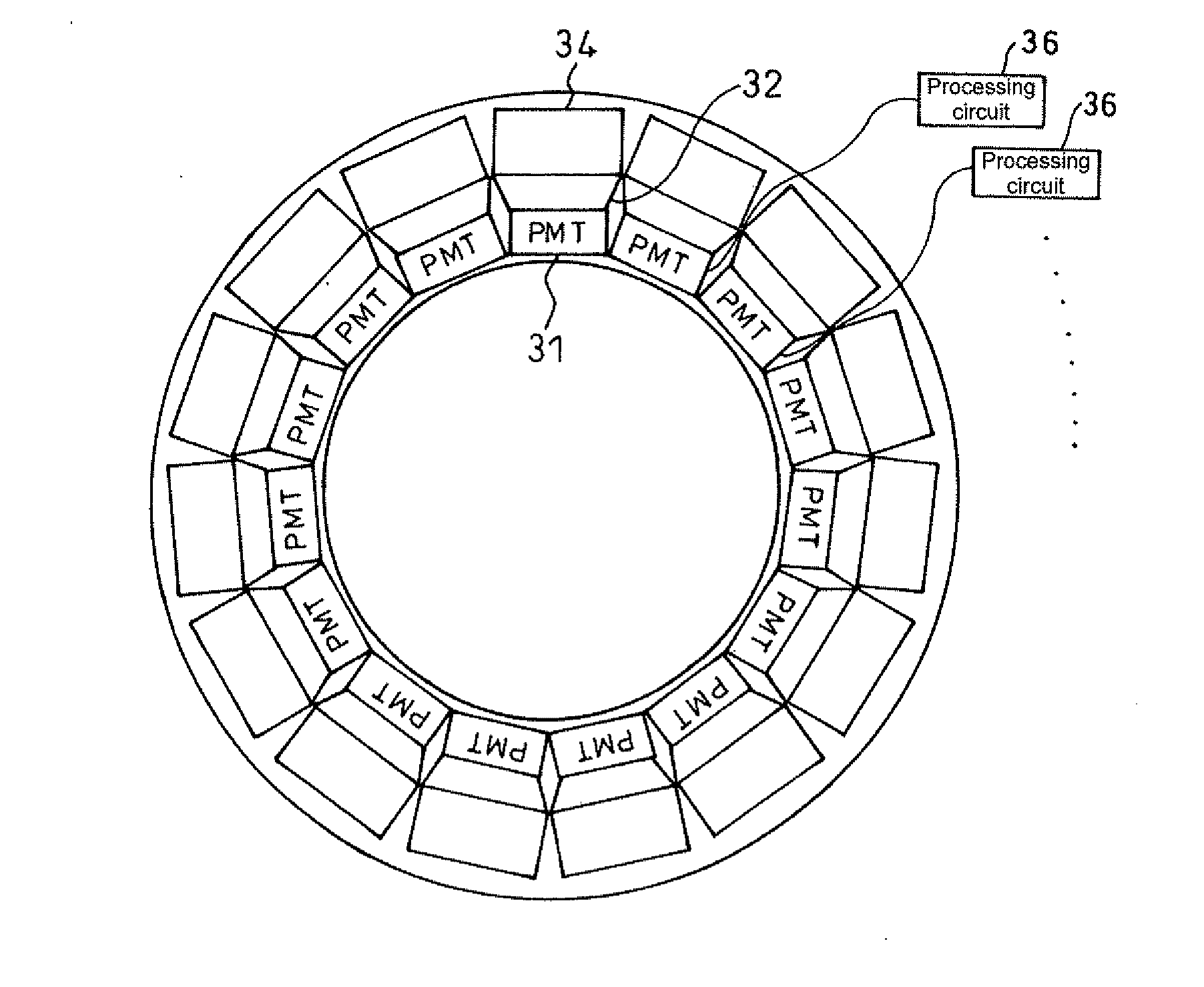
[0056]As shown in FIG. 14, Embodiment 1 of the present invention is a positron emission tomography scanner in which PMTs 31 are arrayed on the side of a patient, which is a radiation source, scintillators 34 are disposed via light guides 32 outside thereof, and processing circuits 36 at a back stage connected with each of the PMTs 31 are used to identify an incident position, incident time and energy of radiation made incident.

[0057]Thereby, it is possible to enhance time resolution and energy resolution.

[0058]In the present embodiment, the light guide 32 is installed and a diameter of the light-emitting light path on the side of the PMT 31 is narrowed down with respect to a diameter of the incident light path on the side of the scintillator 34, thus making it possible to arrange the scintillators 34 and the PMTs 31 without any clearance left.

embodiment 2

[0059]In addition, types of the radiation detector are not limited to a DOI detector. shown in FIG. 15, the scintillators 34 are not stacked in four layers but may be made into a thick one layer, two or three layers, or more than five layers, or the light guide 32 is omitted and the PMT 31 may be directly disposed on the side of a radiation source of the scintillator 34. For example, where the PMT 31 is sufficiently thin or where radiation detectors are not required to be arranged densely, no light guide is needed. Where the light guide is not needed, it is preferably omitted.

embodiment 3

[0060]Further, a combination of the scintillators 34 with the PMTs 31 is not limited to one stage. shown in FIG. 16, the scintillators 34 are changed in constitution for every stage and can be disposed in a multiple stage. Here, it is acceptable that a relationship between the scintillators 34 and the PMTs 31 is on a one to one basis, on a many to one basis, on a one to many basis and on a many to many basis.


PUM


Abstract

A light receiver for detecting incident time is installed on the side of a radiation source of a scintillator (including a Cherenkov radiation emitter), and information (energy, incident time, an incident position, etc.) on radiation made incident into the scintillator is obtained by the output of the light receiver. It is, thereby, possible to identify an incident position and others of radiation into the scintillator at high accuracy.

Description

TECHNICAL FIELD[0001]The present invention relates to a method for detecting radiation, the device thereof, and a positron emission tomography scanner. The present invention relates in particular to a method for detecting radiation which is capable of obtaining information (energy, incident time, an incident position, etc.) on radiation made incident into a scintillator (including a Cherenkov radiation emitter) at high accuracy, the device thereof and a positron emission tomography scanner which utilizes the device.BACKGROUND ART[0002]The positron emission tomography scanner (PET) is a nuclear-medicine imaging equipment utilizing a positron emission nuclide, for use in various applications such as diagnosis of cancers and molecular imaging.[0003]The positron emission nuclide is an isotope such as 18F which is unstable due to an excessively large number of protons in an atomic nucleus as compared with a number of neutrons, having the radioactivity of emitting positrons and neutrinos ...

Claims


Application Information

Patent Timeline
IPC IPC(8): G01T1/20G01T1/161
CPCA61B6/037G01T1/2985
Inventor SHIBUYA, KENGOYAMAYA, TAIGAINADAMA, NAOKONISHIKIDO, FUMIHIKOYOSHIDA, EIJIMURAYAMA, HIDEO
Owner NAT INST FOR QUANTUM & RADIOLOGICAL SCI & TECH
Why Eureka
Try Eureka