Labeled protein and method for obtaining the same
- Summary
- Abstract
- Description
- Claims
- Application Information
AI Technical Summary
Benefits of technology
Problems solved by technology
Method used
Image
Examples
example 1
Preparation of Antibody hu4D5-8 scFv Containing an Affinity Interaction Domain and a Labeling site
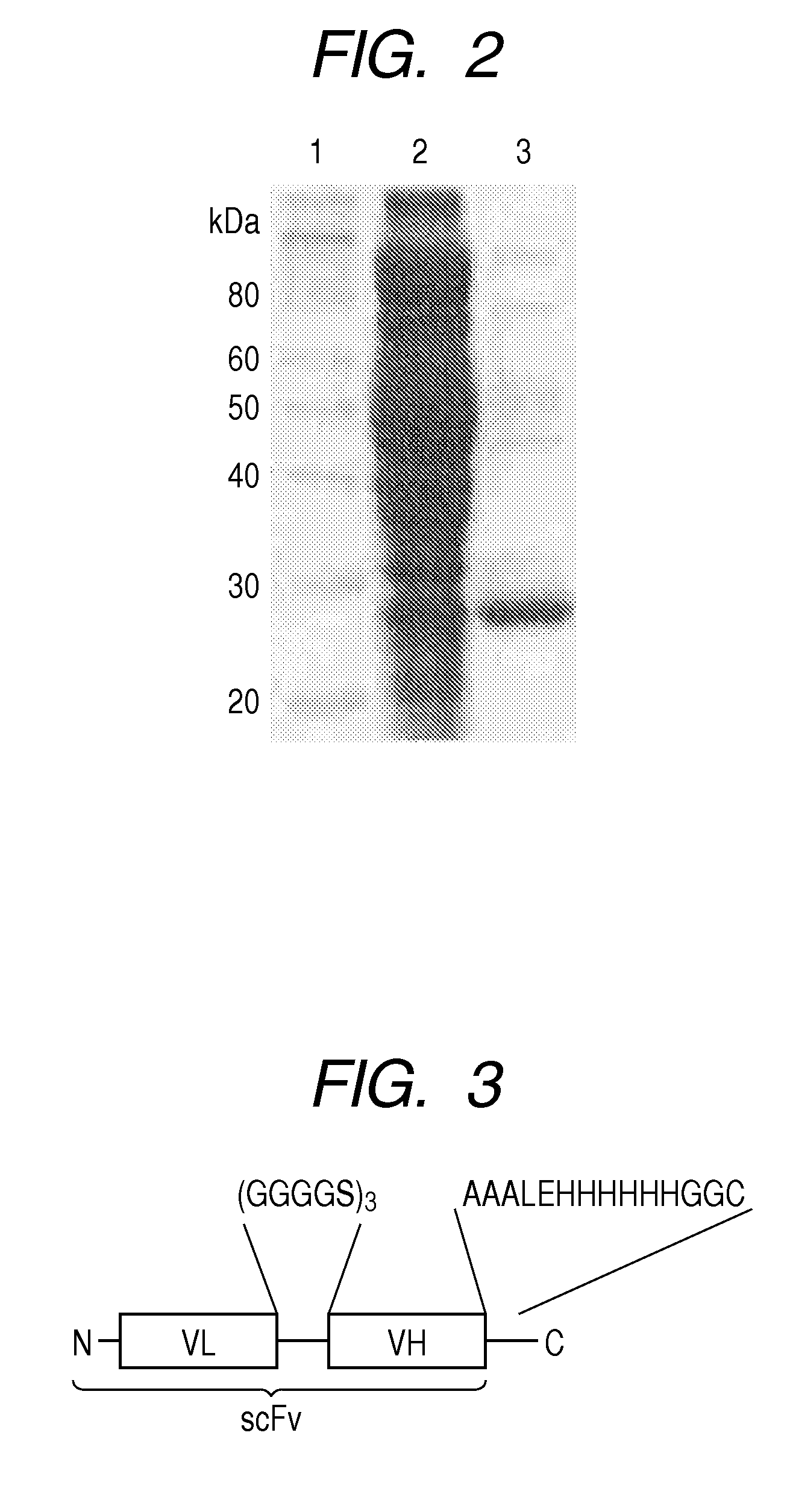
[0075]A gene hu4D5-8 scFv encoding a single-chain antibody (scFv) was prepared based on the gene sequence (hu4D5-8) of the variable region of IgG where HER2 was supposed to bind. First, a cDNA in which VL and VH genes of hu4D5-8 were connected by a cDNA encoding peptide (GGGGS)3 was prepared. The restriction enzyme NcoI recognition site and the restriction enzyme NotI recognition site were introduced at the 5′ terminus and 3′ terminus, respectively. The nucleotide sequence will be shown as follows (the part of the restriction enzyme recognition site is underlined):
(SEQ ID NO: 18)5′-CCATGGATATCCAGATGACCCAGTCCCCGAGCTCCCTGTCCGCCTCTGTGGGCGATAGGGTCACCATCACCTGCCGTGCCAGTCAGGATGTGAATACTGCTGTAGCCTGGTATCAACAGAAACCAGGAAAAGCTCCGAAACTACTGATTTACTCGGCATCCTTCCTCTACTCTGGAGTCCCTTCTCGCTTCTCTGGATCCAGATCTGGGACGGATTTCACTCTGACCATCAGCAGTCTGCAGCCGGAAGACTTCGCAACTTATTACTGTCAGCAACATTATACTACTCCTCCCACGTTCGGACAGGGTAC...
example 2
Labeling with Antibody Dye
[0080]The antibody prepared above was reduced and treated with 20 fold molar amount of tris(2-carboxyethyl)phosphine hydrochloride (TCEP) at 25° C. for 2 hours after buffer replacement with a phosphate buffer containing 5 mM EDTA (2.68 mM KCl / 137 mM NaCl / 1.47 mM KH2PO4 / 1 mM Na2HPO4 / 5 mM EDTA, pH 7.4). The resultant antibody was reacted with 10 fold molar amount of fluorescent dye, Alexa Fluor (registered trademark, distributed by Invitrogen Life Technologies) 750-maleimide at 25° C. for 2 to 4 hours. After 1 hour reaction, unreacted Alexa Fluor (registered trademark, distributed by Invitrogen Life Technologies) 750-maleimide was removed by gel filtration chromatography with Superdex 200 GL 10 / 300 column (manufactured by GE Healthcare UK Ltd.) to obtain a labeled antibody (hereinafter, referred to as a dye-labeled antibody). The labeling index (molar ratio) of the dye to the antibody was 0.4 to 0.6 according to absorbance determination.
example 3
Labeling of an Antibody with Peg
[0081]The antibody prepared above was reduced and treated with 20 fold molar amount of tris(2-carboxyethyl)phosphine hydrochloride (TCEP) at 25° C. for 2 to 4 hours after buffer replacement with a phosphate buffer containing 5 mM EDTA (2.68 mM KCl / 137 mM NaCl / 1.47 mM KH2PO4 / 1 mM Na2HPO4 / 5 mM EDTA, pH 7.4). The resultant antibody was reacted with 10 fold molar amount of 20 kDa PEG-maleimide (distributed by NOF CORPORATION) at 25° C. for 2 to 4 hours. After the reaction, unreacted 20 kDa PEG-maleimide was removed by gel filtration chromatography with Superdex 200 GL 10 / 300 column (manufactured by GE Healthcare UK Ltd.) to obtain a pegylated antibody. Preparation of the Pegylated Antibody can be Confirmed by comparison with the molecular weight of the antibody before pegylation by SDS-PAGE. FIG. 4 illustrates the reducing SDS-PAGE results of the pegylated antibody prepared in the present example and the antibody before pegylation.
PUM
| Property | Measurement | Unit |
|---|---|---|
| Fraction | aaaaa | aaaaa |
| Fluorescence | aaaaa | aaaaa |
| Affinity | aaaaa | aaaaa |
Abstract
Description
Claims
Application Information
 - R&D
- Intellectual Property
- Life Sciences
- Materials
- Tech Scout
- Unparalleled Data Quality
- Higher Quality Content
- 60% Fewer Hallucinations
Browse by: Latest US Patents, China's latest patents, Technical Efficacy Thesaurus, Application Domain, Technology Topic, Popular Technical Reports.
© 2025 PatSnap. All rights reserved.Legal|Privacy policy|Modern Slavery Act Transparency Statement|Sitemap|About US| Contact US: help@patsnap.com


