Ultra-small chitosan nanoparticles useful as bioimaging agents and methods of making same

a technology of nanoparticles and chitosan, applied in the field of biomedical imaging, can solve the problems of insufficient accuracy of image-fusion software, obvious artifacts in such processed data, and millions of american deaths a year

Inactive Publication Date: 2011-01-27
UNIV OF CENT FLORIDA RES FOUND INC
View PDF3 Cites 14 Cited by
  • Summary
  • Abstract
  • Description
  • Claims
  • Application Information

AI Technical Summary

Problems solved by technology

In spite of these advancements in diagnostic imaging capabilities, especially with the modalities of ultrasound, nuclear medicine, nuclear magnetic resonance and spectroscopy, X-ray/CT, optical, endoscopic, and visualization strategies, the hard reality is that millions of Americans die in the United States each year due to poor and late di

Method used


Image

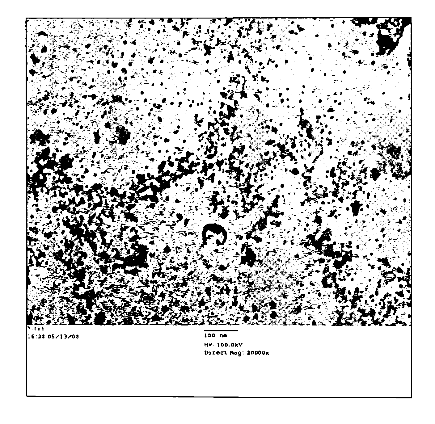
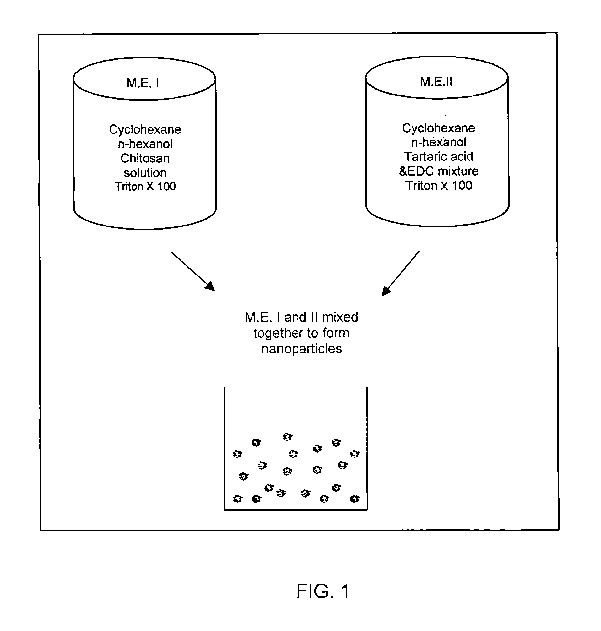
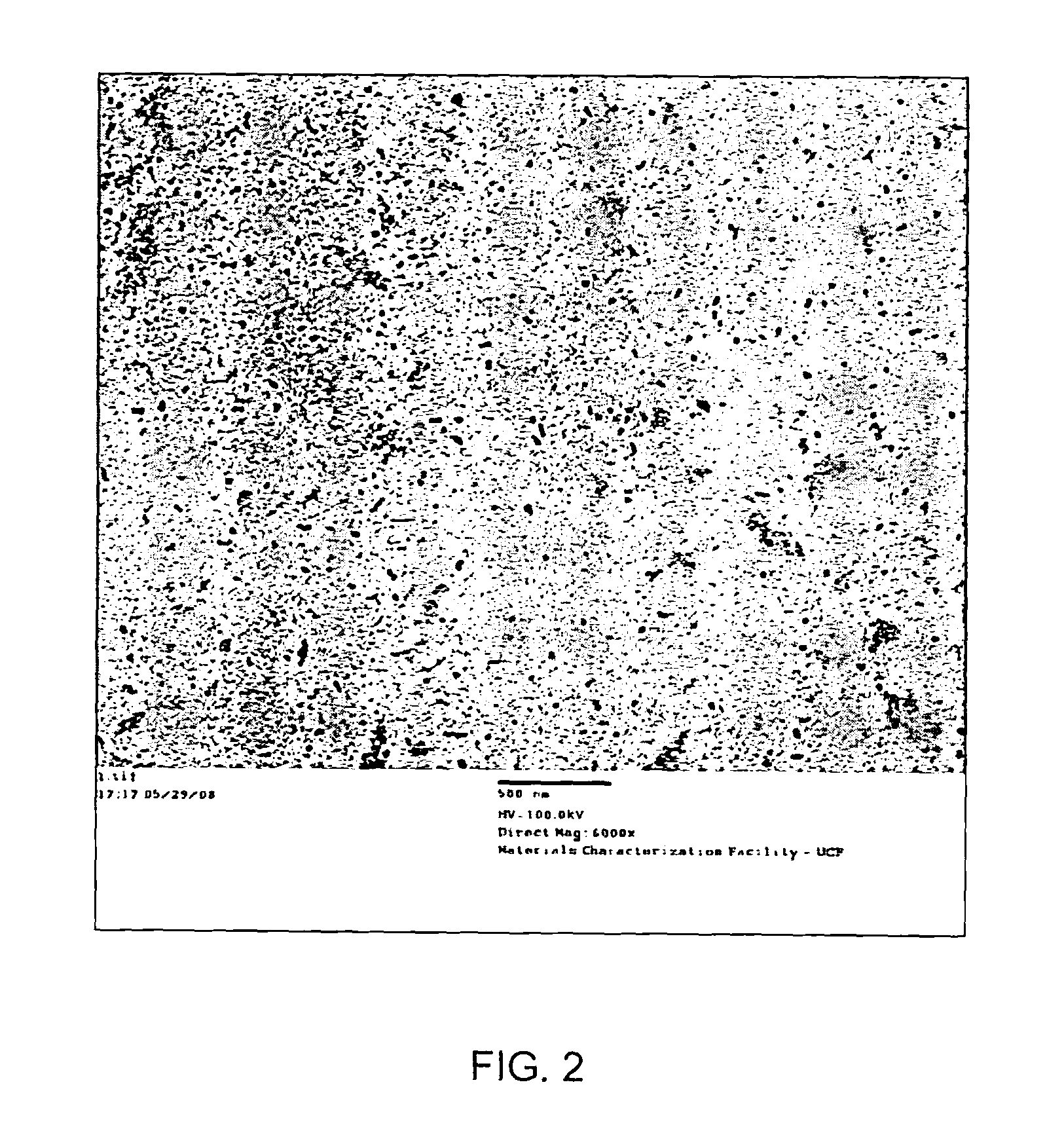
  • Ultra-small chitosan nanoparticles useful as bioimaging agents and methods of making same
  • Ultra-small chitosan nanoparticles useful as bioimaging agents and methods of making same
  • Ultra-small chitosan nanoparticles useful as bioimaging agents and methods of making same

Examples


Embodiment Construction

The present invention will now be described more fully hereinafter with reference to the accompanying drawings, in which preferred embodiments of the invention are shown. Unless otherwise defined, all technical and scientific terms used herein have the same meaning as commonly understood by one of ordinary skill in the art to which this invention pertains. Although methods and materials similar or equivalent to those described herein can be used in the practice or testing of the present invention, suitable methods and materials are described below. Any publications, patent applications, patents, or other references mentioned herein are incorporated by reference in their entirety. In case of conflict, the present specification, including any definitions, will control. In addition, the materials, methods and examples given are illustrative in nature only and not intended to be limiting. Accordingly, this invention may, however, be embodied in many different forms and should not be con...


PUM

PropertyMeasurementUnit
Timeaaaaaaaaaa
Nanoscale particle sizeaaaaaaaaaa
Zeta potentialaaaaaaaaaa

Abstract

A method of making ultra-small chitosan nanoparticles having a size range of approximately 10-20 nm, includes preparing a first microemulsion containing effective amounts of cyclohexane, n-hexanol, chitosan polymer and a nonionic surfactant. A second microemulsion is prepared containing effective amounts of cyclohexane, n-hexanol, tartaric acid, EDC, n-hydroxysuccinimide, and a nonionic surfactant. The method continues by reacting the first and second microemulsions for a time sufficient to form the ultra-small chitosan nanoparticles and recovering the nanoparticles from the reacted microemulsion. The chitosan polymer may be crosslinked and may also be tagged with a fluorescent compound, a radio-opaque compound, a paramagnetic ion, a ligand specific for a predetermined biologic target, a drug, and combinations thereof.

Description

FIELD OF THE INVENTIONThe present invention relates to the field of biomedical imaging and, more particularly, to ultra-small nanoparticles of chitosan useful as bioimaging agents and to methods for making such nanoparticles.BACKGROUND OF THE INVENTIONThe rapidly growing field of biomedical imaging has made substantial contribution in today's healthcare system. This has enabled many advanced diagnostic procedures that are based upon visualization of physiological structures, measurement of biological functions, and evaluation of cellular and molecular events without requiring invasive procedures. In spite of these advancements in diagnostic imaging capabilities, especially with the modalities of ultrasound, nuclear medicine, nuclear magnetic resonance and spectroscopy, X-ray / CT, optical, endoscopic, and visualization strategies, the hard reality is that millions of Americans die in the United States each year due to poor and late diagnosis. This urgently demands a revolutionary brea...

Claims


Application Information

Patent Timeline
IPC IPC(8): C07K2/00C08B37/08C07K16/00
CPCA61K9/5161A61K47/4823A61K49/0002A61K49/0054A61K49/0093B82Y5/00A61K49/0485A61K49/085A61K49/126A61K49/1824A61K49/0442A61K47/61
Inventor SANTRA, SWADESHMUKULTALLURY, PADMAVATHY
Owner UNIV OF CENT FLORIDA RES FOUND INC
Why Eureka
Try Eureka