

Differential enzymatic fragmentation by whole genome amplification
a technology of enzymatic fragmentation and whole genome, which is applied in the field of differential enzymatic fragmentation by whole genome amplification, can solve the problems of not alleviating the caveats associated with the use of methylation-sensitive enzymes, not measuring the dna methylation of sequences in much of the genome, and unavoidable caveats
- Summary
- Abstract
- Description
- Claims
- Application Information
AI Technical Summary
Benefits of technology
Problems solved by technology
Method used
Image

Examples
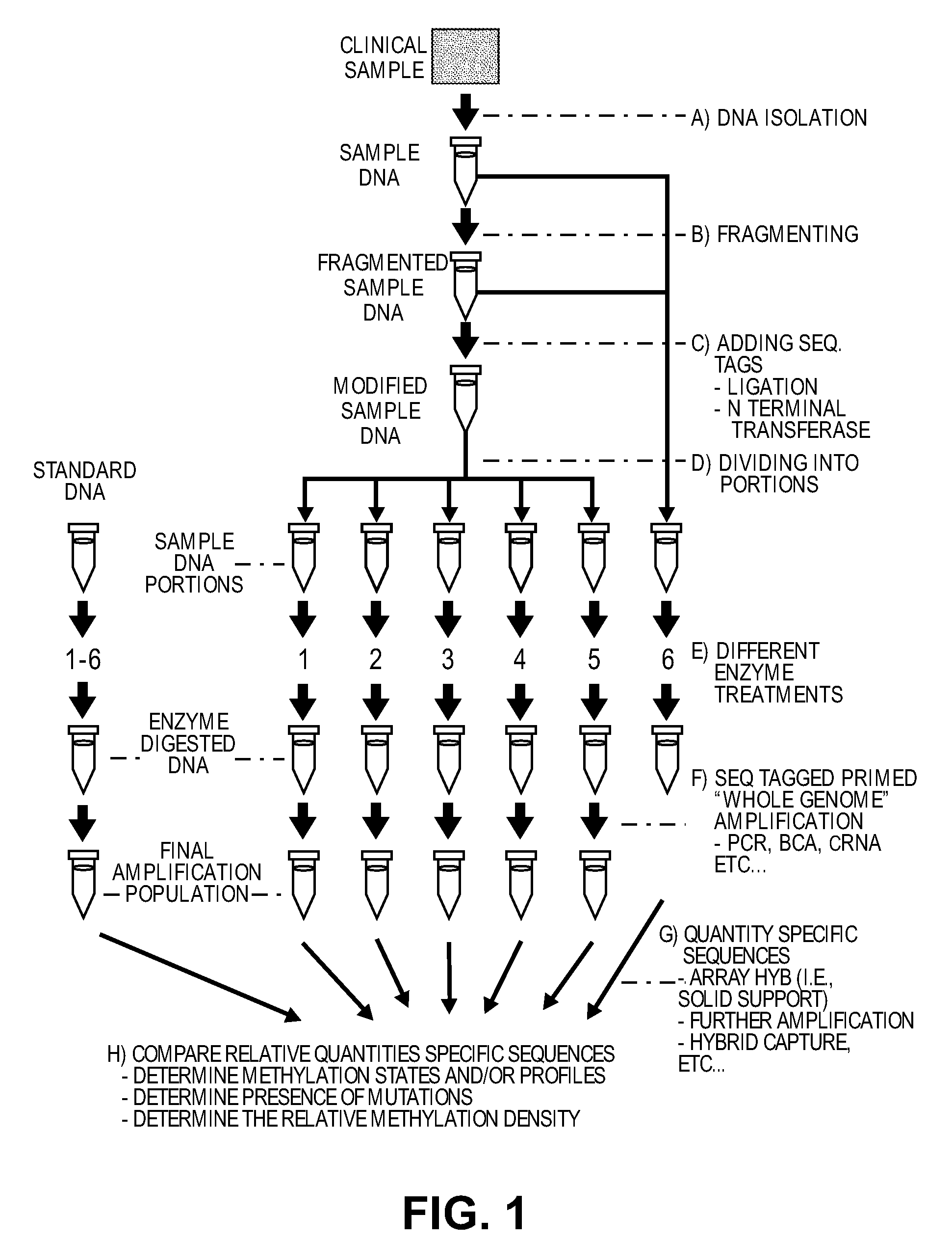
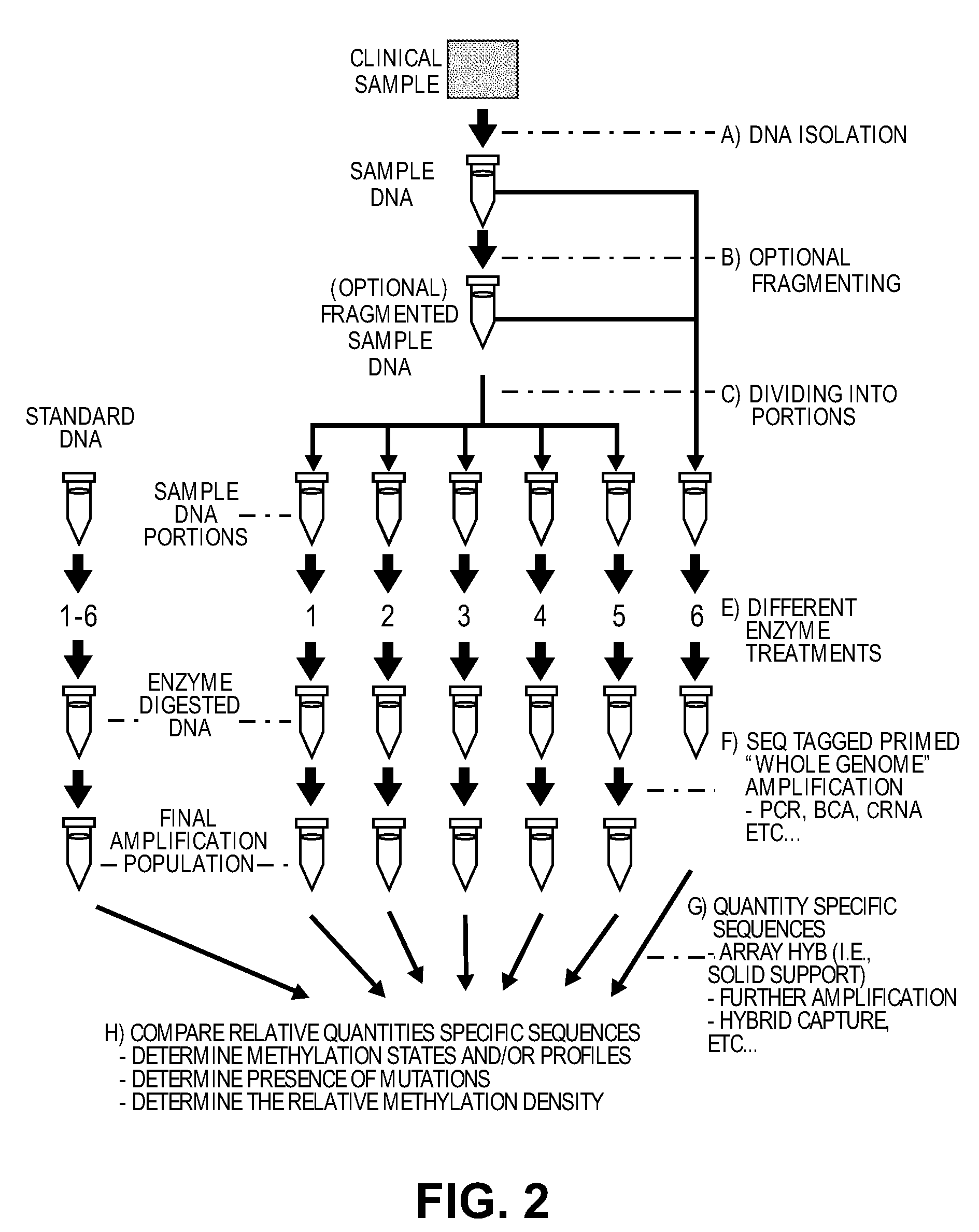
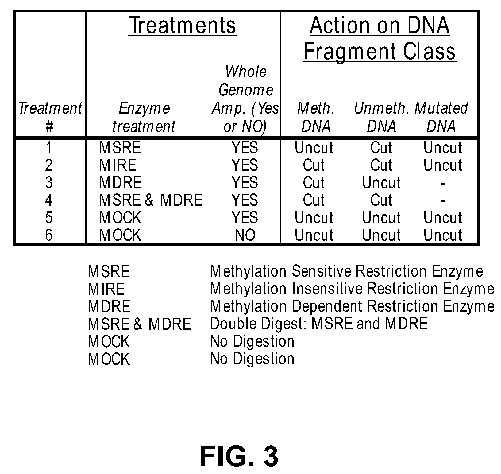
example 1
Generating and Analyzing Six Portions from a First Sample
DNA is obtained from a glioma derived human tumor cell line (ATCC catalogue #2610). 10 ug of the DNA is sheared in a 300 ul volume of water using GeneMachine's hydroshear for 20 cycles at speed code setting 5. An aliquot of sheared DNA is loaded onto an agarose gel and the mode of the ethidium staining is determined to be between 500 and 2,000 bp. The DNA is concentrated on a speed vac to an appropriate concentration just below 1 ug / ul as determined by a spectrophotometer using absorbance readings at 260 and 280 nm.
40 ul are placed into two microcentrifuge tubes. One tube is carried forward and the remainder is frozen at −20 deg C. and would serve as the unamplified control later. 40 ul of the DNA undergoes end-repair using the End-it kit from Epicentre (Madison Wis.) according to the manufacturer's instructions, and wiss cleaned using MinElute Reaction Cleanup Kit (Qiagen #28206).
Two oligos, A and B, are synthesized with the ...
example 2
Utilizing Exonucleases to Remove Digested DNA Products and Lower Background Signal in the Quantifying Step
In this example, a cocktail of exonucleases is used to degrade digested DNA fragments, leaving the undigested DNA fragments intact and available for participation in the amplification reaction.
The procedure set forth in Example 1 is performed on DNA from the same sample. However, prior to the digestion step, the sample is dephosphorylated using shrimp alkaline phosphatase under the conditions specified by USB, in order to protect the ends of the molecules from attack by lambda exonuclease. The phosphatase is then heat inactivated. Note that synthetic polynucleotides that are used as adaptors can offer protection against lambda nuclease attack. Note also that Exonuclease 111 can be used but in this case an overhang of 4 or more nucleotides from 3′ end of DNA created by certain restriction enzyme, by terminal transferase or by synthetic adaptors will work as protection against Exo...
example 3
Comparison of Portions from a First Sample to Portions from a Second Sample
The procedure set forth in Example 1 is repeated on a second sample (Male blood genomic DNA purchased from Novagen, Madison Wis.). Specific sequences are quantified from the six array files of the second sample and are compared to the same specific sequences quantified from six array files generated from the first sample.
PUM
| Property | Measurement | Unit |
|---|---|---|
| average fragment length | aaaaa | aaaaa |
| density | aaaaa | aaaaa |
| fragment length | aaaaa | aaaaa |
Abstract
Description
Claims
Application Information