Biomarkers for Head-And-Neck Cancers and Precancers

a biomarker and headandneck cancer technology, applied in biochemistry apparatus and processes, biocide, pharmaceutical non-active ingredients, etc., can solve the problems of no clinically established biomarkers to facilitate diagnosis or prognosis, and inaccurate cancer risk prediction

Active Publication Date: 2011-05-05
WALFISH PAUL +1
View PDF3 Cites 23 Cited by
  • Summary
  • Abstract
  • Description
  • Claims
  • Application Information

AI Technical Summary

Benefits of technology

[0013]The protein expression profiles of HNSCCs and OPLs were compared with non-cancerous head-and-neck tissues (controls) using the iTRAQ-labelling technique in combination with multidimensional LC-MS / MS analysis. In the iTRAQ technology, primary amines are tagged, thereby potentially allowing the tagging of most tryptic peptides. The multiplexing ability afforded by the iTRAQ reagents, which are available in four different tags, was ideally suited for the present study, as it provided a means to perform a proteomic analysis of both paired and non-paired non-cancerous (histologically normal) head-and-neck tissues, while simultaneously comparing them against the cancer samples. This strategy helps to identify proteins that might be differentially expressed due to manifestation of field cancerization (25-27) in clinically normal mucosa, and may be useful in designing strategies for risk prediction of disease recurrence or second primary tumor development.
[0014]Some of the overexpressed proteins that were identified in the tissues by the iTRAQ technology and LC MS / MS analysis were confirmed by immunohistochemistry and Western blotting. These approaches ensured that the selected proteins demonstrated a consistent pattern of overexpression in HNSCCs and OPLs, and greatly increased confidence in the observations stemming from iTRAQ analysis. Apart from their potential utility as biomarkers for HNSCC and OPLs, these proteins also provide valuable insight into the previously unknown molecular networks and mechanisms that govern the normal-to-malignant conversion of epithelium.
[0098]Thus, the present invention relates to a method for diagnosing and monitoring a head-and-neck disease (e.g., OPL or head-and-neck cancer, HNSCC or related condition) in a sample from a subject including isolating nucleic acids, preferably mRNA, from the sample; and detecting OPL or HNSCC marker polynucleotides associated with the disease in the sample. The presence of different levels of OPL or HNSCC marker polynucleotides in the sample compared to a standard or control may be indicative of head-and-neck disease, disease stage, and / or a negative or positive prognosis (e.g., longer progression-free and overall survival).

Problems solved by technology

The presence of dysplastic areas in the oral epithelium is associated with a likely progression to cancer; however, it is not an accurate predictor of cancer risk (6, 7).
The major challenge in oral tumorigenesis is the identification of proteins that may serve as markers to differentiate the high-risk leukoplakic lesions from more benign lesions for early intervention to reduce the morbidity associated with this devastating disease.
Currently, there are no clinically-established biomarkers to facilitate the diagnosis or prognosis of head-and-neck cancer and oral leukoplakia.

Method used


Image

  • Biomarkers for Head-And-Neck Cancers and Precancers
  • Biomarkers for Head-And-Neck Cancers and Precancers
  • Biomarkers for Head-And-Neck Cancers and Precancers

Examples


example 1

Samples and Reagents

[0441]Head-and-neck cancer and oral leukoplakia tissues were retrieved from an in-house, dedicated, research head-and-neck tissue bank, with approval from the Human Ethics Committee of All India Institute of Medical Sciences, New Delhi, India. With patient consent, biopsies / excised tissue specimens of oral leukoplakia and surgically resected specimens of HNSCCs, and paired non-cancerous tissues (each taken from a distant site) were collected and banked from patients undergoing treatment at the Department of Otorhinolaryngology, All India Institute of Medical Sciences. Normal tissues with no evidence of cancer (non-paired noncancerous controls) were collected from patients attending the Dental Outpatient Department of All India Institute of Medical Sciences for tooth extraction, after consent of the patients. After excision, tissues were flash-frozen in liquid nitrogen within 20 min of devitalization and stored at −80° C. until further use; one tissue piece was co...

example 2

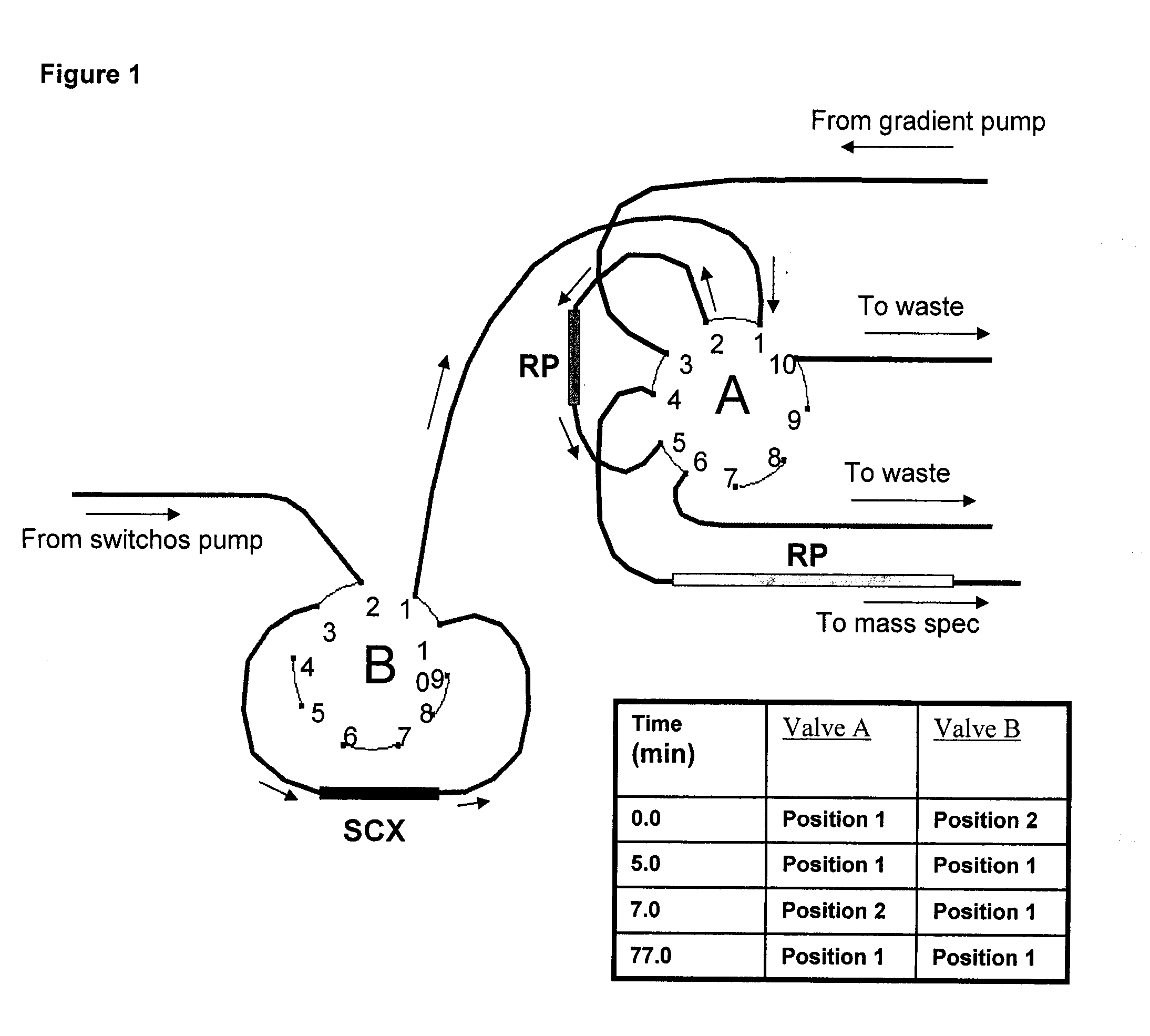
Strong Cation Exchange (SCX) Separation Conditions

[0445]For the offline 2D LC-MS / MS analysis, each set of labeled samples was first separated by SCX fractionation using an HP1050 high-performance liquid chromatograph (Agilent, Palo Alto, Calif., U.S.) with a 2.1-mm internal diameter (ID)×100-mm length polysulfoethyl A column packed with 5 μm beads with 300 Å pores (The Nest Group, Southborough, Mass.) as described previously (21). A 2.1-mm ID×10-mm length guard column of the same material was fitted immediately upstream of the analytical column. Separation was performed as previously described (21). Briefly, each pooled sample set was diluted with the loading buffer (15 mM KH2PO4 in 25% acetonitrile, pH 3.0) to a total volume of 2 ml and the pH adjusted to 3.0 with phosphoric acid. Samples were then filtered using a 0.45-μm syringe filter (Millipore, Cambridge, ON, Canada) before loading onto the column. Separation was performed using a linear binary gradient over one hour. Buffer A...

example 3

LC-MS / MS Run Conditions

[0447]The SCX fractions from 6 to 30 were analyzed by nanoLC-MS / MS using the LC Packings Ultimate instrument (Amsterdam, The Netherlands) fitted with a 10-μl sample loop. Samples were loaded, using a μl pick-up mode, onto a 5-mm RP C18 pre-column (LC Packings) at 50 μl / min and washed for 4 min before switching the precolumn inline with the separation column. The separation column used was either a 75-μm ID×150-mm length PepMap RP column from LC Packings packed with 3 μm C18 beads with 100 Å pores, or an in-house equivalent packed with similar beads (Kromasil; The Nest Group, Southborough, Mass.). The flow rate used for separation on the RP column was 200 nl / min with the following gradient:

Time (min)01015125145150160162188% B5515356080805Stop

[0448]Samples were analyzed on a QSTAR Pulsar i mass spectrometer (Applied Biosystems / MDS SCIEX, Foster City, Calif.) in information-dependent acquisition (IDA) mode with the scan cycles set up to perform a 1-s MS scan foll...


PUM

PropertyMeasurementUnit
median timeaaaaaaaaaa

Abstract

The invention provides markers and methods for detecting head-and-neck precancers, (including OPLs), cancers and related disease conditions in a subject. The invention also provides localization and imaging methods for head-and-neck precancers (including OPLs) and cancers, along with kits for carrying out methods of the invention. The invention further provides therapeutic applications for head-and-neck precancers (including OPLs) and cancers which employ head-and-neck precancer and cancer markers, polynucleotides encoding the markers, and binding agents for the markers.

Description

RELATED APPLICATIONS[0001]This application claims the benefit of Canadian Patent Application No. 2,618,163, filed Feb. 7, 2008 and Canadian Patent Application No. 2,643,611, filed Nov. 11, 2008.FIELD OF THE INVENTION[0002]The invention relates to markers for head-and-neck cancers, including oral cancers.[0003]The invention also relates to markers for head-and-neck precancers—including, but not limited to, oral leukoplakia with hyperplasia or dysplasia. The invention further relates to methods for assessing the status of head-and-neck tissue and oral tissue, and methods for the detection, diagnosis, prediction, and therapy of head-and-neck disease. In one aspect, the invention relates to biomarkers of head-and-neck squamous cell carcinoma and biomarkers of head-and-neck precancers (including, but not limited to, oral leukoplakia with hyperplasia or dysplasia), and methods for detecting, diagnosing, predicting, and treating these and related conditions. In a further aspect, the invent...

Claims


Application Information

Patent Timeline
Patent Type & Authority Applications(United States)
IPC IPC(8): A61K49/00C12Q1/68G01N33/68A61P35/00A61K47/32A61K47/10
CPCC12Q1/6886G01N33/57484C12Q2600/158G01N33/57407A61P35/00
Inventor SIU, K. W. MICHAELRALHAN, RANJUMATTA, AJAYDESOUZA, LEROI V.
Owner WALFISH PAUL
Why Eureka
Try Eureka