Compositions and Methods for Spatial Separation and Screening of Cells
a cell and spatial separation technology, applied in the field of cell spatial separation and cell screening, can solve the problems of difficult evolution of enzymes that do not provide a selectable phenotype, current methods do not provide a facile or generalized strategy for engineering diverse enzymes, and achieve rapid and large-scale production of glycoconjugates, and improve substrate selectivity
- Summary
- Abstract
- Description
- Claims
- Application Information
AI Technical Summary
Benefits of technology
Problems solved by technology
Method used
Image
Examples
example 1
The Development of a New Technique for Screening a Library of Mutant Enzymes for Improved Catalytic Activity or Altered Substrate Specificity
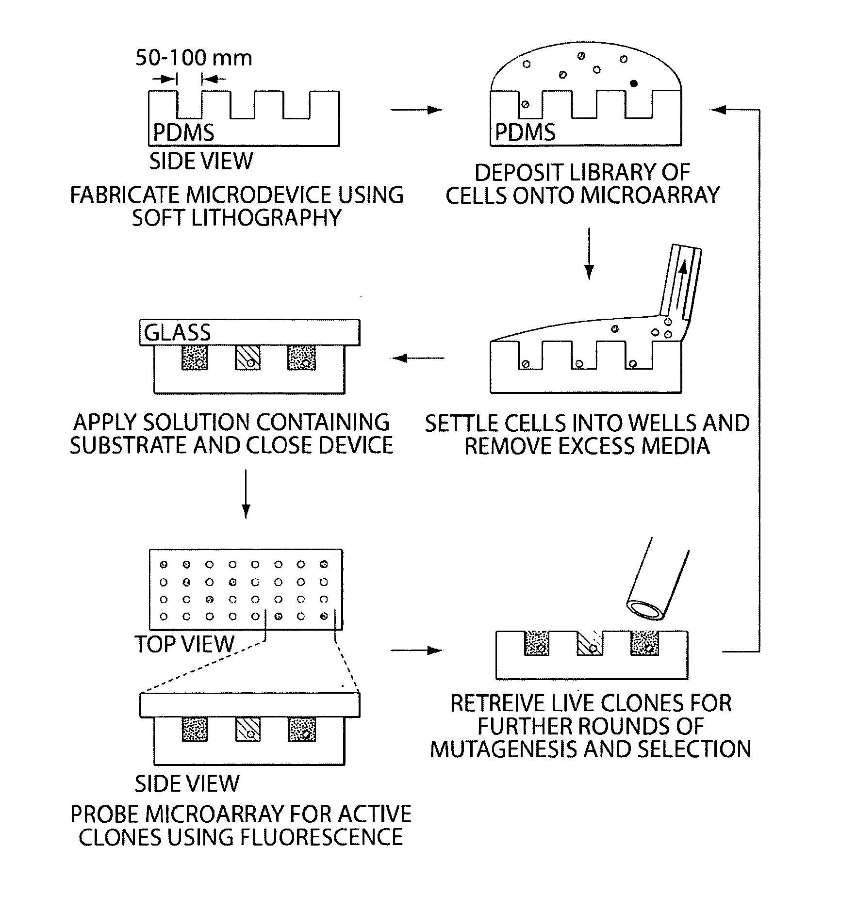
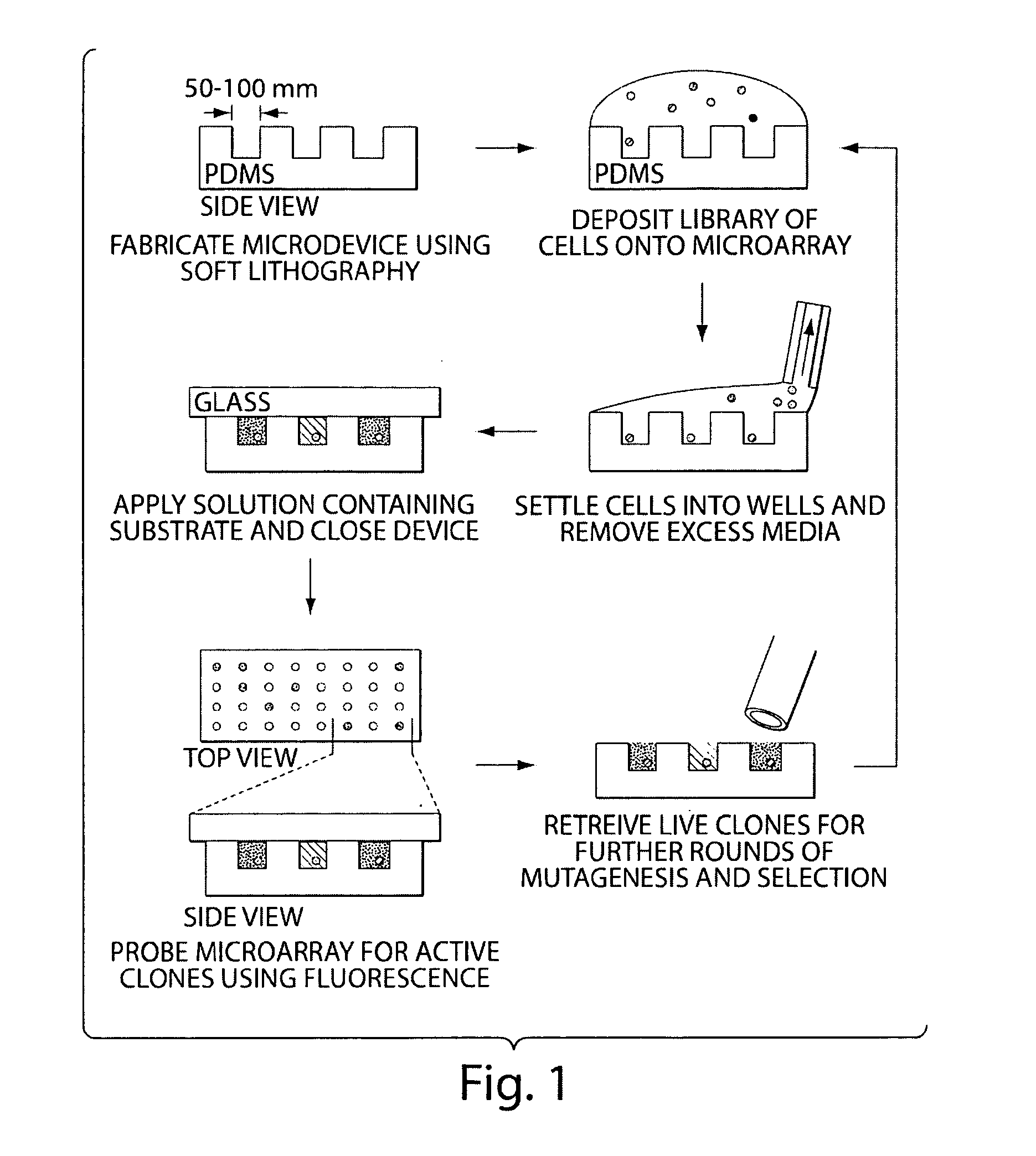
[0065]The following experiment consists of (1) illustration of a technique for the spatial separation of a library of yeast cells secreting an enzyme of interest, and (2) enrichment of cells expressing an active protease from an inactive variant to determine the sensitivity of the technique. Briefly, a library of yeast cells capable of secreting a protein of interest is loaded into microwells 50 microns in diameter so that each well contains, on average, one library member. Each compartment in the device is interrogated in parallel with enzyme substrates; successful enzyme turnover yields a fluorescence signal. Feasibility of the technique is demonstrated with a protease.
[0066]Microfabricated arrays of wells have been used for diverse biological applications. Microwells have proven useful to study enzymology at the single molecule level, and we...
example 2
The Evolution of a Mutant GTase with Improved Catalytic Activity
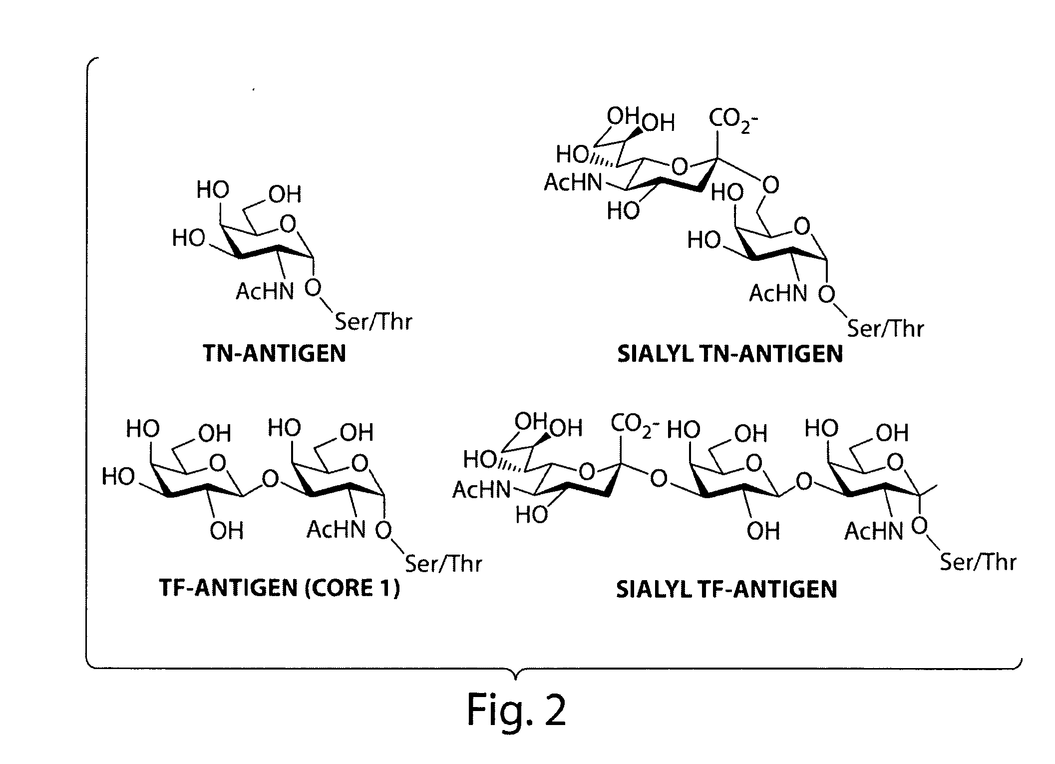
[0071]The following experiment consists of evolution of ppGalNAcTase mutants with increased catalytic efficiency and altered substrate specificity. Microdevices are used to screen for mutants of ppGalNAcTase-T1 having improved catalytic efficiency. ppGaINAcTase-T1 is responsible for the transfer of alpha-GalNAc to Ser / Thr residues to form the Tn-antigen—a tumor-associated carbohydrate epitope. Mutants identified in this screen are used for the in vitro synthesis of the Tn-antigen.
[0072]A recent crystal structure of murine ppGaINAcTase-T1 shows that this protein folds to form distinct catalytic and lectin domains (Fritz et al., 2004 Proc Natl Acad Sci, 101(43):15307-15312). Error-prone PCR is used to create random libraries of ppGalNAcTase-T1 mutagenized within the catalytic domain. A library of transformants is spatially segregated as previously described and screened using fluorescent substrates (FIG. 4).
Design and Syn...
example 3
Enzyme Turnover in Microwells—Trypsin Cleavage Assay
[0078]The following experiment demonstrates detection of enzyme activity in a cell-free microwell system. A method for detecting enzyme turnover in microwells via a trypsin cleavage assay is diagramed in FIG. 11. Increasing concentrations (0.05 μg / ml, 0.5 μg / ml, and 5 μg / ml) of trypsin were incubated with 10 μg / ml FTC-casein for 1 hour in microwells. As shown in FIG. 12, the intensity of the observed fluorescent signal was dependent on the concentration of trypsin in the microwells. In a separate experiment, 0.5 μg / ml of trypsin was incubated with 10 μg / ml FTC-casein in microwells, and photomicrographs were taken at 1 and 18 hours. As shown in FIG. 13, the intensity of the observed fluorescent signal was dependent on the time of incubation. Microwells have been used previously to study isolated enzymes in microwells. See, JP2004309405A1; and Rondelez et al., 2005 Nat Biotechnol, 23(3):361-365.
PUM
| Property | Measurement | Unit |
|---|---|---|
| molecular weight | aaaaa | aaaaa |
| molecular weight | aaaaa | aaaaa |
| diameter | aaaaa | aaaaa |
Abstract
Description
Claims
Application Information
 - R&D
- Intellectual Property
- Life Sciences
- Materials
- Tech Scout
- Unparalleled Data Quality
- Higher Quality Content
- 60% Fewer Hallucinations
Browse by: Latest US Patents, China's latest patents, Technical Efficacy Thesaurus, Application Domain, Technology Topic, Popular Technical Reports.
© 2025 PatSnap. All rights reserved.Legal|Privacy policy|Modern Slavery Act Transparency Statement|Sitemap|About US| Contact US: help@patsnap.com


