

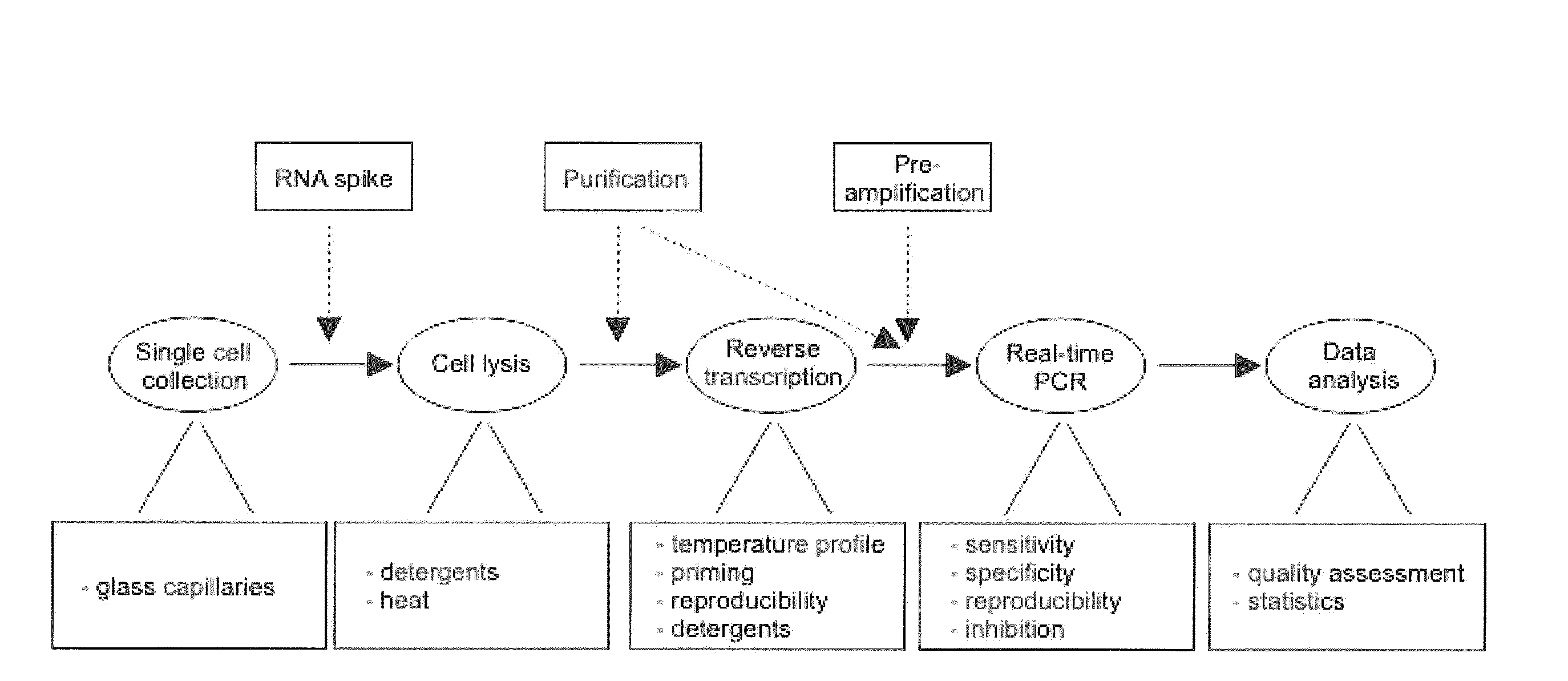
Lysis and reverse transcription for mRNA quantification
a reverse transcription and mrna technology, applied in the field of mrna measurement, can solve the problems of inability to assume the behavior of individual cells in data obtained from a population of cells, and inability to discriminate between. , to achieve the effect of avoiding evaporation and low sample volum
- Summary
- Abstract
- Description
- Claims
- Application Information
AI Technical Summary
Benefits of technology
Problems solved by technology
Method used
Image

Examples
example 1
Preparation and Culture of Cells
[0084]Pancreatic islets were prepared from healthy female National Maritime Research Institute (NMRI) mice aged 3-4 months (Bomholtgaard, Ry, Denmark) and fed a normal diet ad libitum. The mice were sacrificed by cervical dislocation, and pancreatic islets were isolated by collagenase P digestion (Roche, Basel, Switzerland) followed by manual collection of islets. All experimental procedures involving animals were approved by the ethical committee of Lund University. To prepare dispersed single cells the collected islets were gently shaken at low extracellular Ca2+ concentration to dissolve the structure of the islet [21]. Dispersed cells were plated on plastic 35 mm Petri dishes (Nuns, Roskilde, Denmark) in RPMI 1640 medium (SVA, Uppsala, Sweden) supplemented with 10% FCS, 100 U mL−1 penicillin, and 10 μgmL−1 streptomycin (all from Invitrogen, Carlsbad, Calif., USA) in the presence of various concentrations of glucose (Sigma-Aldrich, St. Louis, Mo., ...
example 2
Single Cell Collection
[0086]Attached dispersed cells were washed twice with a buffer containing 138 mM NaCl, 5.6 mM KCl, 1.2 mM MgCl2, 2.6 mM CaCl2, 5 mM HEPES (pH 7.4 with NaOH) and 3-20 mM glucose (same glucose concentration as in culture) to remove dead and loose debris for cell collection with patch-clamp pipettes. The dish, containing adhered cells and approximately 1 mL buffer, was mounted in a standard inverted light-microscope (Zeiss Axiovert 135, Oberkochen, Germany). Borosilicate glass capillaries (Hilgenberg GmbH, Malsfeld, Germany) with outer diameter of 1.6 mm and wall thickness of 0.16 mm were pulled to pipettes using a patch-clamp pipette puller (Heka PIP5, Lambrecht, Germany). The diameter of the tip was approximately 10 μm on average, substantially wider than standard patch-clamp pipettes and large enough to allow passage of an intact cell. The glass pipette was mounted on a hydraulic micromanipulator (Narishige, Tokyo, Japan) on the microscope. By controlling the p...
example 3
Lysis and Purification
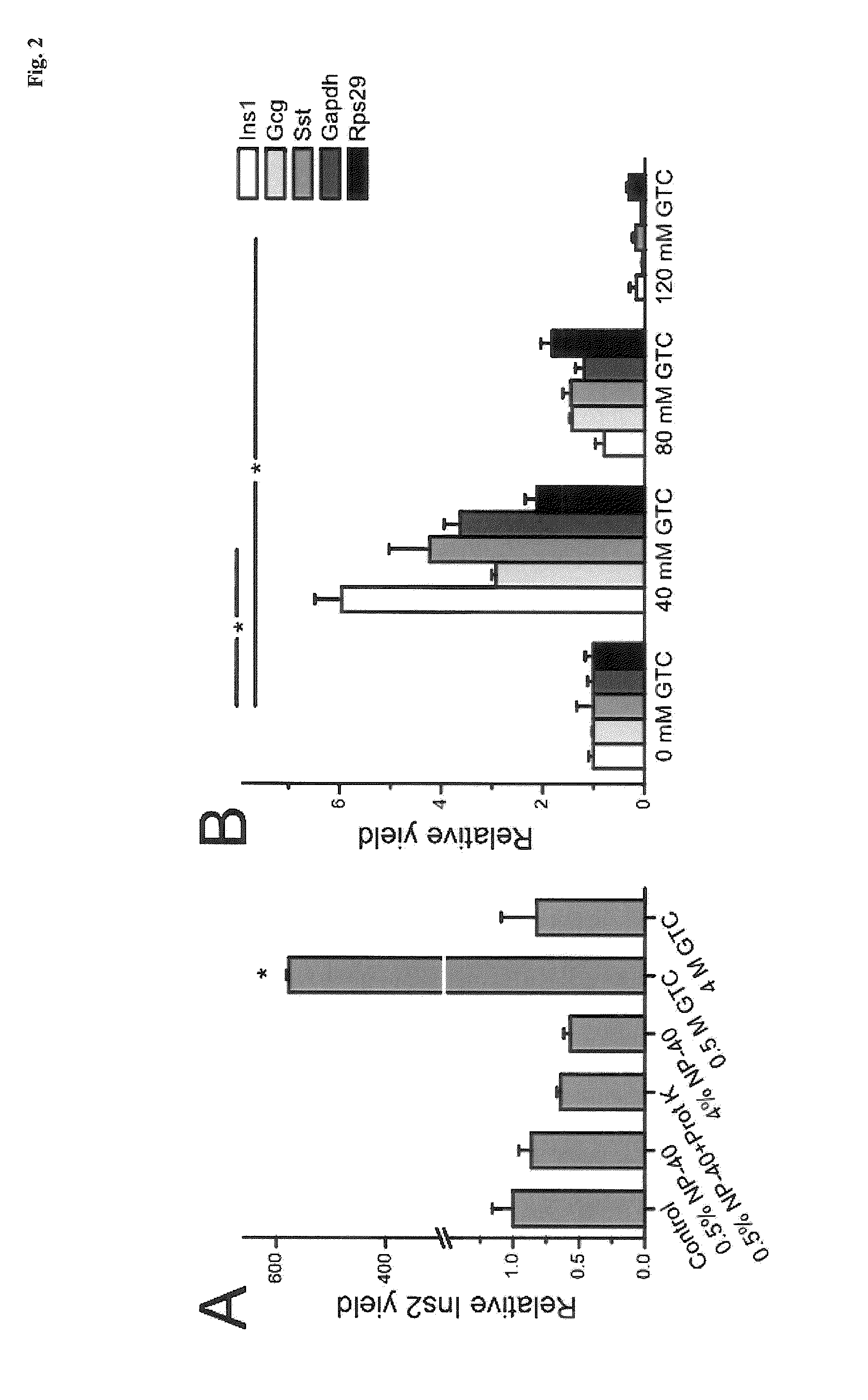
[0087]Islet lysis: Single pancreatic islets of roughly the same size and about 1000 cells were placed in 10 μl of various lysis buffers. The detergents Nonidet P-40 (NP-40, a.k.a. Igepal CA-630, Sigma-Aldrich) and guanidine thiocyanate (GTC, Sigma-Aldrich) were used. Samples were incubated at 60° C. or 80° C. for 15 minutes (60° C. for samples containing 0.4 mg / ml proteinase K (Invitrogen)) followed by 5 min incubation at 95° C. and frozen at −25° C. for subsequent analysis. Samples were diluted 1:20 prior reverse transcription to minimize possible inhibitory effects.
[0088]Single cell lysis: In single cell experiments, the glass pipettes were emptied in 0.2 ml plastic tubes containing 2 μl of lysis solution. The emptying required a custom-made device, consisting of a tube holder lined up with a coarse micromanipulator on which the pipette was mounted. The glass pipette was carefully flushed with lysis buffer a few times to make sure the cell entered the tube. I...
PUM
| Property | Measurement | Unit |
|---|---|---|
| V/V | aaaaa | aaaaa |
| temperature | aaaaa | aaaaa |
| temperature | aaaaa | aaaaa |
Abstract
Description
Claims
Application Information