Delivery device for localized delivery of a therapeutic agent

a delivery device and localized delivery technology, applied in the field of delivery of therapeutic agents, can solve the problems of reducing the accuracy of instrument placement, requiring careful monitoring, and requiring less than optimal injection, infusion, inflation or sample collection, etc., to reduce the loss of therapeutic agents, reduce the delivery and/or application of therapeutic agents, and reduce the effect of loss

Inactive Publication Date: 2011-06-09
BOSTON SCI SCIMED INC
View PDF17 Cites 123 Cited by
  • Summary
  • Abstract
  • Description
  • Claims
  • Application Information

AI Technical Summary

Benefits of technology

[0015]Depending on the embodiment, a device and / or method as disclosed herein can have advantages including reduced loss of therapeutic agent during and / or after the procedure, reduced delivery and / or application of therapeutic agent at undesired times or to undesired locations, simplicity of design, reduced procedural complications, improved ease of use, and / or improved overall performance during and / or after the procedure. These and other features and advantages of the disclosed devices and methods are described in, or apparent from, the following detailed description of various exemplary embodiments.

Problems solved by technology

These procedures often require careful, time-consuming monitoring of the placement of the instrument tip within the body.
Even with such care, however, limitations on the quality of the available images and obstruction of views by surrounding tissues or fluids can degrade the accuracy of placement of the instrument.
Such difficulties can result in less than optimal injection, infusion, inflation or sample collection.
Moreover, even if positioned properly, the instrument might be aligned with areas in which performance of the medical procedure would not be desired, such as where an asymmetric plaque deposit inside a blood vessel would render infusion delivery or angioplasty ineffective or potentially dangerous.

Method used


Image

  • Delivery device for localized delivery of a therapeutic agent
  • Delivery device for localized delivery of a therapeutic agent
  • Delivery device for localized delivery of a therapeutic agent

Examples


example 1

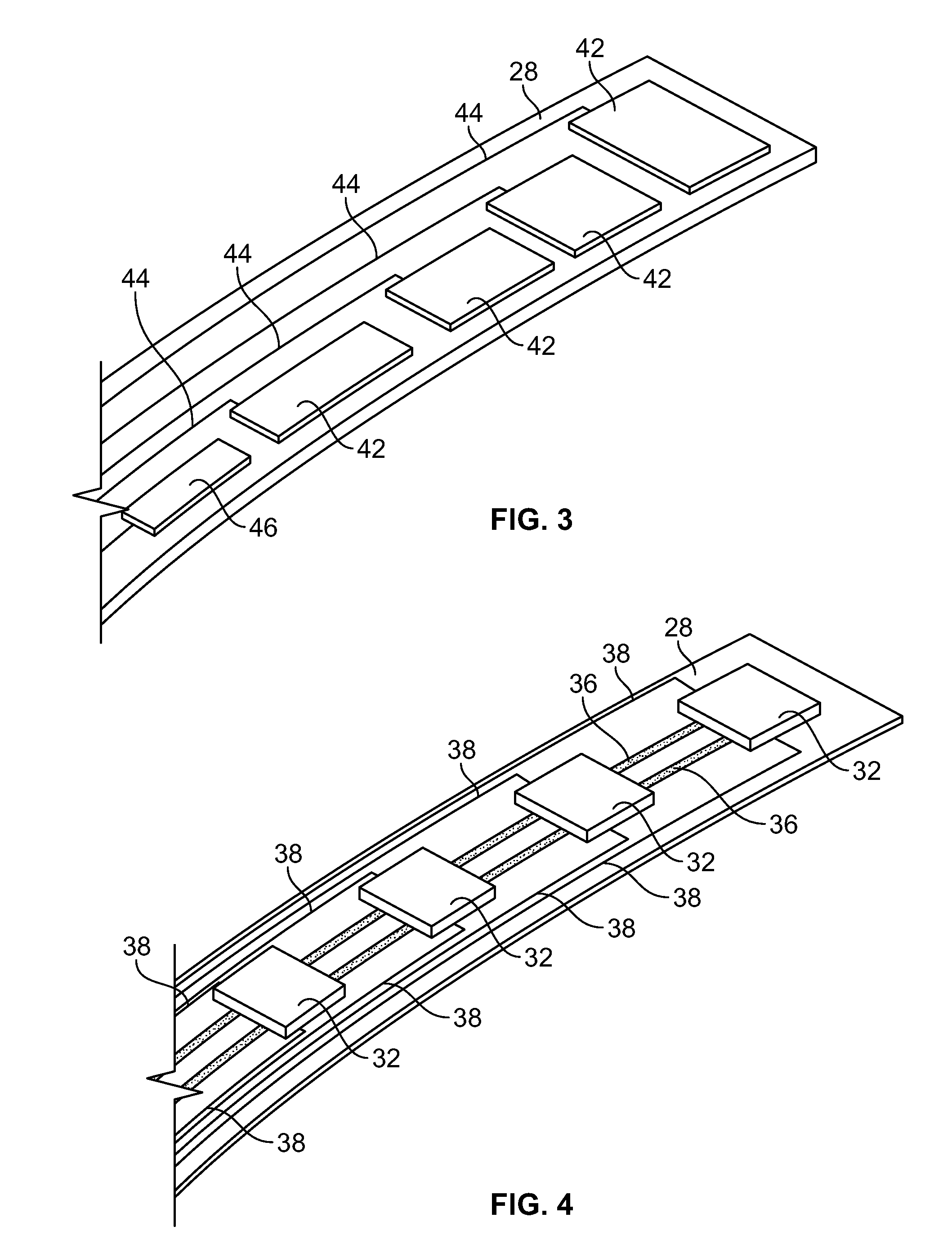
[0091]A device as illustrated in FIGS. 3-6 can be made by mounting Hall sensors and electroactive polymer on a strip, and mounting the strip onto a balloon surface. First, a flexible polymer strip is made. Nylon strips (VESTAMID®) can be extruded and cut having dimensions 1 meter long (approximately the length of the catheter) by 2 mm wide and a thickness of 20 micrometers. The strips are cleaned with HNO3 for 10 minutes and rinsed with deionized water. On one side, 10 parallel conductive lines (100 micrometers wide and 2 micrometers high with a spacing of 50 micrometers) are printed using an aqueous silver nanoparticle dispension SP100 (PChem Associates Inc., Bensalem, Pa.) and a MD-K-130 printing system from Microdrop (Microdrop Technologies GmbH, Muehlenweg 143, D-22844 Norderstedt, Germany). The conductive lines are for power supply to the sensors (2 conductive lines) as well as signal retrieval (8 conductive lines). On the opposite side of the strip, a number of lines as well a...

example 2

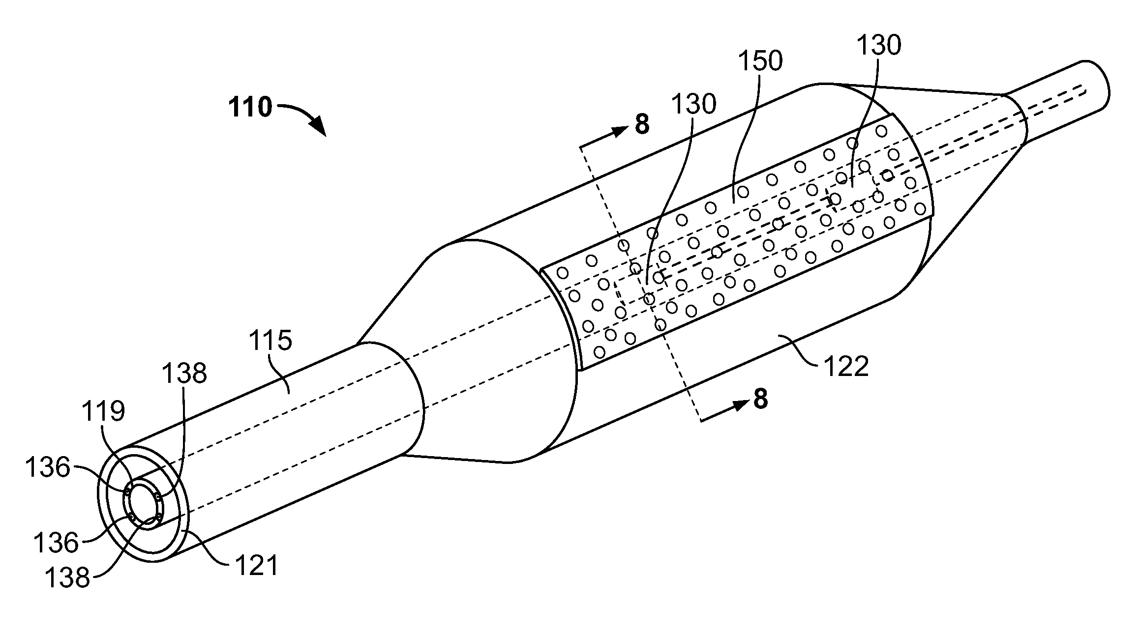
[0096]A device as illustrated in FIGS. 7-10 can be made by first making a polyimide inner tube with four copper conducting wires inserted in the wall. Micro Hall sensors can be obtained from Cryomagnetics Oak Ridge, Tenn. (http: / / www.cryomagnetics.com / hall-effect-sensor.php). The type HSU-1 comes without packaging with a sensing area of 100 micrometers squared. The surrounding ceramic area can be further reduced in size by laser ablating this material away using a 193 nm excimer laser to a final size of 200 by 200 micrometers. The wires of the Hall sensors are soldered to the wires of the inner tube after removing 10 mm of the distal end of the polymer wall of the inner tube using the same excimer laser. Two square cavities are ablated out of the inner tube 10 mm and 20 mm proximal to the distal end with a depth of 0.04 inches to fit both sensors. Both holes are aligned axially. The sensors and wires are folded backwards over the polymer inner tube whereby the sensors are placed bac...

example 3

[0099]A device as illustrated in FIGS. 14-15 is constructed by first making a polyimide inner tube with four conducting copper wires inserted in the wall as described in Example 2. Micro Hall sensors are attached to the inner tube in the same manner as in Example 2.

[0100]A tri-wing shaped soft silicon rubber piece (http: / / www.appliedsilicone.com / products-index.html, component part 40088) is cast and attached to an outer tube by using Loctite® 4981™ Super Bonder® Medical Device Adhesive. The rubber tri-shape has tipped wings such that upon retrieval in the delivery catheter they all will fold in the same direction. The three wings will make three channels (spaces between the wings), and one of them is closed by a silicon rubber membrane in place. In the valley of the closed chamber, one or more holes are punctured for the drug delivery ports.

[0101]The inner tube is fed through the outer tube and silicon wing shape and aligned such that the Hall sensors are located underneath the clos...


PUM


Abstract

Therapeutic agent delivery devices and methods for delivering a therapeutic agent to a target location as well as methods for determining the location of a lesion on a vessel wall are disclosed. Various embodiments disclose an expandable member comprising a drug delivery matrix for selectively delivering a therapeutic agent to a lesion on a vessel wall. The drug delivery matrix may comprise one or more sensors and an electroactive polymer for releasing the therapeutic agent. Other embodiments disclose an expandable member comprising a plurality of radially-expanding flexible walls forming a plurality of channels for selectively delivering therapeutic agent to a target area adjacent one or more of the channels. Detecting a lesion may comprise using a plurality of Hall effect sensors disposed on a distal end of a catheter.

Description

CROSS REFERENCE TO RELATED APPLICATION[0001]The present application claims priority to U.S. provisional application Ser. No. 61 / 267,944 filed Dec. 9, 2009, the disclosure of which is incorporated herein by reference in its entirety.TECHNICAL FIELD[0002]The present disclosure relates to the delivery of a therapeutic agent, for example to the interior walls of a vessel such as a blood vessel, via a therapeutic agent delivery device, and to detection of lesions on the walls.BACKGROUND INFORMATION[0003]The deployment in the body of medications and other substances, such as materials useful in tracking biological processes through non-invasive imaging techniques, is an oft repeated and advantageous procedure performed during the practice of modern medicine. Such substances may be deployed through non-invasive procedures such as endoscopy and vascular catheterization, as well as through more invasive procedures that require larger incisions into the body of a patient.[0004]In conventional...

Claims


Application Information

Patent Timeline
Patent Type & Authority Applications(United States)
IPC IPC(8): A61M25/10A61B5/05A61F2/958
CPCA61B5/01A61M2205/3303A61B5/14503A61B5/14539A61B5/4839A61B2562/0223A61B2562/043A61M25/10A61M2025/0024A61M2025/0057A61M2025/0058A61M2025/105A61M2205/0266A61M2205/0283A61B5/02007A61B5/0037
Inventor WEBER, JANHARRISON, KENTFLANAGAN, AIDEN
Owner BOSTON SCI SCIMED INC
Why Eureka
Try Eureka