Method of evaluating the integrity of the plasma membrane of cells by detecting glycans found only intracellularly
- Summary
- Abstract
- Description
- Claims
- Application Information
AI Technical Summary
Problems solved by technology
Method used
Image
Examples
example1
Detection of Cells Damaged By EDTA Treatment By Anti-Glycan Antibodies
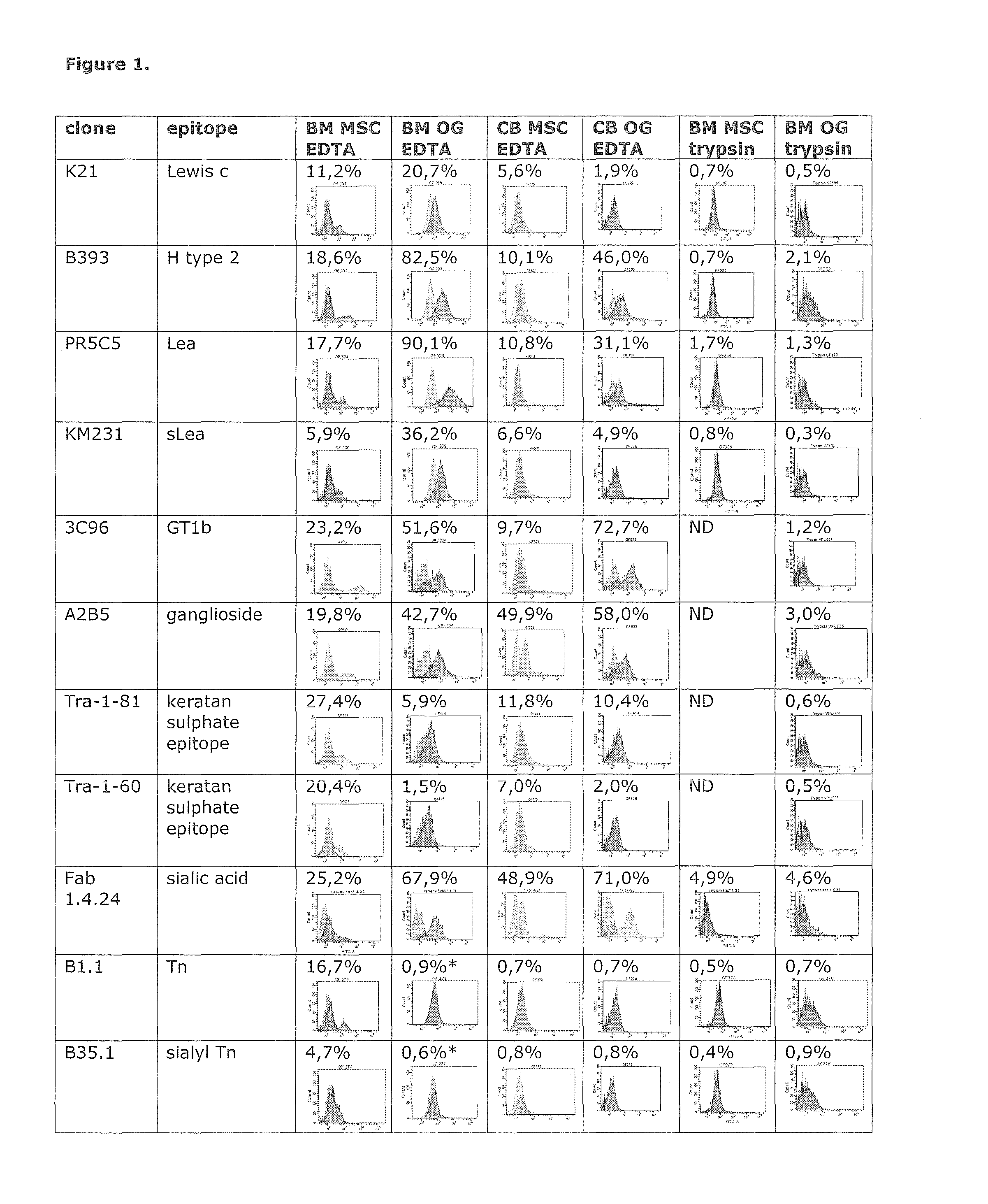
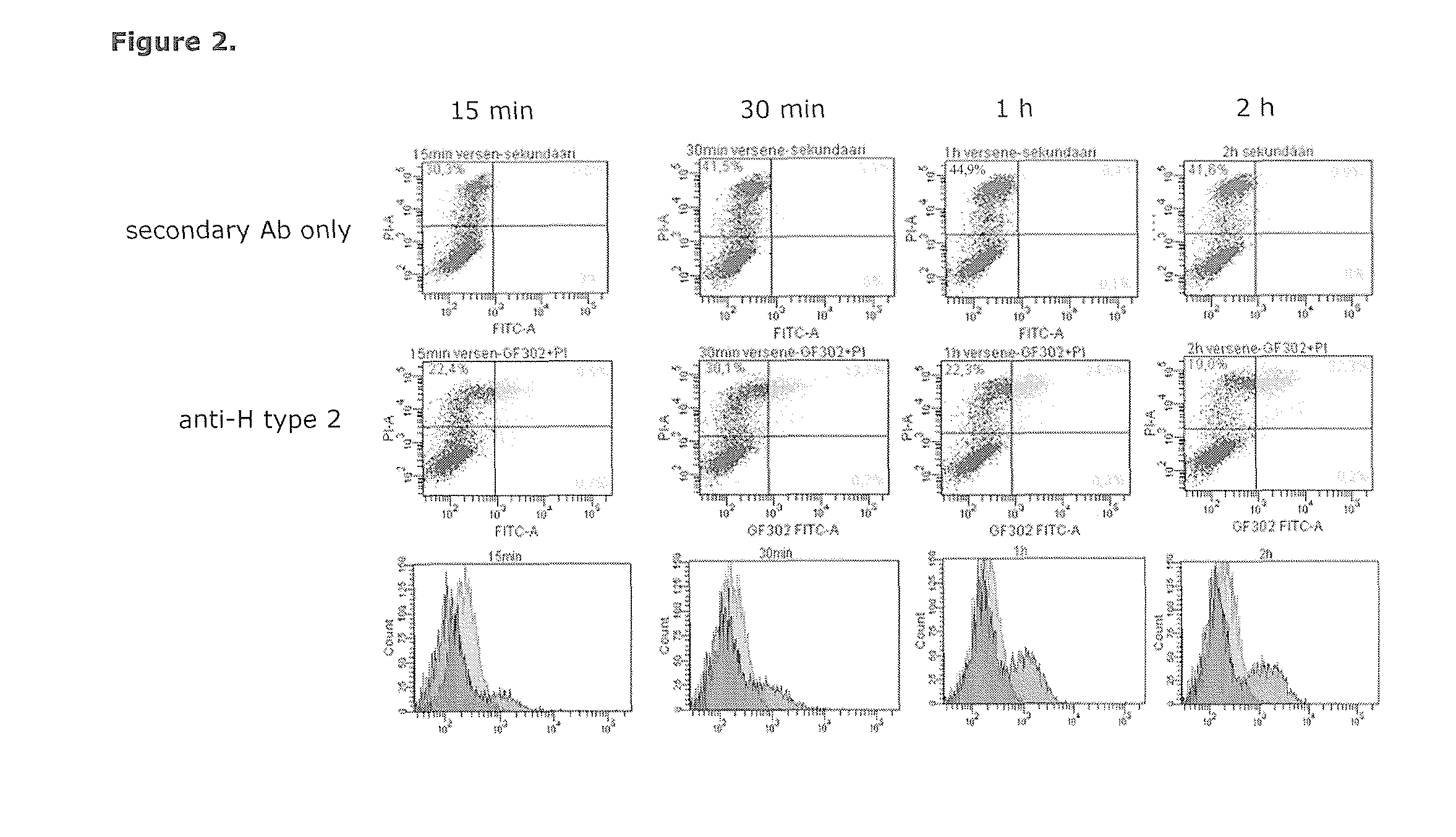
[0123]EDTA (Versene) is commonly used to detach cells from culture plates instead of trypsin, especially when there is the need to preserve cell surface proteins intact. In this example was is shown that 2 mM EDTA damages cell membranes so that the cells became propidium iodide permeable. Furthermore, it was shown that certain anti-glycan antibodies recognize epitopes that were accessible only in these damaged cells.
[0124]Materials and Methods Cell samples. Bone marrow (BM) derived mesenchymal stem cells (MSC:s). BM MSC:s were obtained as described by Leskelä et al. (2003). Briefly, bone marrow obtained during orthopaedic surgery was cultured in Minimun Essential Alpha-Medium supplemented with 20 mM HEPES, 10% fetal calf serum, penicillin-streptomycin and 2 mM L-glutamine (all from Gibco). After a cell attachment period of 2 days the cells were washed with PBS, subcultured further by plating the cells at a density...
example2
Detection of Necrotic and Permeabilized Cells By Anti-Glycan Antibodies
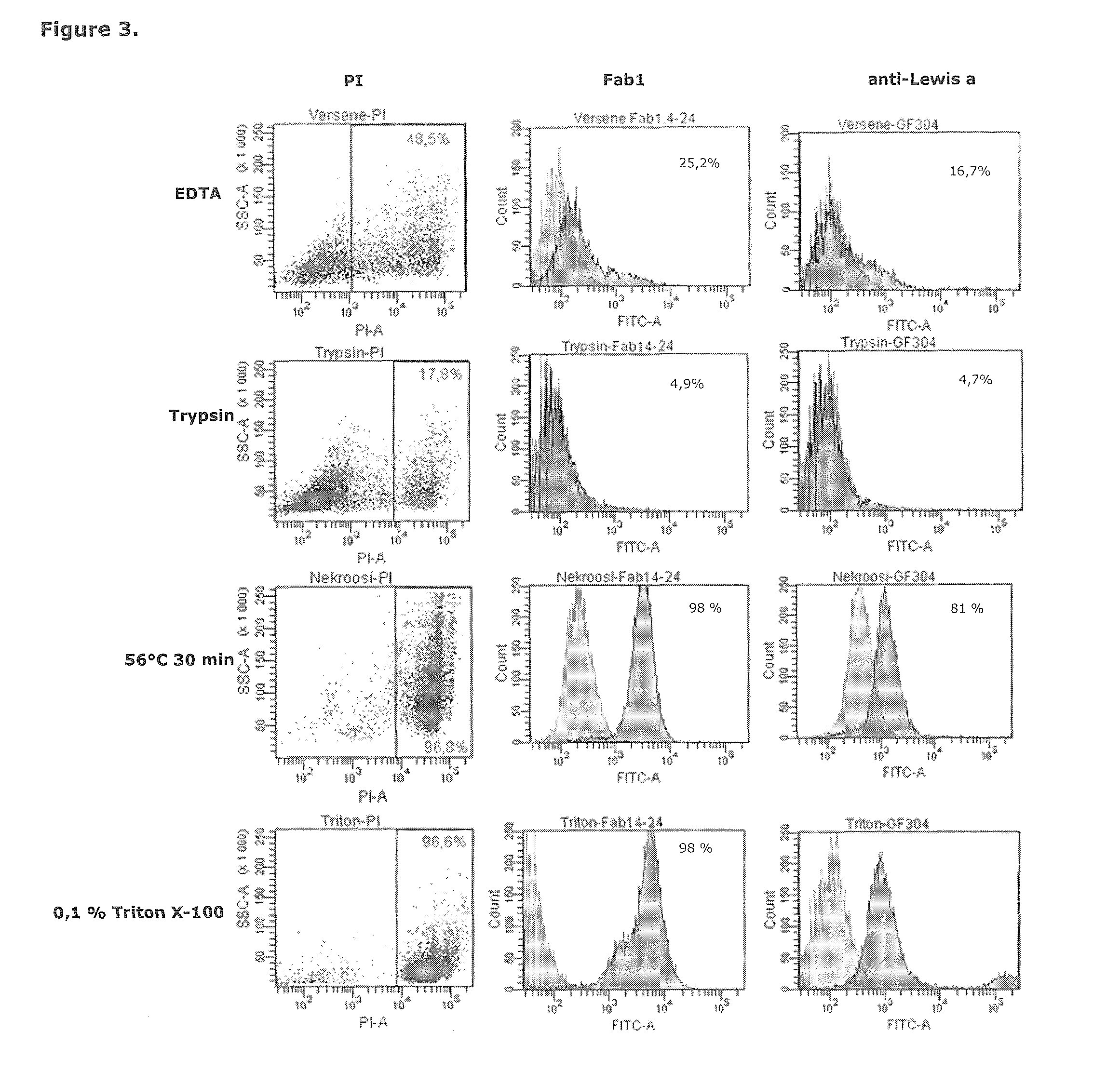
[0135]Cells were permeabilized by Triton X-100 or induced to undergo necrosis by heating at 56° C. for 30 min and analyzed by a set of glycan antibodies. Certain glycan antibodies were shown to bind to cells damaged by detachment methods, permeabilized cells and necrotic cells, and not to intact cells.
[0136]Materials and Methods Cell samples and antibodies were obtained as in Example 1; detachment of cells from culture plates and flow cytometry were carried out as in Example 1.
[0137]Induction of necrosis. Necrosis was induced by incubating the cells (detached with trypsin) in culture medium at +56° C. for 30 min.
[0138]Permeabilization of cells. Cells (detached with trypsin) were permeabilized by incubating them with 0.1% Triton X-100, 0.3% BSA, 2 mM EDTA-PBS for 10 min at room temperature.
[0139]Results and Discussion To extend the observation that membrane damage in EDTA treated cells exposes carbohydrate epitope...
example 3
Proliferation of Mesenchymal Stem Cell Subpopulations Sorted on the Basis of Glycan Antibody Binding
[0144]Materials and methods Cells, cell culture, antibodies and FACS as described in Example 1.
[0145]Results and Discussion Bone marrow mesenchymal stem cells were labeled with anti-Tn and sorted by FACS. Sorting is shown in FIG. 4a. The sorted subpopulations were plated separately at 1500 cells / cm2 (14 000 cells per one well on a six well plate) in culture medium and let to attach and proliferate for three days. The Tn negative cells attached to the substratum and started proliferate, where as the Tn positive cells failed to attach or proliferate (FIG. 4b), suggesting that the Tn positive subpopulation does not consist of viable cells.
PUM
| Property | Measurement | Unit |
|---|---|---|
| Temperature | aaaaa | aaaaa |
| Temperature | aaaaa | aaaaa |
| Fraction | aaaaa | aaaaa |
Abstract
Description
Claims
Application Information
 - R&D
- Intellectual Property
- Life Sciences
- Materials
- Tech Scout
- Unparalleled Data Quality
- Higher Quality Content
- 60% Fewer Hallucinations
Browse by: Latest US Patents, China's latest patents, Technical Efficacy Thesaurus, Application Domain, Technology Topic, Popular Technical Reports.
© 2025 PatSnap. All rights reserved.Legal|Privacy policy|Modern Slavery Act Transparency Statement|Sitemap|About US| Contact US: help@patsnap.com


