BIOMARKERS FOR IgA NEPHROPATHY AND APPLICATIONS THEREOF

a biomarker and nephropathy technology, applied in the field of biomarkers for iga nephropathy, can solve the problems of not being diagnosed or delayed, renal biopsy entails risk for serious bleeding complications, and has a major negative impact on the diagnosis and prognosis of patients with glomerular disorder

Inactive Publication Date: 2011-07-28
NAT DEFENSE MEDICAL CENT
View PDF1 Cites 9 Cited by
  • Summary
  • Abstract
  • Description
  • Claims
  • Application Information

AI Technical Summary

Benefits of technology

[0016]FIG. 7 shows the renal terminal deoxynucleotidyl transferase dUTP nick end labeling (TUNEL) analysis of the Prg-IgAN model in a time-course manner. (A) to (D) represent the kidney tissues on day 0, 3, 14, or 21 showing cell apoptosis. Original magnification is ×400 each. (E) represents the scoring of the apoptotic cells. **p<0.01 represents statistical significance compared to the normal control (day 0).
[0017]FIG. 8 shows the renal ISH results for the IgAN patients with unfavorable prognosis factors (UPF). (A) represents the results for Tmsb4, Serpine2, OPN, BTNL2, S100A8, and CysC. Arrows indicate parietal epithelial cells, and arrowheads indicate podocytes. Original magnification is ×400 each. (B) to (G) represent the semi-quantitative analysis of cellular mRNA expression. The scoring was performed for the three major components: parietal epithelial cells (solid bars), podocytes (open bars) and mesangial cells (hatched bars). Each bar represents the mean±SE. *p<0.05, **p<0.01 and **p<0.005 represent statistical significance compared to normal control. The symbol “#” means data not detectable.
[0018]FIG. 9 shows the renal IHC results for IgAN patients with UPF. (A)

Problems solved by technology

To date, a renal biopsy is required for diagnosis and prognosis of IgA nephropathy, but many patients might be reluctant to accept the invasive procedure, which might cause not diagnosed or delayed diagnosed until the clinical features are o

Method used


Image

  • BIOMARKERS FOR IgA NEPHROPATHY AND APPLICATIONS THEREOF
  • BIOMARKERS FOR IgA NEPHROPATHY AND APPLICATIONS THEREOF
  • BIOMARKERS FOR IgA NEPHROPATHY AND APPLICATIONS THEREOF

Examples


example 1

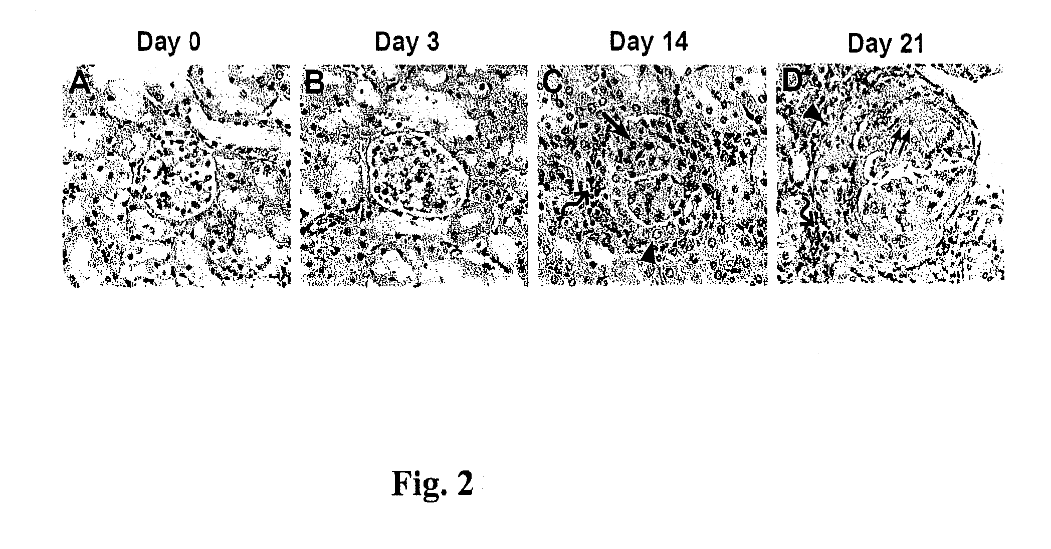
Establishment of a Prg-IgAN Animal Model and the Clinical and Pathological Evaluation Thereof

[0051]Prg-IgAN was induced in B-cell-deficient (BCD) mice by daily injection of purified IgA anti-phosphorylcholine and pneumococcal C-polysaccharide (PnC) as described previously (Kidney Int 2006; 70: 283-297). To confirm the establishment of the IgAN animal model, clinical and pathological evaluation was conducted as below.

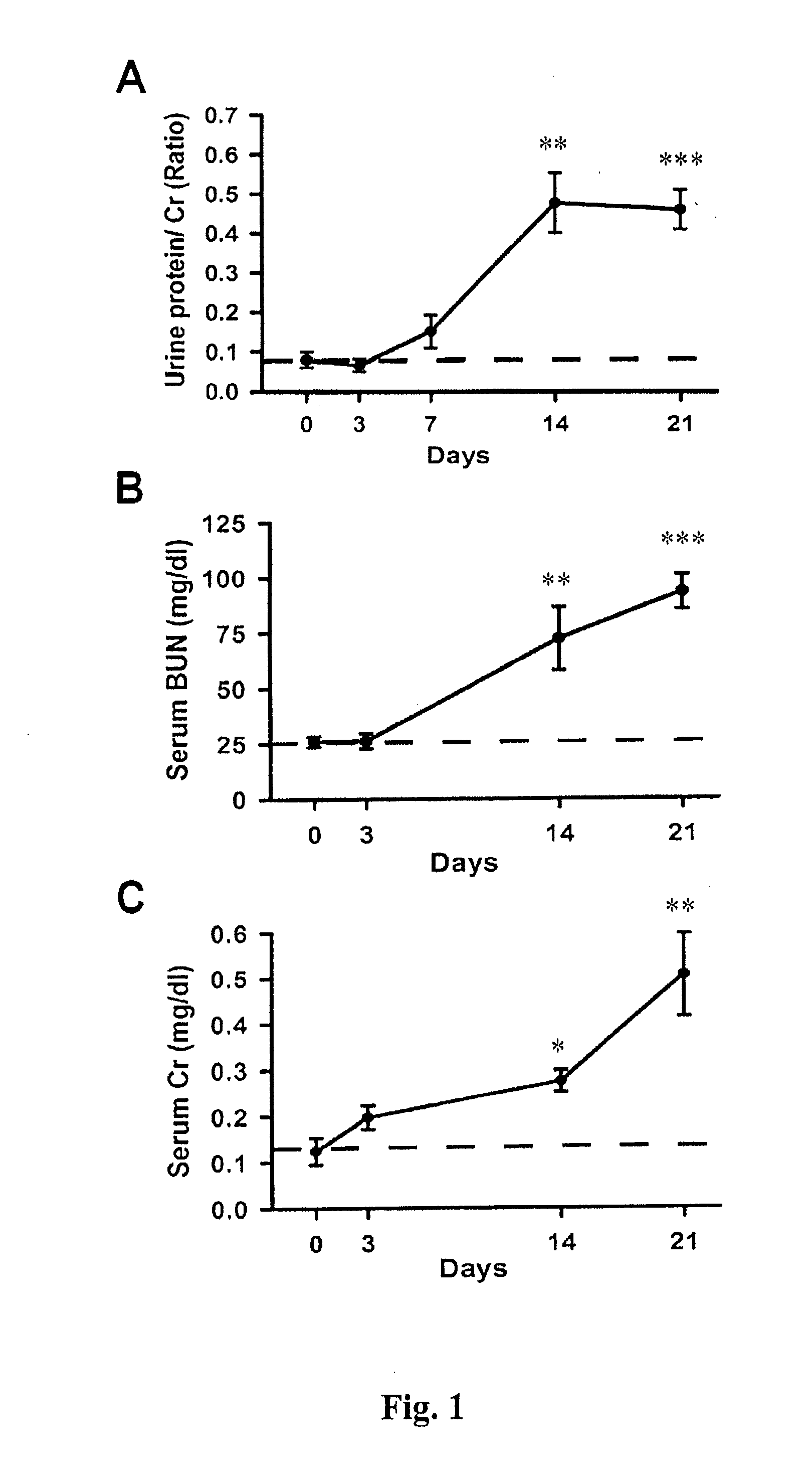
[0052]Urine and blood samples were collected from the mice at different time points, which were analyzed for proteinuria and blood urea nitrogen (BUN) and creatinine (Cr) levels by using a urease assay and a picric acid method respectively (Nephron 1998; 78: 440-452). BCD mice treated with saline only were used as normal controls.

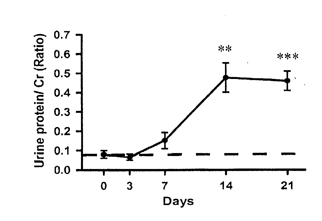
[0053]As shown in FIG. 1A, a significant increase of Cr-corrected urine protein levels (0.48±0.07) was observed in the Prg-IgAN mice at day 14 compared with basal levels (0.08±0.02) (p<0.01), and the protein levels remained a range of high level...

example 2

Gene Expression Profiling

[0056]To characterize the profile of altered gene expressions in the glomeruli of the Prg-IgAN model, a combined laser capture microdissection (LCM) and cDNA microassay analysis was conducted. Briefly, LCM was performed to obtain glomerular sections from normal controls and the Prg-IgAN mice at day 21 according to the protocols as previously described (Reprod Biol Endocrinol 2007; 5: 18; and Methods Mol Biol 2009; 466: 73-82). For each sample, approximately 150 glomeruli were harvested from at least three consecutive sections. Subsequently, cDNA microarray analysis was performed as described previously (Nephrol Dial Transplant 2006; 21: 288-298). In total, 8,500 mouse gene spots were screened for the Prg-IgAN model at day 21 versus normal controls.

[0057]As a result, totally 918 up-regulation genes (Prg-IgAN at day 21 / normal control ratio□2) in the glomerulus were identified. Highly expressed genes of interest with the ratio□10 (totally 39 genes), including T...

example 3

mRNA Expression of Candidate Genes

[0058]To determine whether these upregulated genes in the glomerulus were associated with the progression of IgAN, a time-course (days 0, 3, 14, and 21) mRNA expression analysis by RT-PCR was followed in isolated glomeruli from the Prg-IgAN model. The glomeruli samples of the Prg-IgAN mice were isolated with a sieving technique as described previously (Nephrol Dial Transplant 2006; 21: 1794-1802), and then subjected to total RNA extraction with Trizol reagent (Life Technologies, MD, USA) according to the manufacturer's instruction. Subsequently, real-time RT-PCR was conducted based on the RNA samples with gene-specific primers as shown in Table 1.

TABLE 1Primer sequences used for real-time RT-PCR inanimal samplesGene(mouse)Primer sequencesBTNL25′-CTCTGGGCCAGGAGAAAAC-3′SEQ ID NO: 135′-TGAGCCTCTCATCAGAAGGAA-3′SEQ ID NO: 14CysC5′-TACAACAAGGGCAGCAACGA-3′SEQ ID NO: 155′-GCACCCTTCTGCGAGATGAA-3′SEQ ID NO: 16GAPDH5′-TCCGCCCCTTCTGCCGATG-3′SEQ ID NO: 175′-CACG...


PUM

PropertyMeasurementUnit
Fractionaaaaaaaaaa
Fractionaaaaaaaaaa
Fractionaaaaaaaaaa

Abstract

The present invention provides a method for diagnosis or prognosis of IgA nephropathy in a subject based on detection of the expression level of one or more biomarker genes selected from the group consisting of thymosin β4 (Tmsb4), serine or cysteine proteinase inhibitor clade E member 2 (Serpine2), secreted phosphoprotein 1 (OPN), butyrophilin-like-2 (BTNL2), S100 calcium binding protein A8 (S100A8), Cystatin C (CysC), and any combination thereof.

Description

FIELD OF THE INVENTION[0001]The preset invention relates to biomarkers for IgA nephropathy and applications thereof.BACKGROUND OF THE INVENTION[0002]IgA nephropathy is the most frequent type of glomerular disorders worldwide, up to 60% of the patients eventually progressing to either end-stage renal disease or chronic renal failure after 20 years of follow-up. Although the pathogenesis of IgA nephropathy is still largely unknown, an initial response of intrinsic glomerular cells to IgA immune complexes, including abnormal glycosylated IgA1 as “neoantigen”, plays a primary role, whereby triggering release of cytokines and growth factors in the glomerulus affected. To date, a renal biopsy is required for diagnosis and prognosis of IgA nephropathy, but many patients might be reluctant to accept the invasive procedure, which might cause not diagnosed or delayed diagnosed until the clinical features are outward or a disease progression has already developed. Unfortunately, renal biopsy a...

Claims


Application Information

Patent Timeline
IPC IPC(8): C12Q1/68G01N33/53G01N33/00
CPCC12Q1/6883C12Q2600/158C12Q2600/156G01N2800/347G01N33/6893
Inventor CHEN, ANNKA, SHUK-MAN
Owner NAT DEFENSE MEDICAL CENT
Why Eureka
Try Eureka