Method for treating keratoconus by UV radiation and a device for carrying out said method (variants)

a technology of keratoconus and uv radiation, which is applied in the field of keratoconus treatment by uv radiation and a device for carrying out said method, can solve the problems of traumatic method and affect the damage of endothelial cells, and achieve the effect of reducing the adverse effects of exposur

Inactive Publication Date: 2011-08-04
ANISIMOV SERGEY IGOREVICH
View PDF13 Cites 41 Cited by
  • Summary
  • Abstract
  • Description
  • Claims
  • Application Information

AI Technical Summary

Benefits of technology

[0006]The technical result achieved when working of the invention is to reduce the adverse effects of exposure.

Problems solved by technology

However, this method is traumatic because of the adverse effects of UV radiation on the cornea.
However, irradiation of the cornea by full aperture UV source, providing uniform illumination of the entire cornea area, can cause photo-burn, and the imperfect cornea thickness, smaller than the depth of UV radiation exposure, can affect damage of endothelial cells with the progression of complications such as corneal dystrophy.

Method used


Image

  • Method for treating keratoconus by UV radiation and a device for carrying out said method (variants)
  • Method for treating keratoconus by UV radiation and a device for carrying out said method (variants)
  • Method for treating keratoconus by UV radiation and a device for carrying out said method (variants)

Examples


example 1

Patient A., Aged 26

[0025]Diagnosis: keratoconus of both eyes. Visual acuity before surgery:

right eye—0,01 sph—4,0 D, cyl—4,0 D ax 85°

left eye—0,2 sph—1,5 D, cyl—1,5 D ax 95°

[0026]Intensive disease progression is present over the past six months.

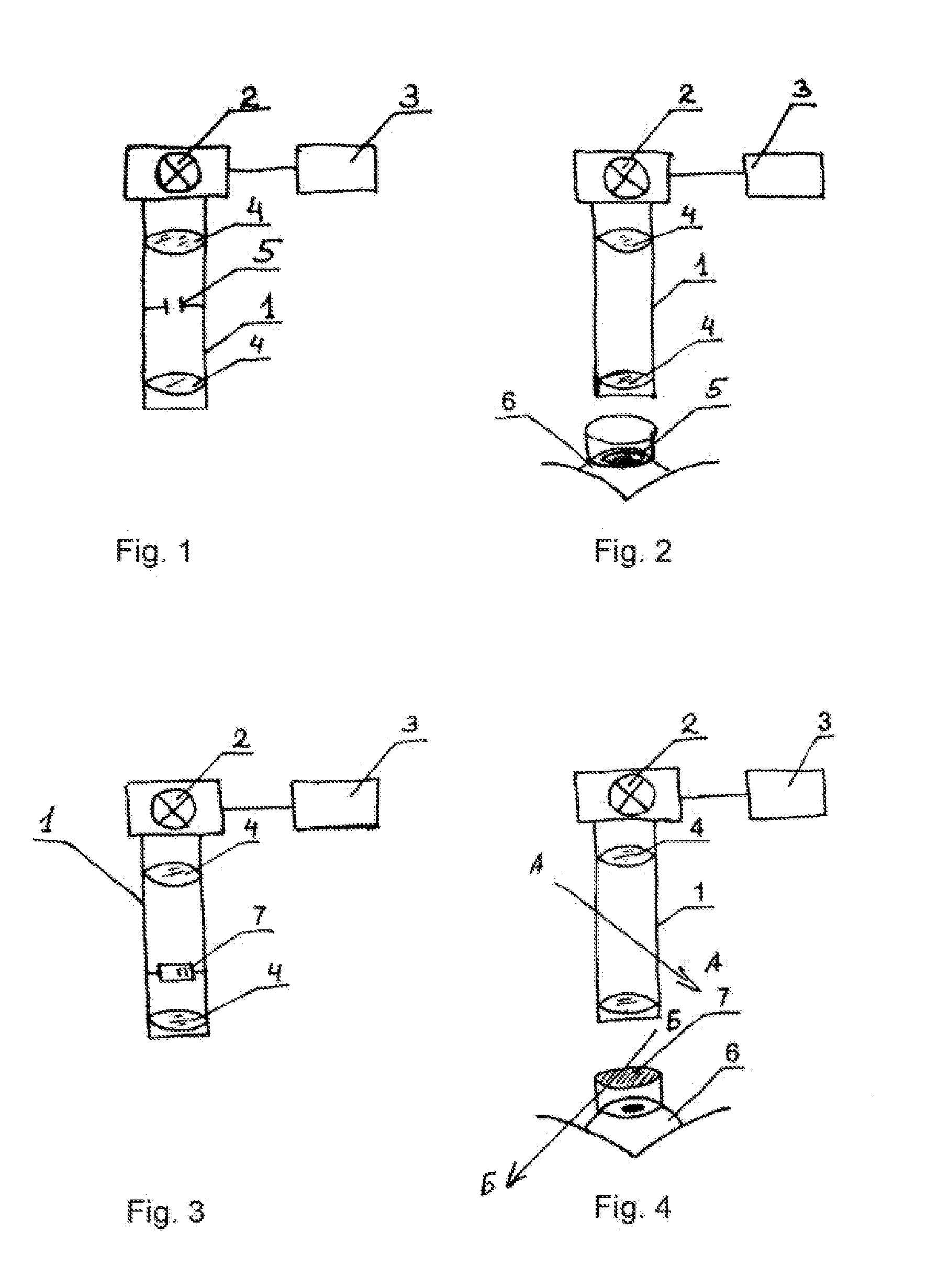
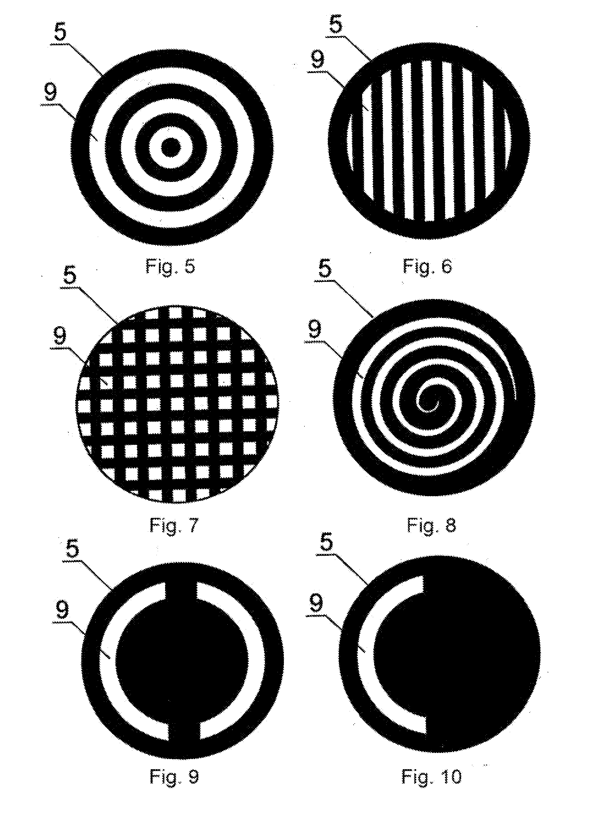
[0027]Surgery was carried out according to the suggested method. After the local anesthesia photomediator instillation is provided, namely the solution of 1% riboflavin within 30 minutes. Then on the corneal surface the diaphragm image of concentric rings formed by the UV source was focused. The exposition was 1 minute.

[0028]The next day after surgery—right eye calm, zero degree reaction, no soreness, and no signs of cornea damage seen on slit lamp examination. Control examination after 1 year showed no disease progression in the right eye. The same method was used for the treatment of the left eye.

example 2

Patient B., 28 Years Old

[0029]Diagnosis: keratoconus of both eyes. Visual acuity before surgery:

right eye —0,01 sph—2,0 D, cyl—5,0 D ax 65°

left eye—0,1 sph—1,5 D, cyl—3,5 D ax 105°

[0030]Intensive disease progression is present over the past six months.

[0031]Surgery was carried out according to the suggested method. After the local anesthesia photomediator instillation is provided, namely the solution of 1% riboflavin within 30 minutes, on the surface of the cornea aperture in the form of parallel lines was set, it was formed by the UV source, the direction of the lines was chosen to be parallel to the astigmatism axis, i.e., parallel to the meridian 65 degrees. Diaphragm was fixed with a vacuum ring. The exposition was 30 minutes.

[0032]The next day after surgery—right eye calm, zero degree reaction, no soreness, and no signs of cornea damage seen on slit lamp examination. Control examination after 1 year showed no disease progression in the right eye. The same method was used for th...

example 3

Patient Sh., Aged 38

[0033]Diagnosis: keratoconus of both eyes. Visual acuity before surgery:

right eye—0,01 sph—0,5 D, cyl—4,0 D ax 75°

left eye—0,2 cyl—3,75 D ax 80°

[0034]Deterioration of vision during the last year.

[0035]Surgery was carried out according to the suggested method. Cornea polarization plane was defined with the polarimeter. Local anesthesia was applied. After photomediator instillation, namely the solution of 1% riboflavin within 30 minutes, the polarization plane of the device polarizer deployed at 90 degrees to the cornea polarization plane. On the right eye cornea surface was focused spot formed by the UV source. The exposition was 40 minutes.

[0036]The next day after surgery—right eye calm, zero degree reaction, no soreness, and no signs of cornea damage seen on slit lamp examination. Control examination after 1 year showed no disease progression in the right eye.


PUM


Abstract

The invention relates to ophthalmology and is used for treating keratoconus. The inventive method comprises forming one or more radiation areas of different shape, comprising concentric circles, arcs, parallel lines, cells, grids, spirals or other geometrical figures. In the other variant, the method comprises polarizing UV radiation and adjusting the depth of the UV radiation action by directing the UV radiation polarization plane at an angle of 1-180° to the plane of light polarization by the cornea, thereby using the effect of light polarization by the cornea. The device for treating keratoconus comprises, positioned on the optical axis common with an UV radiation source optical focusing elements and a diaphragm. The diaphragm is situated on the optical axis common with the UV radiation source and is made in the form of a mask with alternating transparent and shaded elements in the form of concentric circles, arcs, parallel lines, cells, grids, spirals or other geometrical figures. In the other variant, the device comprises, situated on the optical axis common with the UV radiation source, the optical focusing elements, the diaphragm and a polarisor.

Description

(i) SCOPE OF USE[0001]The invention relates to medicine, more specifically to ophthalmology and can be used for treating keratoconus by UV radiation.(ii) PRIOR KNOWLEDGE[0002]It is already known a method and a device for treating keratoconus by UV radiation that affects the cornea with full aperture (Wollensak G., Spörl E., Seiler T. Treatment of keratoconus by collagen cross linking. Der Ophthalmologie: Zeitschrift der Deutschen Ophthalmologischen Gesellschaft, 2003, January, 100(1): p. 44-49).[0003]However, this method is traumatic because of the adverse effects of UV radiation on the cornea.[0004]The influence of UV radiation on the cornea results collagen molecules photopolymerization. Photopolymerization is activated by non-toxic water-soluble photomediators, and their radiation absorption at depth should protect the deep tissue of the cornea, in particular, endothelial cells against damage. Such photomediator may be riboflavin, in particular, its 1% solution. The process of co...

Claims


Application Information

Patent Timeline
Patent Type & Authority Applications(United States)
IPC IPC(8): A61B18/18
CPCA61F9/0079A61F2009/00872A61N2005/073A61N5/0616A61F2009/00893
Inventor ANISIMOV, SERGEY IGOREVICH
Owner ANISIMOV SERGEY IGOREVICH
Why Eureka
Try Eureka