Electrode member for specific detection of analyte using photocurrent

Inactive Publication Date: 2011-08-11
TOTO LTD
View PDF8 Cites 10 Cited by
  • Summary
  • Abstract
  • Description
  • Claims
  • Application Information

AI Technical Summary

Benefits of technology

[0016]The present invention can advantageously improve the sensitivity of detection of

Problems solved by technology

Further, damage to genital systems, nervous systems and the like by exogenous endocrine disrupting chemicals (environmental hormones) including dioxins is recognized as social problems, leading to a demand for the development of a simple method that can detect the e

Method used


Image

  • Electrode member for specific detection of analyte using photocurrent
  • Electrode member for specific detection of analyte using photocurrent
  • Electrode member for specific detection of analyte using photocurrent

Examples


example 1

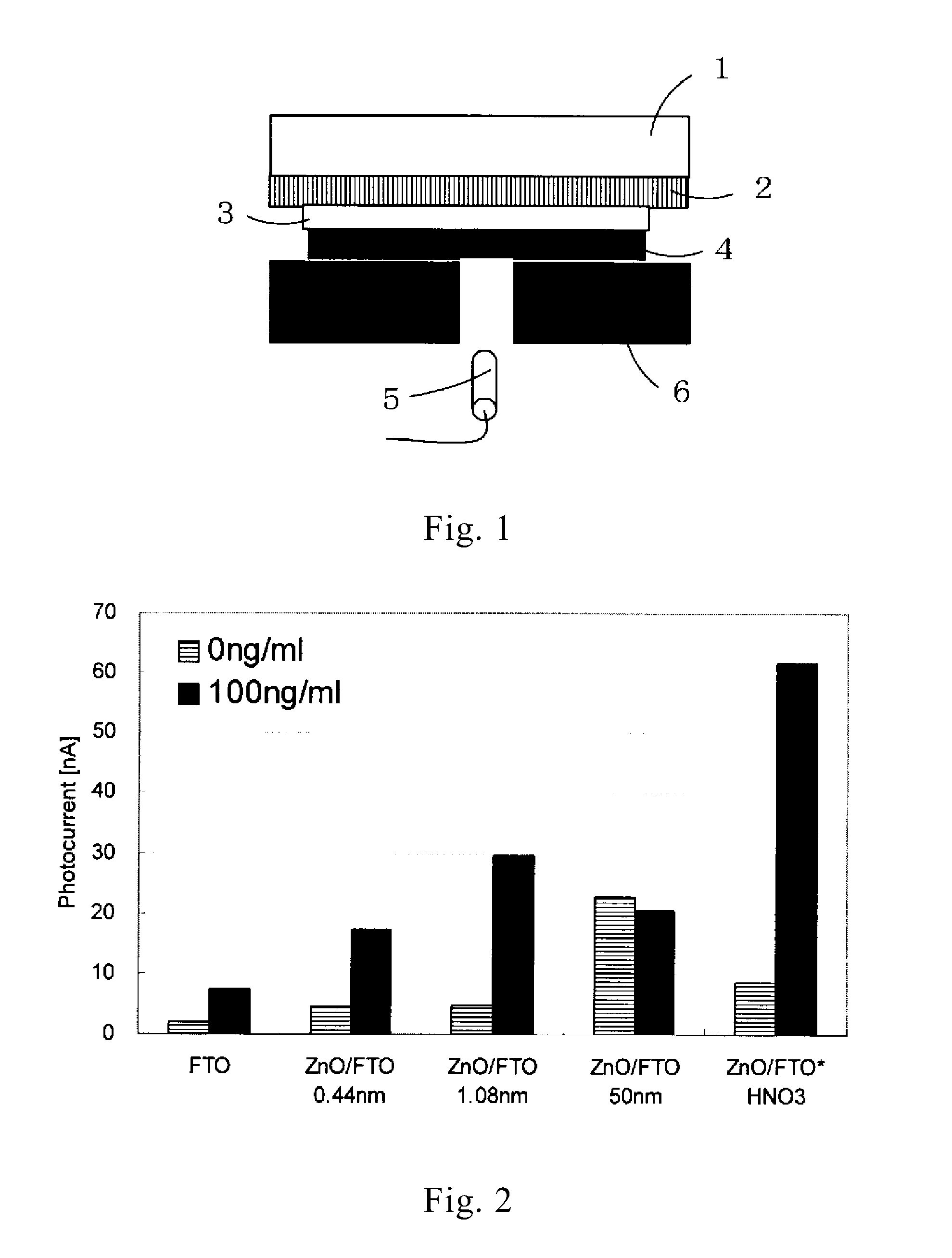
Specific Detection of Protein with Photocurrent Using Different Working Electrodes

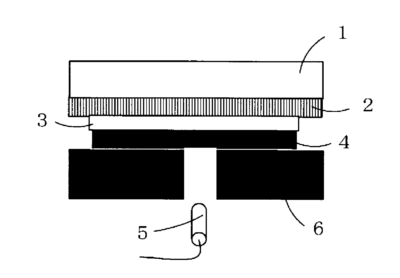
[0065]Preparation of Working Electrode

[0066]FTO Electrode

[0067]A fluorine-doped tin oxide (F—SnO2:FTO) coated glass (manufactured by AI Special Glass Company, U film, sheet resistance: 12 Ω / cm2, shape: 50 mm×26 mm) was provided as a substrate for a working electrode.

[0068]ZnO / FTO Electrode

[0069]ZnO was sputtered on the FTO electrode to a thickness of 0.44 nm (sputtering time 20 sec, 50 W, sputter rate 1.3 nm / min), to a thickness of 1.08 nm (sputtering time 50 sec, 50 W, sputter rate 1.3 nm / min), and to a thickness of 50 nm (sputtering time 8 min, 100 W, sputter rate 6.25 nm / min) to provide electrodes (film thickness: estimated roughly from sputter rate).

[0070]ZnO / FTO* Electrode

[0071]ZnO was sputtered on the FTO electrode to a thickness of 50 nm (200 W, sputtering time 8 min, sputter rate 6.25 nm / min) to provide an electrode (film thickness: estimated roughly from sputter rate). This electrode was ultra...

example 2

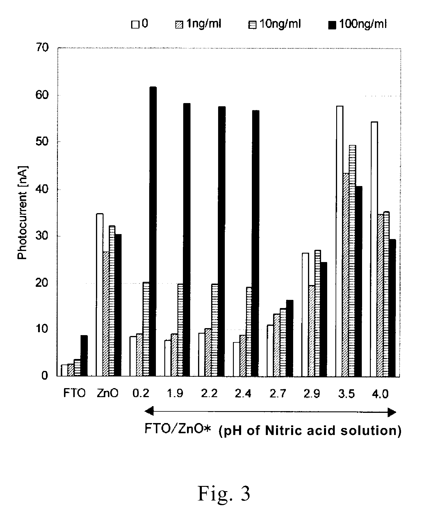
Partial Removal by Nitric Acid Treatment

[0079]Preparation of Electrodes

[0080]A fluorine-doped tin oxide (F—SnO2:FTO) coated glass (manufactured by AI Special Glass Company, U film, sheet resistance: 12 Ω / cm2, shape: 50 mm×26 mm) was provided as a substrate for a working electrode. Electrodes (ZnO / FTO) were prepared by sputtering ZnO to a thickness of 50 nm on the electrode in the same manner as in Example 1. The electrodes were ultrasonically cleaned with acetone, ultrapure water, and acetone successively in that order each for one min and were then immersed in respective nitric acid solutions having different concentrations. The nitric acid solutions used were prepared to have the following eight pH values (concentrations). The nitric solutions prepared and the immersion times of the electrodes are shown in Table 1. After immersion in the solutions, the electrodes were cleaned by thorough rinsing with ultrapure water.

TABLE 11pHNitric acid concentrationImmersion time0.21M3 min1.910m...

example 3

Surface Elementary Analysis of Electrode

[0085]Nine types in total of electrodes, i.e., ZnO / FTO electrodes (ZnO film thickness: 0.44 nm, 1.03 nm, 50 nm) and ZnO / FTO* electrodes (nitric acid treatment concentration: 1 M, 5 mM, 3.2 mM, 2 mM, 0.1 mM) prepared in Example 1 and the FTO electrode, were subjected to a surface elementary analysis by an X-ray photoelectric analysis. The analysis was carried out with an X-ray photoelectron spectroscopic device (manufactured by ULVAC-PHI, model PHI 1800) under conditions of X-ray source MgKα (100 W) and analysis area 0.8×2.0 mm. The results were as shown in Table 2.

[0086]It was found from Table 2 that the amount of zinc present on the surface of the electrodes varied depending upon zinc sputtering time and pH of the nitric acid solution.

TABLE 2pH ofpH of nitricnitric acidacid solutionsolutionCOZnSnFTO—23.152.70.024.2FTO / ZnO (0.44 nm, untreated)—21.452.34.222.1FTO / ZnO (1.08 nm, untreated)—23.749.510.516.3FTO / ZnO (50 nm, untreated)—26.445.927.60....


PUM


Abstract

Disclosed is an electrode member for specific detection of an analyte using a photocurrent. The electrode member has at least a conductive substrate and an electron-accepting substance provided on said conductive substrate. The aforementioned electron-accepting substance consists at least of a first substance layer that is made of a semiconductor and a second substance that is made of a semiconductor of a kind different from that of the aforementioned semiconductor, a metal or a metal oxide, and is carried on the surface of said first substance layer. With the electrode member, improved detection sensitivity for the test substance and improved measurement precision can be achieved with specific detection of an analyte using a photocurrent.

Description

RELATED APPLICATION[0001]This application is based upon and claims the benefit of priority from Japanese Patent Applications No. 2008-223350, filed on Sep. 1, 2008, the contents of which are incorporated herein by reference.TECHNICAL FIELD[0002]The present invention relates to an electrode member for specific detection of analytes having a specific binding capability such as nucleic acids, exogenous endocrine disrupting chemicals, or antigens through the utilization of photocurrent, and a process for producing the electrode member.BACKGROUND ART[0003]Attempts have recently been made to develop testing systems that can simply detect at low cost hormones and proteins which can predict symptoms and progress of diseases of humans. Further, damage to genital systems, nervous systems and the like by exogenous endocrine disrupting chemicals (environmental hormones) including dioxins is recognized as social problems, leading to a demand for the development of a simple method that can detect...

Claims


Application Information

Patent Timeline
IPC IPC(8): H01L31/0224H01L31/18B82Y99/00
CPCY02E10/542H01G9/2027Y02P70/50
Inventor NAKAMURA, MASAKOBEKKI, MAKOTONARITA, JYUNYASONEZAKI, SHUJIOHARA, HITOSHI
Owner TOTO LTD
Why Eureka
Try Eureka