Methods And Devices For Delivering Appropriate Minimally-Invasive Extraocular Radiation

a technology of extraocular radiation and radiation delivery method, applied in the field of minimally invasive, can solve the problems of progressive damage to the retina, difficult or impossible reading, driving, recognizing faces and other detailed tasks, and severe retinal complications in the study results

Inactive Publication Date: 2011-08-25
SALUTARIS MEDICAL DEVICES
View PDF99 Cites 27 Cited by
  • Summary
  • Abstract
  • Description
  • Claims
  • Application Information

AI Technical Summary

Benefits of technology

[0026]The radiation shield may comprise a base having a groove disposed in a top surface near a side edge. A lid may be pivotally or removably attached to the base, wherein the lid forms an inner cavity. The lid can move between at least an open position and a closed position r

Problems solved by technology

ARMD attacks the center region of the retina (i.e., macula), responsible for detailed vision and damages it, making reading, driving, recognizing faces and other detailed tasks difficult or impossible.
In wet AR

Method used


Image

  • Methods And Devices For Delivering Appropriate Minimally-Invasive Extraocular Radiation
  • Methods And Devices For Delivering Appropriate Minimally-Invasive Extraocular Radiation
  • Methods And Devices For Delivering Appropriate Minimally-Invasive Extraocular Radiation

Examples


example 1

Surgical Procedure

[0196]The following example describes a surgical procedure using a device of the present invention.

[0197]1. Create buttonhole incision in conjunctiva and Tenon's Capsule.

[0198]2. Gently separate Tenon's Capsule from sclera by injecting Balance Salt Solution (BSS) or lidocaine without epinephrine.

[0199]3. Insert the distal tip of the cannula into the subtenon space using the previously made incisions.

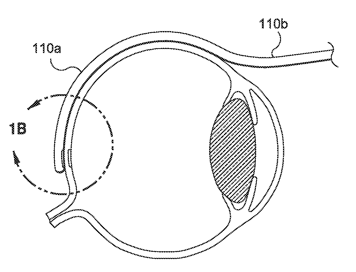
[0200]4. Continue to insert the cannula until the distal tip is located at the posterior pole of the eye (See FIG. 1A).

[0201]5. Once this gross position is achieved, activate the light tip of the cannula by activating the power of the light source.

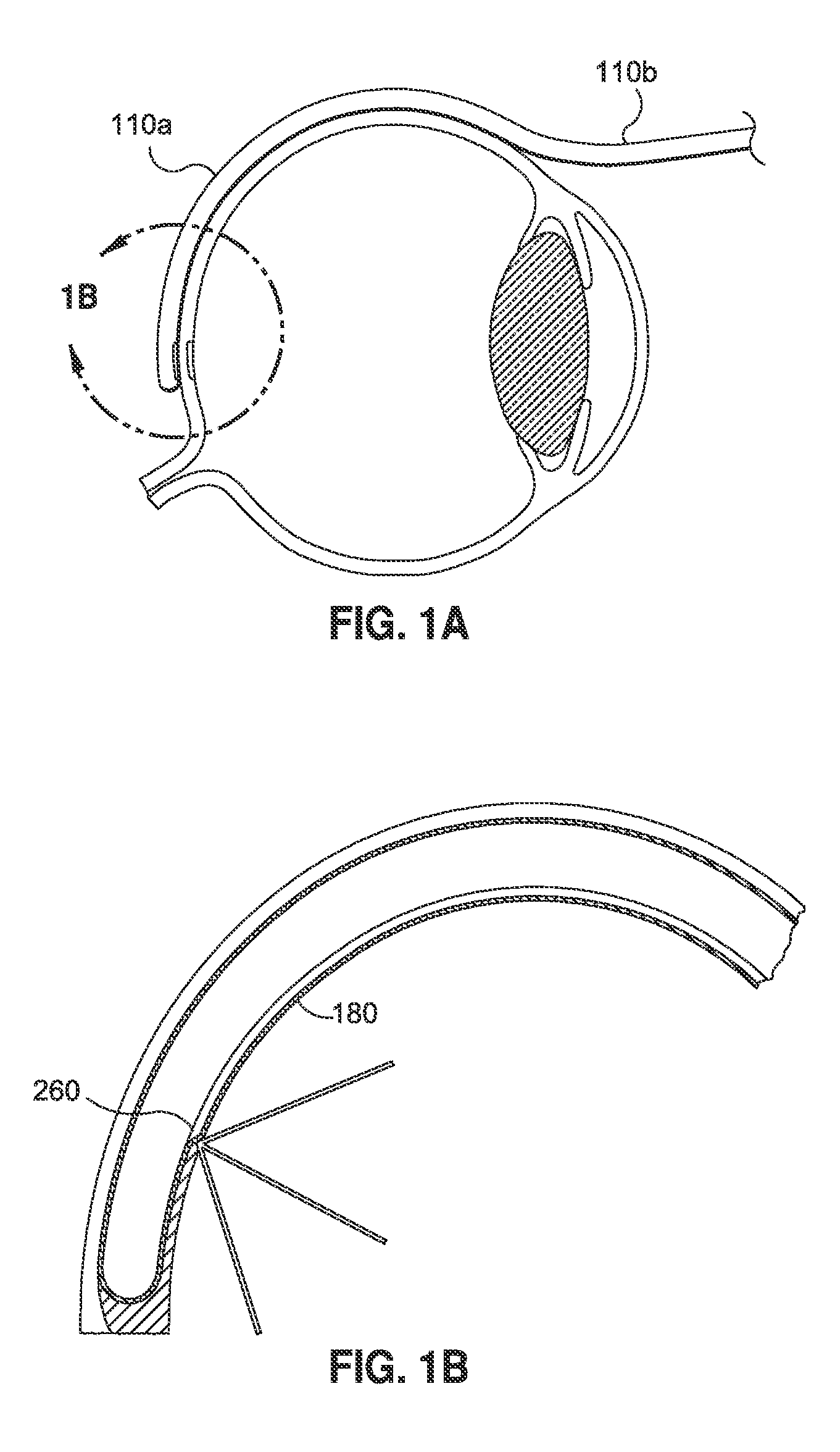
[0202]6. View the lighted tip through an indirect ophthalmoscope and adjust the tip position for treatment of the defect (see FIG. 1B).

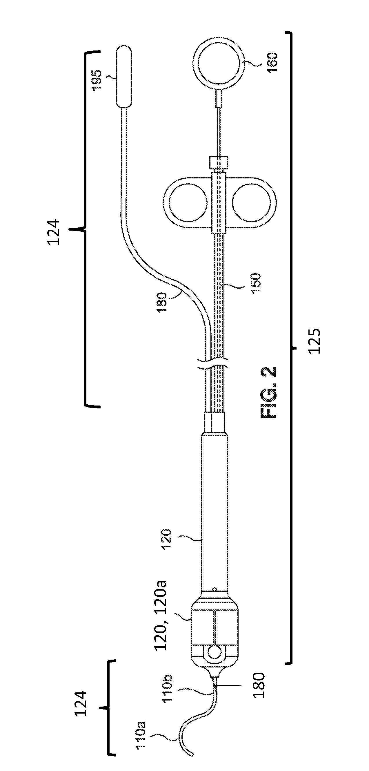
[0203]7. Brachytherapy administration: Deploy the radioactive seed. Full deployment of the seed may be verified visually in the handle 120 of the device (e.g., viewing a visual marker on the pl...

example 2

Calibration of RBS Placement

[0207]The following example describes an example of verifying / calibrating RBS placement using GafChromic® film (e.g., procedure for checking deployment of the seed, adjusting the travel of the seed in the device to center it under the light source fiberoptic termination). Testing was performed within an acrylic test box (e.g., radiation shield with inner cavity).

[0208]1. A pen mark was placed on the GafChromic® film. The film and device (loaded with a radioisotope seed) were placed into the box.

[0209]2. The cannula tip light was centered on the pen mark. The seed was then deployed. In approximately 5 seconds, a small dark exposure was noted.

[0210]3. Examination showed that the exposure was not centered on the pen mark. Thus, the device actuator stop-collar position was adjusted with an Allen wrench.

[0211]4. The cannula tip light was again centered on another mark on the film. The seed was then re-deployed. In approximately 5 seconds, a small dark exposure...

example 3

Testing of Sterilization of Film

[0212]The following example describes a test of the competency of ethylene oxide (EO) sterilized GafChromic® film.

[0213]1. Previously GafChromic® film was packaged and EO sterilized in a sterile pouch.

[0214]2. The GafChromic® film was inspected inside its sterile pouch. The film appeared without evidence of damage. It was noted that the EO indicator strip had turned positive.

[0215]3. Testing was performed within an acrylic test box (radiation shield with inner cavity). The device cannula was placed over the outside of the sterilization pouch. The seed was advanced. In approximately 5 seconds, a small dark exposure was noted.

[0216]4. Following the procedure the sterile package was opened and the film directly inspected. No discoloration nor other damage was noted. It was concluded that the GafChromic® film retained its competency for this purpose following EO sterilization.


PUM


Abstract

The present invention also features a brachytherapy system comprising a spiral cut tube having a first end and a second end; a radioactive brachytherapy source (RBS) disposed on the first end of the spiral cut tube; and a handle and a generally hollow cannula disposed on the handle, wherein a channel is disposed in the handle aligned with the hollow cannula, and the spiral cut tube and RBS are adapted to slide within the channel and the hollow cannula.

Description

CROSS REFERENCE[0001]This application claims priority to U.S. provisional application Ser. No. 61 / 257,232 filed Nov. 2, 2009 and U.S. provisional application Ser. No. 61 / 376,115 filed Aug. 23, 2010, the specifications of which are incorporated herein by reference in its entirety.FIELD OF THE INVENTION[0002]The present invention is directed to minimally-invasive methods and devices for introducing radiation to the posterior portion of the eye for treating and / or managing eye conditions including but not limited to macula degeneration.BACKGROUND OF THE INVENTION[0003]Several diseases and conditions of the posterior segment of the eye threaten vision. Age related macular degeneration (ARMD), choroidal neovascularization (CNV), retinopathies (e.g., diabetic retinopathy, vitreoretinopathy), retinitis (e.g., cytomegalovirus (CMV) retinitis), uveitis, macular edema, and glaucoma are several examples.[0004]Age related macular degeneration (ARMD) is the leading cause of blindness in the elde...

Claims


Application Information

Patent Timeline
IPC IPC(8): A61N5/00
CPCA61M25/00A61M35/00A61M2210/0612A61B2019/5445A61N2005/1025A61N5/1048A61N5/1017A61B2090/3945
Inventor DICARLO, PAULMEADE, ERICDEGON, ROBERTHAMILTON, RUSSELL J.
Owner SALUTARIS MEDICAL DEVICES
Why Eureka
Try Eureka