Monitoring urodynamics by trans-vaginal nirs

a trans-vaginal nirs and urodynamic technology, applied in the field of nirs, can solve problems such as abnormal bladder function, incomplete bladder emptying, overactive bladder,

Active Publication Date: 2011-09-15
THE UNIV OF BRITISH COLUMBIA
View PDF8 Cites 16 Cited by
  • Summary
  • Abstract
  • Description
  • Claims
  • Application Information

AI Technical Summary

Benefits of technology

[0019]In accordance with an alternative aspect of the invention, a method is provided for transvaginal NIRS monitoring of a target tissue, such as non-invasive monitoring of a human patient. To record periodic or continuous monitoring signals, the probe of the invention may be inserted into a vaginal lumen defined by vaginal walls. A near infrared light signal source may be operated, in communication with an emitter, to control the emission of near infrared light by the emitter, and a near infrared light detector, in communication with the collector, may be operated to monitor the collected light signal. An output device may be operated to display information that is indicative of an activity of the target tissue, such as a urogenital muscle.
[0020]In an alternative aspect, the invention provides an instrument for non-invasive transvaginal monitoring of a target tissue, such as urodynamic muscle activity. The instrument may include a probe body, as discussed above, with means provided for communicating with the probe body, comprising means for communicating a near infrared light signal to the emitter; and, means for communicating the collected light signal ex vivo. The means for communicating with the probe body may for example include a near infrared light signal source, in communication with the emitter to control the emission of near infrared light by the emitter; and, a near infrared light detector, in communication with the collector to detect the collection of near infrared light b

Problems solved by technology

Abnormal muscle activity in the urinary bladder or urethral sphincter may also cause other problems, for instance overactive bladder or incomplete bladder emptying.
Abnormality of the detrusor muscles of the bladder, resulting in abnormal bladder function such as overactive bladder or urinary incontinence, may be caused by impairments, for instance, to the detrusor muscle activity or to the neurological connections to the detrusor muscle (Semins and Chancellor 2004).
Conventional urodynamics procedures do not provide direct measurements of bladder muscle activity.
Other imaging modalities have

Method used


Image

  • Monitoring urodynamics by trans-vaginal nirs
  • Monitoring urodynamics by trans-vaginal nirs
  • Monitoring urodynamics by trans-vaginal nirs

Examples


example

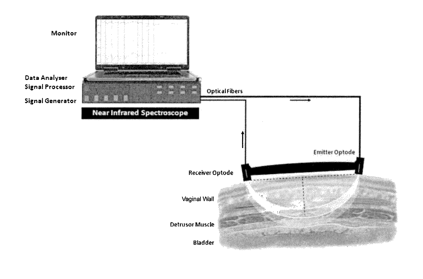
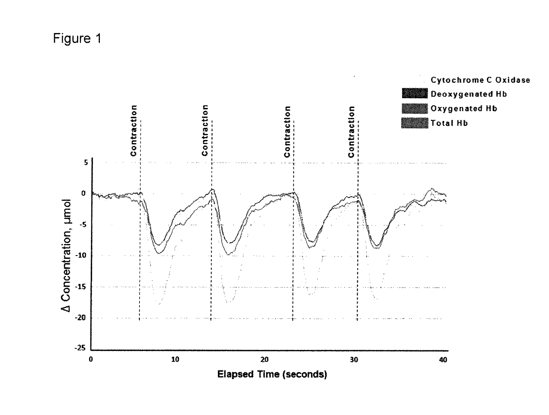
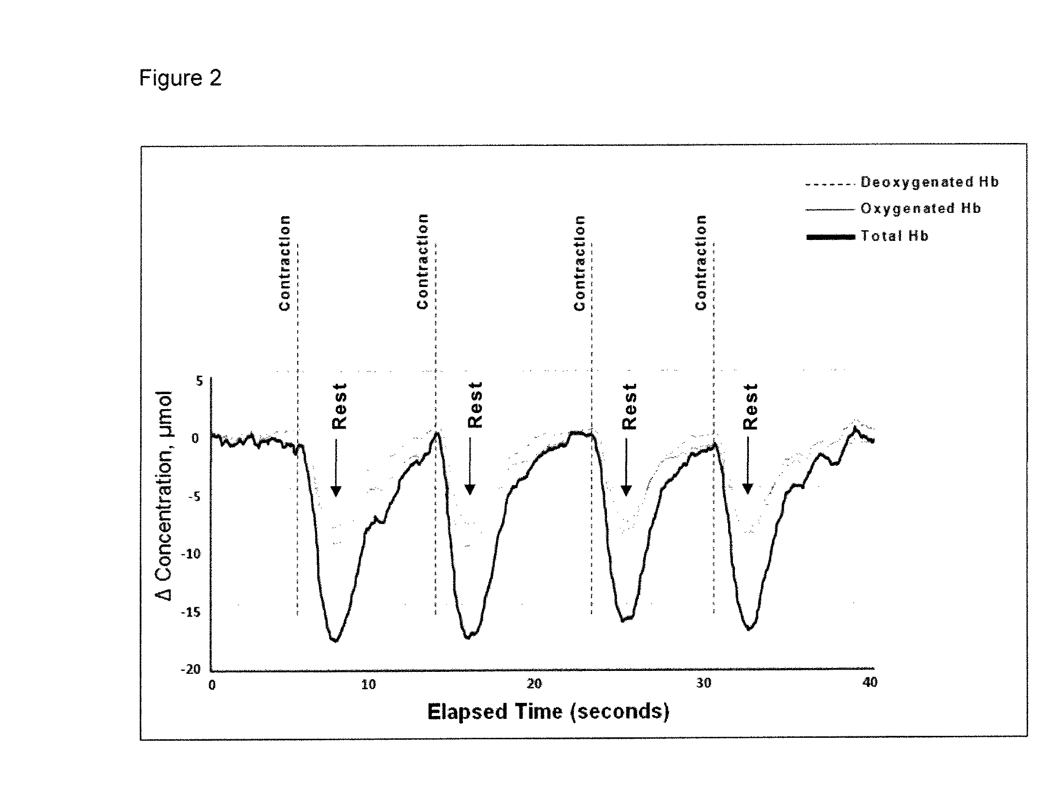
[0058]Biofeedback Monitoring of Urethral Sphincter via the Vagina using a NIRS Apparatus

[0059]Urethral sphincter tissue was monitored in a female subject using an intravaginal NIRS apparatus (FIG. 1). The apparatus contained a NIRS emitter and NIRS collector / detector within a probe housing designed to be inserted into the female vagina. The NIRS emitter and NIRS collector / detector were connected to a NIRS device via fiber optic cables. The NIRS device used to control the NIRS emitter and collector / detector was the Artinis Medical Systems (Netherlands) Oxymon Mk III. Four contractions of the urinary sphincter during urinary sphincter muscle exercises were detectable using this system. The speed of recovery was observed to lengthen with each subsequent contraction, which may be a result of muscle fatigue.

[0060]The size of the emitter and receiver optodes was as follows: 3 mm wide, 8 mm long, and 4 mm deep, and the fine glass fiber cables were small enough to incorporate into a probe h...


PUM


Abstract

The invention relate to the demonstration herein that it is feasible to use a transvaginal NIRS probe to interrogate functioning urological tissues, such as the urethral sphincter, the bladder detrusor muscle, and pelvic floor musculature, to obtain clinically relevant information. The present invention accordingly provides methods and devices for transvaginal monitoring or imaging of the urological tissues, such as the urethral sphincter and/or the bladder, and/or pelvic floor musculature, using NIRS.

Description

FIELD OF THE INVENTION[0001]The present invention relates to the field of physiological near-infrared spectroscopy (NIRS). The invention more specifically relates to the use of NIRS in diagnostic urological applications.BACKGROUND[0002]Urinary incontinence, or involuntary leakage of urine, is a condition which affects a significant portion of the population, but is especially prevalent in women (Melville et al. 2005). Urinary incontinence may present in different forms, such as stress incontinence and urge incontinence, or a mixture of forms. Continence relies on a number of factors, including relaxation of the detrusor muscles around the bladder wall, and proper activity of the urethral sphincter muscles and structures (including blood vessels) around the urethra (Smith et al. 2006). Abnormal muscle activity in the urinary bladder or urethral sphincter may also cause other problems, for instance overactive bladder or incomplete bladder emptying.[0003]Abnormality of the detrusor mus...

Claims


Application Information

Patent Timeline
IPC IPC(8): A61B6/00
CPCA61B5/0075A61B5/0084A61B2503/40A61B5/202A61B5/4519A61B5/0086
Inventor STOTHERS, LYNNMACNAB, ANDREW J.SHADGAN, BABAK
Owner THE UNIV OF BRITISH COLUMBIA
Why Eureka
Try Eureka