Method and system for mapping tissue status of acute stroke

a tissue status and acute stroke technology, applied in the field of acute stroke tissue status mapping, can solve the problems of inability to accurately delineate the penumbra and infarct regions of inability to apply the same cbv or cbf threshold values to gray matter and white matter, and inability to accurately delineate the penumbra and infarct regions by applying the same cbv or cbf threshold values. the effect of normal decon

Active Publication Date: 2011-09-22
APOLLO MEDICAL IMAGING TECH
View PDF44 Cites 12 Cited by
  • Summary
  • Abstract
  • Description
  • Claims
  • Application Information

AI Technical Summary

Benefits of technology

[0017]The method may further include the step of applying motion correction after loading the data to compensate for motion artefacts due to the movement over time of the subject, such as a tissue or organ. The method may further comprise the s

Problems solved by technology

It practice, it is difficult to measure the AIF from a blood vessel immediately feeding the tissue of interests.
However, the CBF and CBV values in normal gray matter are higher (usually about 2 to 3 times) than those in normal white matter, applying the same CBV or CBF threshold values to gray matter and white matter may not be able to delineate the penumbra and infarct regions accurately.
Further, in the case of major vessel disease such as acute stroke, a measured global AIFg from a large artery is often associated with a delay and dispersion effect before it reaches the ischemic tissue, causing overestimation of the MTT and underestimation of the CBF calculated by the normal deconvolution technique.
Hence method using MTT or CBF thresholds for identifying the ischemic lesion has the pote

Method used


Image

  • Method and system for mapping tissue status of acute stroke
  • Method and system for mapping tissue status of acute stroke
  • Method and system for mapping tissue status of acute stroke

Examples


Embodiment Construction

[0036]Although the following detailed description contains many specifics for the purposes of illustration, anyone of ordinary skill in the art will readily appreciate that many variations and alterations to the following exemplary details are within the scope of the invention. Accordingly, the following preferred embodiment of the invention is set forth without any loss of generality to, and without imposing limitations upon, the claimed invention.

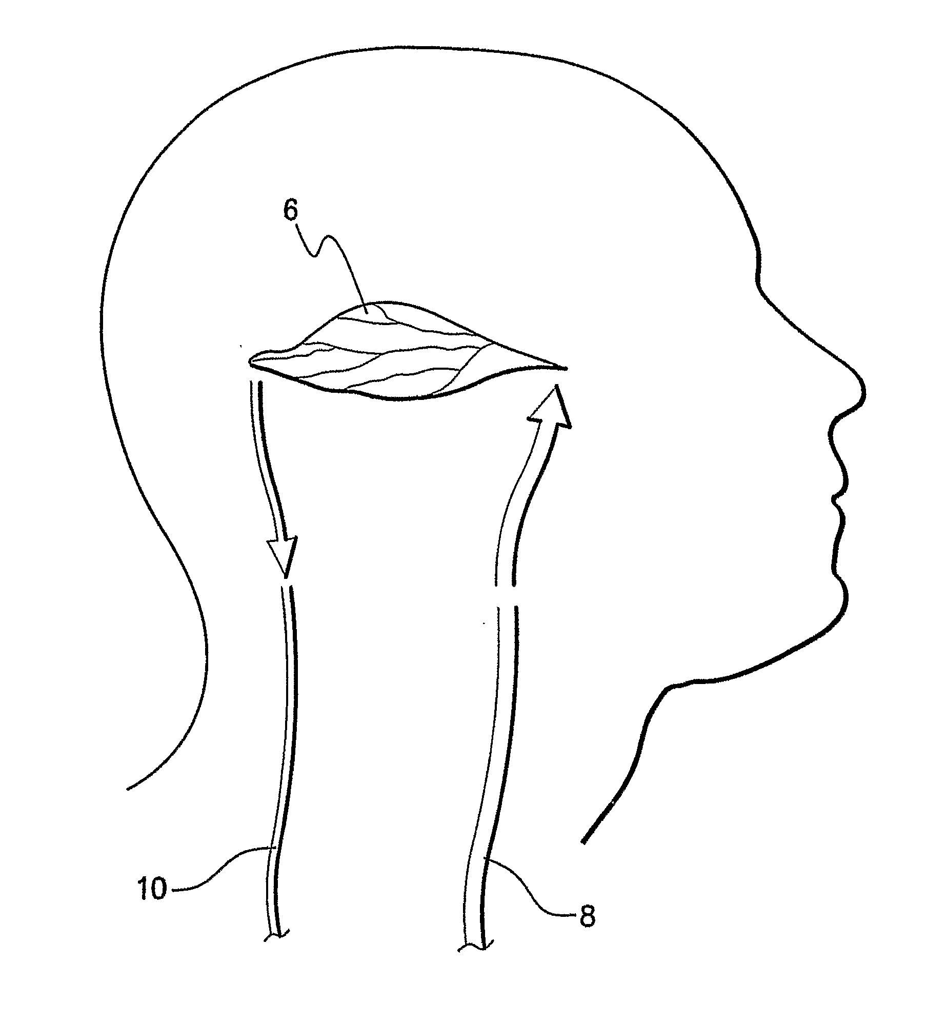
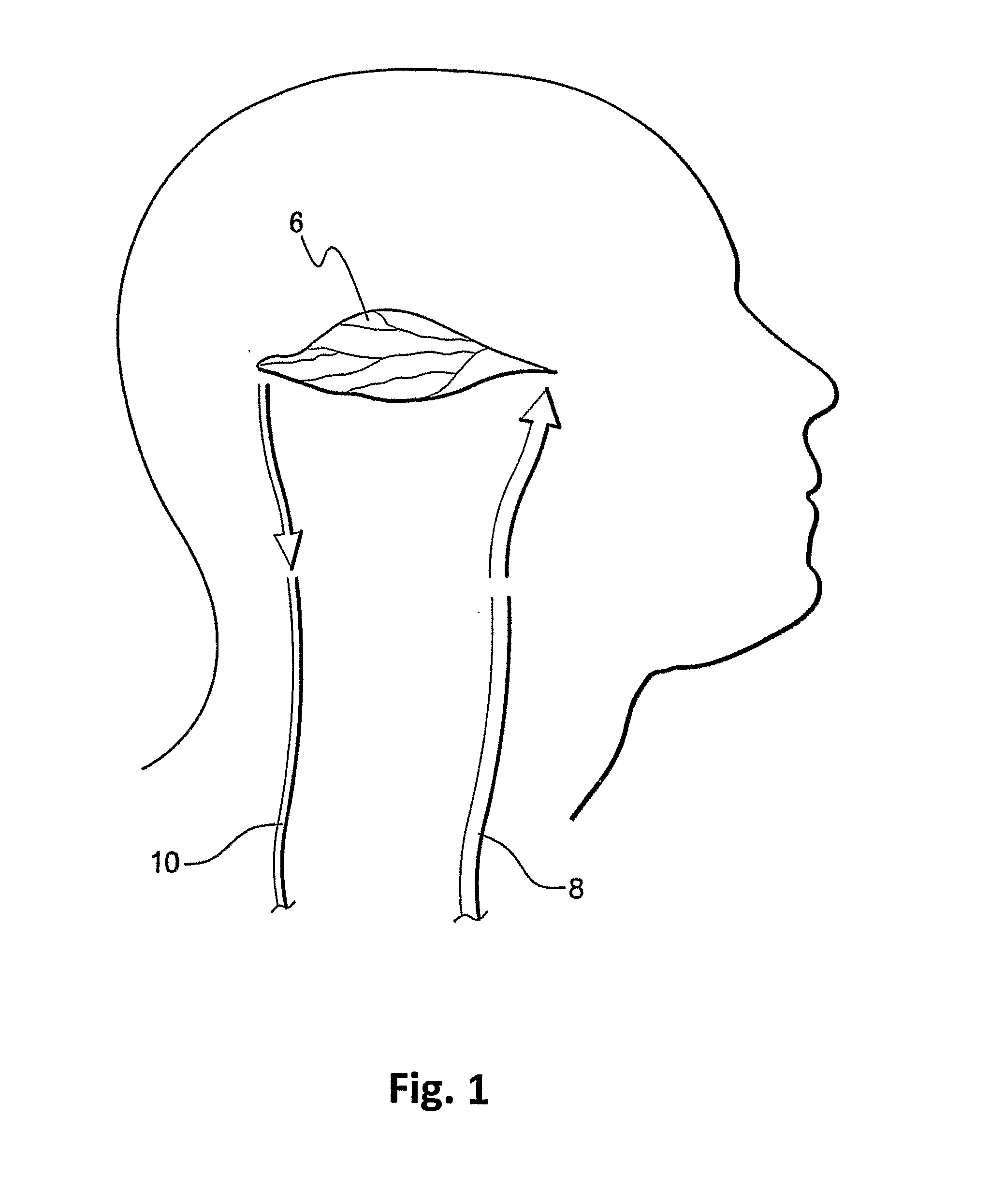
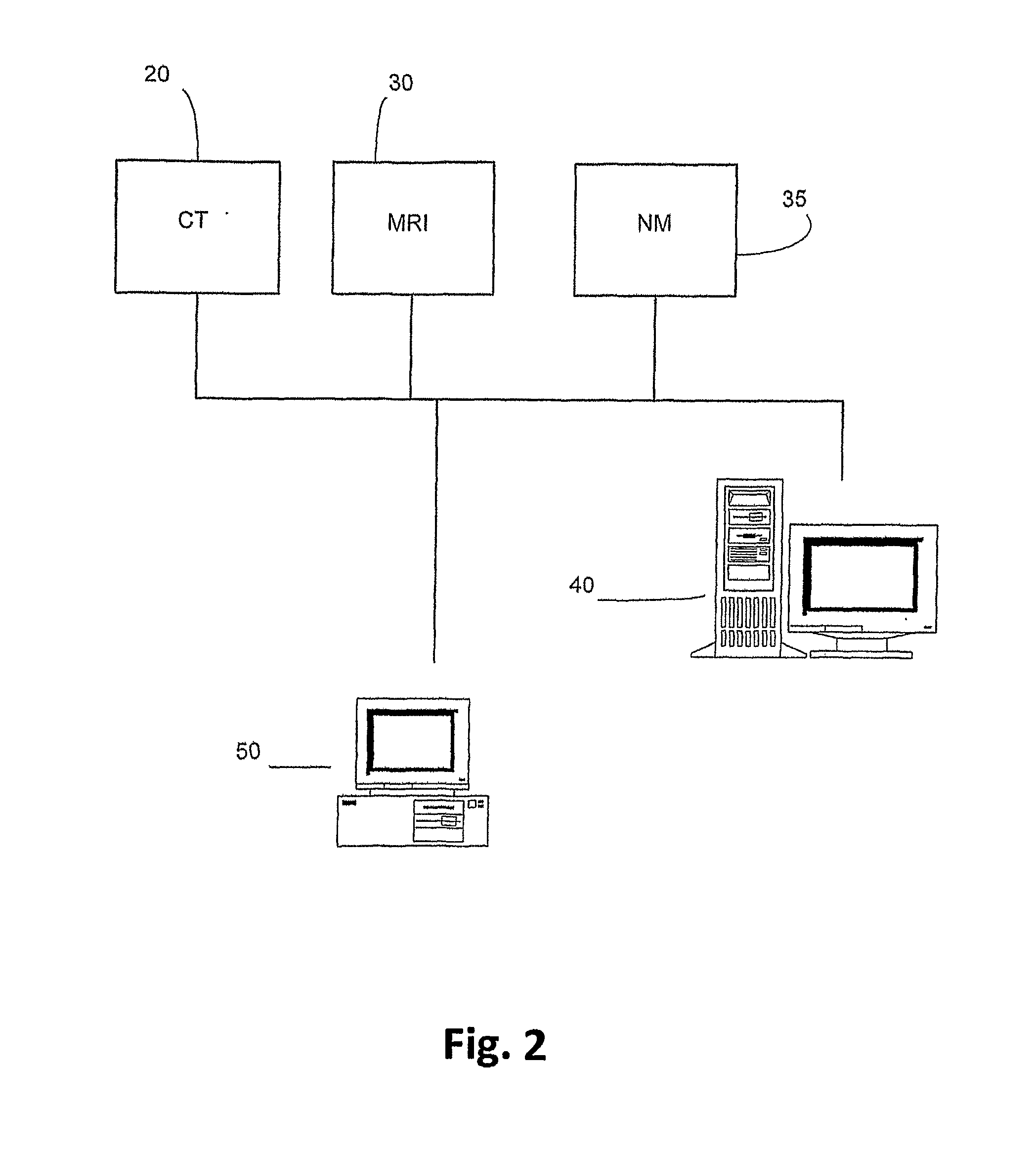
[0037]The present invention is particularly applicable to CT, MRI and MN imaging systems. A bolus of contrasting agents is introduced via a needle into a patient at, for example, the arm of the patient. However the bolus can be input to any other part of the patient. A region of interest (ROI) may be a tissue 6 in a part of the patient's brain as shown in FIG. 1. Alternatively, the ROI may be a pixel or a plurality of pixels, where many pixels represent a calculated image to produce one or more perfusion maps. Blood circulating throughout...


PUM


Abstract

The current invention provides a method of identifying a ischemic lesion. The method includes loading perfusion imaging data into an electronic memory element and deriving perfusion maps from the perfusion imaging data, where the perfusion maps include a cerebral blood volume (CBV) map and an arterial delay time (DT) map, which utilize arterial delay and dispersion effects. Ischemic pixels are determined from the perfusion imaging data, where the DT is greater than a predetermined first threshold value and the CBV is below a second threshold value and the infarct portion of the ischemic lesion is determined, where DT is greater than a predetermined third threshold value and/or the CBV is below a forth threshold value. A cluster analysis is applied to all of the determined ischemic lesion and infarct pixels and the penumbra is then determined, where mismatch regions between the ischemic lesion and the infarct core define the penumbra.

Description

FIELD OF THE INVENTION[0001]This invention relates to a method and system of identifying the ischemic lesion, particularly for identifying the penumbra and infarct regions reflecting the tissue status of acute stroke.BACKGROUND[0002]Stroke is one of the major diseases causing death and disability. While stroke can be hemorrhagic and ischemic, the majority of stroke is ischemic due to a sudden blockage of blood supply to whole or part of the brain. During a stroke, the viability of the ischemic lesion depends on the level and duration of compromised blood flow. Within hours of the onset of a stroke, the ischemic lesion can consist any of the two types of tissues: (1) an infarct core of irreversibly damaged tissue; and (2) a penumbra region with reduced blood flow that is at risk of further infarction, but the tissue is salvageable by quick restoration of blood supply to the region. The process of measuring blood perfusion hence identifying the infarct core and penumbra in acute strok...

Claims


Application Information

Patent Timeline
IPC IPC(8): G06K9/00
CPCA61B5/055A61B5/416A61B6/507A61B6/5217A61B6/5247A61B6/501G06T7/0012G06T2207/10072G06T2207/30104G06T7/11G06T7/136G16H50/30
Inventor YANG, QING
Owner APOLLO MEDICAL IMAGING TECH
Why Eureka
Try Eureka