
Stem cells derived from the carotid body and uses thereof

a stem cell and carotid body technology, applied in the field of stem cells derived from the carotid body isolating and characterization, to achieve the effect of improving the treatment methodology and reducing damag

Inactive Publication Date: 2011-10-13
UNIV DE SEVILLA
View PDF2 Cites 0 Cited by
  • Summary
  • Abstract
  • Description
  • Claims
  • Application Information

AI Technical Summary

Benefits of technology

[0011]Now, the inventors discovered, surprisingly, the presence of stem cells in the adult peripheral nervous system (PNS), more concretely in the carotid body (CB). Said adult stem cells, contrary to what might be thought initially, are not glomus cells (or type I cells), but sustentacular cells or type II cells. As shown in the examples in this description, said adult stem cells have the capacity to divide (proliferation), renew themselves by means of cell division over long periods of time (self-renewal) and, under certain physiological or experimental conditions, the capacity to be induced to differentiate into other types of specific differentiated cells (differentiation). Moreover, the inventors developed an in-vitro method for the expansion of the CB that makes it possible to obtain cellular aggregates capable of expressing and secreting trophic factors, neurotransmitters, neuromodulators, enzymes, etc. Transplants with said adult stem cells or cellular aggregates obtained by said method of expansion could be useful in clinical practice, in particular in the treatment of neurodegenerative diseases.
[0012]As already mentioned in the Background of the Invention, autografts with cells obtained from the carotid body for the treatment of neurodegenerative diseases have been used for the treatment of degenerative diseases, since it is known that in these pathologies, deficiency of some molecules (for example, neurotransmitters, growth factors, etc.) can be compensated by implanting cells capable of expressing and secreting said molecules. In any case, autografts with glomus cells obtained from the CB have proved inadequate for mitigating the damage caused by diseases, such as Alzheimer's disease or Parkinson's disease, therefore it is necessary to improve said treatments or find alternatives to them. The experiments described in detail below show that the glomus cells obtained by the method provided by this invention, as well as being capable of obtaining a larger amount of them, makes it possible to improve the methodologies of treatment by autografts of cells from the CB that are currently in existence.

Problems solved by technology

In any case, autografts with glomus cells obtained from the CB have proved inadequate for mitigating the damage caused by diseases, such as Alzheimer's disease or Parkinson's disease, therefore it is necessary to improve said treatments or find alternatives to them.

Method used


Image

  • Stem cells derived from the carotid body and uses thereof
  • Stem cells derived from the carotid body and uses thereof
  • Stem cells derived from the carotid body and uses thereof

Examples


example 1

Induction of Growth of the Carotid Body by Hypoxia and Generation of New Glomus Cells Derived from the Neural Crest

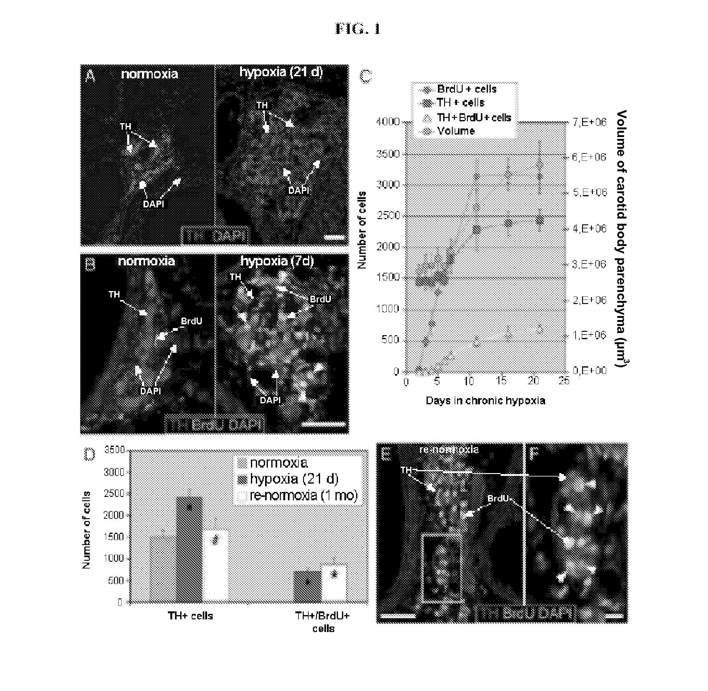
[0189]Exposure of wild-type C57BL / 6 mice to hypoxia (10% O2) in an isobaric atmosphere for 3 weeks induced an increase in size of the carotid body of approximately 2 to 3 times relative to the initial size, owing to dilatation and multiplication of blood vessels, as well as expansion of the parenchyma, with an increase in the number of TH+ glomus cells (positive for tyrosine hydroxylase) (FIG. 1A).

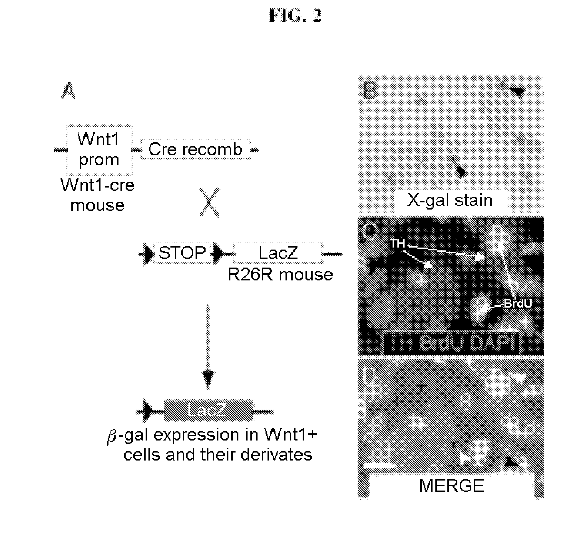
[0190]For analysis of the proliferation and formation of new glomus cells, the C57BL / 6 mice were treated with BrdU and were then kept for several days in a normoxic (21% O2) or hypoxic (10% O2) environment. No TH+ and BrdU+ cells were observed in the normoxic animals, indicating absence of proliferation (FIG. 1B, left). In contrast, after some days in hypoxia, even before growth of the CB was apparent, numerous TH+ and BrdU+ cells were observed (arrows in FIG. 1B right), indic...

example 2

Identification of Self-Renewing and Pluripotent Stem Cells of the Carotid Body

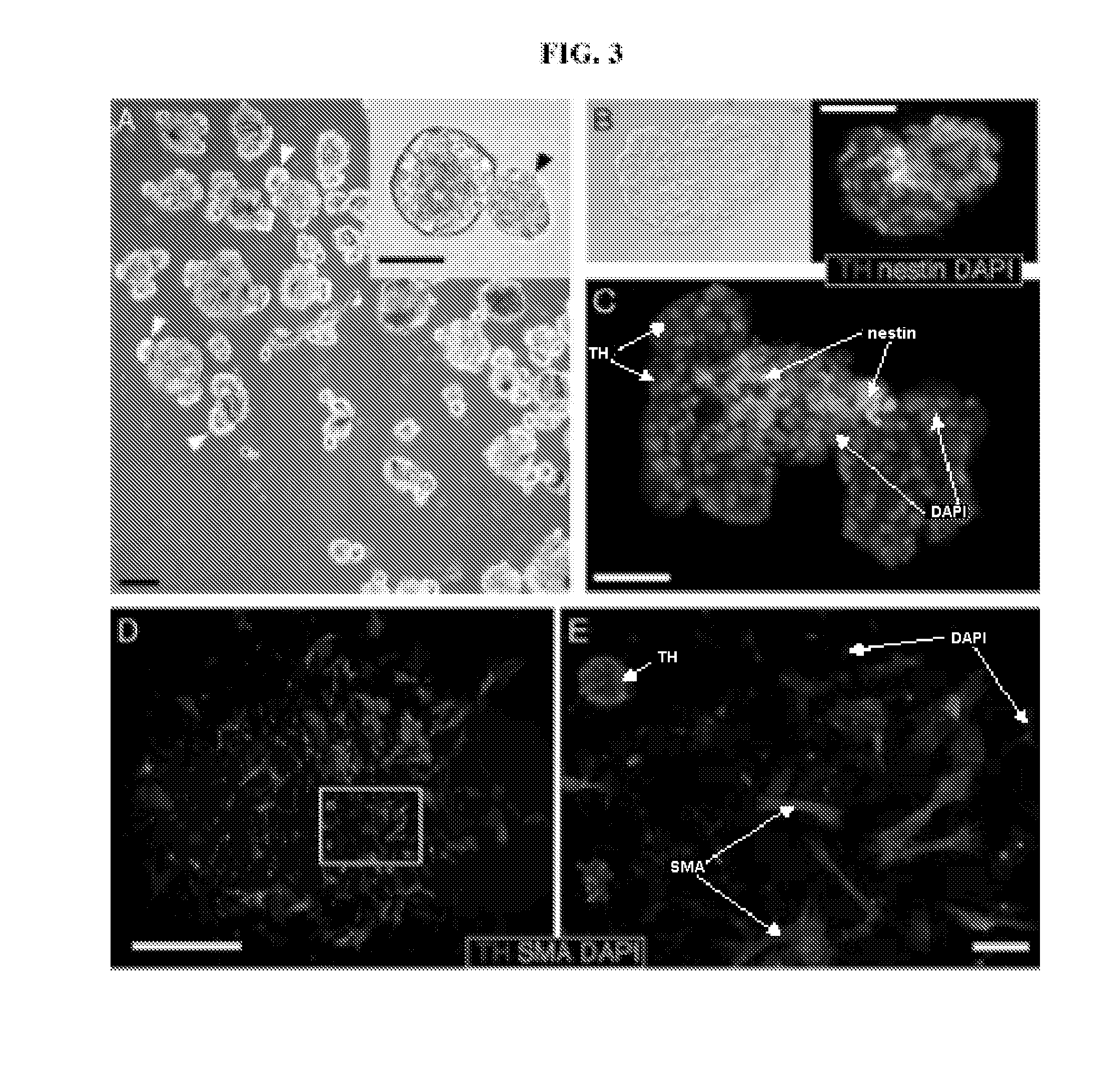
[0193]For identification of the precursors that give rise to the glomus cells in adult rodents exposed to hypoxia, the carotid bodies of young Wistar rats (>P20) were dispersed enzymatically and the cells were plated in neural crest (NC) culture media that contained bFGF, IGF-I and EGF (Morrison et al., 1999, Cell 96, 737-749). Using culture plates treated for non-adhesion, growth was induced in conditions of flotation and neurospheres were thus obtained. Clonogenic proliferation was stimulated by culture of the cells in moderate hypoxia (3% O2) (Morrison et al., 2000, J Neurosci 20, 7370-7376), a condition that mimics the stimulation by hypoxia of the growth of the CB in vivo.

[0194]In these experiments, 1.04±0.4% (n=6 preparations) of the cells plated gave rise to neurospheres (FIG. 3A). After 10 days in culture the diameter of the neurospheres was 85±34 micrometres (n=112), a value comparable to that des...

example 3

The Sustentacular Cells of Glial Type are the Stem Cells of the CB

[0199]Until now, it has always been thought that in certain circumstances the glomus cells are capable of entering a mitotic cycle (Nurse and Vollmer, 1997 Dev Biol 184, 197-206; Wang and Bisgard, 2002 Microsc Res Tech 59, 168-177). However, it is well established that the TH+ glomus cells can, after dissociation, be maintained in culture for several days (López-Barneo et al., 1988 Science 241, 580-582; Villadiego et al., 2005 J Neurosci 25, 4091-4098). To verify whether proliferation of the glomus cells gave rise to the CB neurospheres, an immunocytochemical analysis was carried out in different stages of formation of neurospheres (FIG. 4A). Initially, the neurospheres were small and were formed by groups of TH−, nestin+ and other nestin− cells. This stage is soon followed by the appearance of small germinal nuclei (protrusions) with some TH+ cells at the periphery of the growing neurospheres. The number of TH+ cells...


PUM

PropertyMeasurementUnit
partial pressureaaaaaaaaaa
pressureaaaaaaaaaa
concentrationaaaaaaaaaa

Abstract

Adult stem cells obtained from the carotid body, characterized in that they are positive for the phenotypic marker GFAP (glial fibrillary acidic protein) and negative for the phenotypic markers TH (tyrosine hydroxylase) and nestin, are described. These stem cells can undergo proliferation, self-renewal and differentiation to progenitor cells and differentiated cells. Said stem cells, progenitor cells and differentiated cells, expanded by any method, can be used in the treatment of neurodegenerative diseases such as Alzheimer's disease or Parkinson's disease.

Description

FIELD OF THE INVENTION[0001]The present invention relates to the isolation and characterization of stem cells derived from the carotid body (CB), to methods for their expansion and differentiation, and to their potential applications both in research and in therapy, for example in the treatment of neurodegenerative diseases such as Alzheimer's disease or Parkinson's disease.BACKGROUND OF THE INVENTION[0002]Neural or paraneural cells that secrete neurotransmitters, neuromodulators or enzymes have been employed for the treatment of neurodegenerative diseases, as an alternative to drug therapy. These cells have been used experimentally, although with limited success so far, in clinical studies.[0003]At the end of the 1970s and beginning of the 1980s, the beneficial effect of intrastriatal transplants of adrenal medulla, which possesses dopamine-secreting chromaffin cells, was observed in experimental models of Parkinson's disease in rats.[0004]The therapeutic effect of paraneural adren...

Claims


Application Information

Patent Timeline
Patent Type & Authority Applications(United States)
IPC IPC(8): A61K35/12C12N5/071C12N5/10A61P25/28C12P21/00A61K9/00A61P25/16A61P25/00C12N5/074C12Q1/02
CPCA61K35/12C12N5/0618C12N2506/08C12N2501/11C12N2501/115C12N2501/105A61P25/00A61P25/16A61P25/28A61K35/30C12N5/0622C12N5/0623C12R2001/91C12N1/00
Inventor LOPEZ BARNEO, JOSEPARDAL, RICARDOORTEGA-SAENZ, PATRICIADURAN, ROCIOBONILLA HENAO, VICTORIA EUGENIAORDONEZ FERNANDEZ, ANTONIOTOLEDO ARAL, JUAN JOSE
Owner UNIV DE SEVILLA
Learn More