
Molecular marker for cancer stem cell

a cancer stem cell and molecular marker technology, applied in the direction of immunoglobulins, peptides, drugs against animals/humans, etc., can solve the problems of difficult to specifically recognize and treat cancer stem cells, low proportion of cancer stem cells in tumor tissue, etc., and achieve tumorigenicity high relevance, discrimination of cancer stem cells, and versatile molecular markers for cancer stem cells

Active Publication Date: 2011-10-27
SUMITOMO DAINIPPON PHARMA CO LTD +1
View PDF2 Cites 15 Cited by
  • Summary
  • Abstract
  • Description
  • Claims
  • Application Information

AI Technical Summary

Benefits of technology

[0046]The molecular marker provided by the present invention can discriminate between cancer stem cells contained in cancer cells and cancer cells other than cancer stem cells. Furthermore, since the discrimination may be carried out by detection of a single molecular marker, cancer stem cells can be discriminated very easily.
[0047]Moreover, since the molecular marker of the present invention may be used in common for a plurality of cancer stem cells such as lung cancer cells, breast cancer cells, and colorectal cancer cells, it is very effective as a versatile molecular marker for cancer stem cells.
[0048]Furthermore, since the molecular marker of the present invention is not detected at all or is hardly detected in normal cells, it is useful for the discrimination of cancer stem cells and, moreover, for the discrimination of tumor tissue containing cancer stem cells.
[0049]Moreover, the molecular marker of the present invention has high relevance to tumorigenicity and is useful in cancer stem cell immunotherapy, molecular targeted therapy or gene transfer therapy targeted thereat, as well as the screening of drugs, peptides, etc. that can be used in cancer therapy.

Problems solved by technology

However, the proportion of cancer stem cells in tumor tissue is very low (Non-Patent Document 4), and it is very difficult to specifically recognize and treat cancer stem cells.
The development of techniques for detecting cancer stem cells and new therapies targeting cancer stem cells are important issues in cancer care.

Method used


Image

  • Molecular marker for cancer stem cell
  • Molecular marker for cancer stem cell
  • Molecular marker for cancer stem cell

Examples


example 1

Experimental Example 1

Isolation of Cancer Cell Sp Fraction

a) Preparation of Reagent

[0124]A 5% anti-fetal bovine serum (FCS)-containing DMEM medium was prepared, and warmed to 37° C. 50 mM Verapamil was prepared, and diluted to 5 mM with 5% FCS+DMEM. Hoechst 33342 was adjusted to 250 μg / mL with 5% FCS+DMEM. DNAseI was adjusted to 1 mg / mL with DDW, and subjected to filtration sterilization using a 0.2 μm filter.

b) Preparation of Cells for Flow Cytometry (FACS)

[0125]Cells were suspended in 4 mL of 5% FCS+DMEM, and the number of cells was counted. Furthermore, the cell concentration was adjusted to 1×10⁶ counts / mL by adding 5% FCS+DMEM, and 1 mL was sampled in a Falcon tube for verapamil (+). The verapamil (+) cells and the remaining cells (for verapamil (−)) were incubated in a water bath at 37° C. for 10 minutes. After incubation, a verapamil solution was added to the verapamil (+) such that the final concentration of verapamil became 50 to 75 μM, and a Hoechst 33342 solution was then...

example 2

Experimental Example 2

Tumorigenicity Experiment

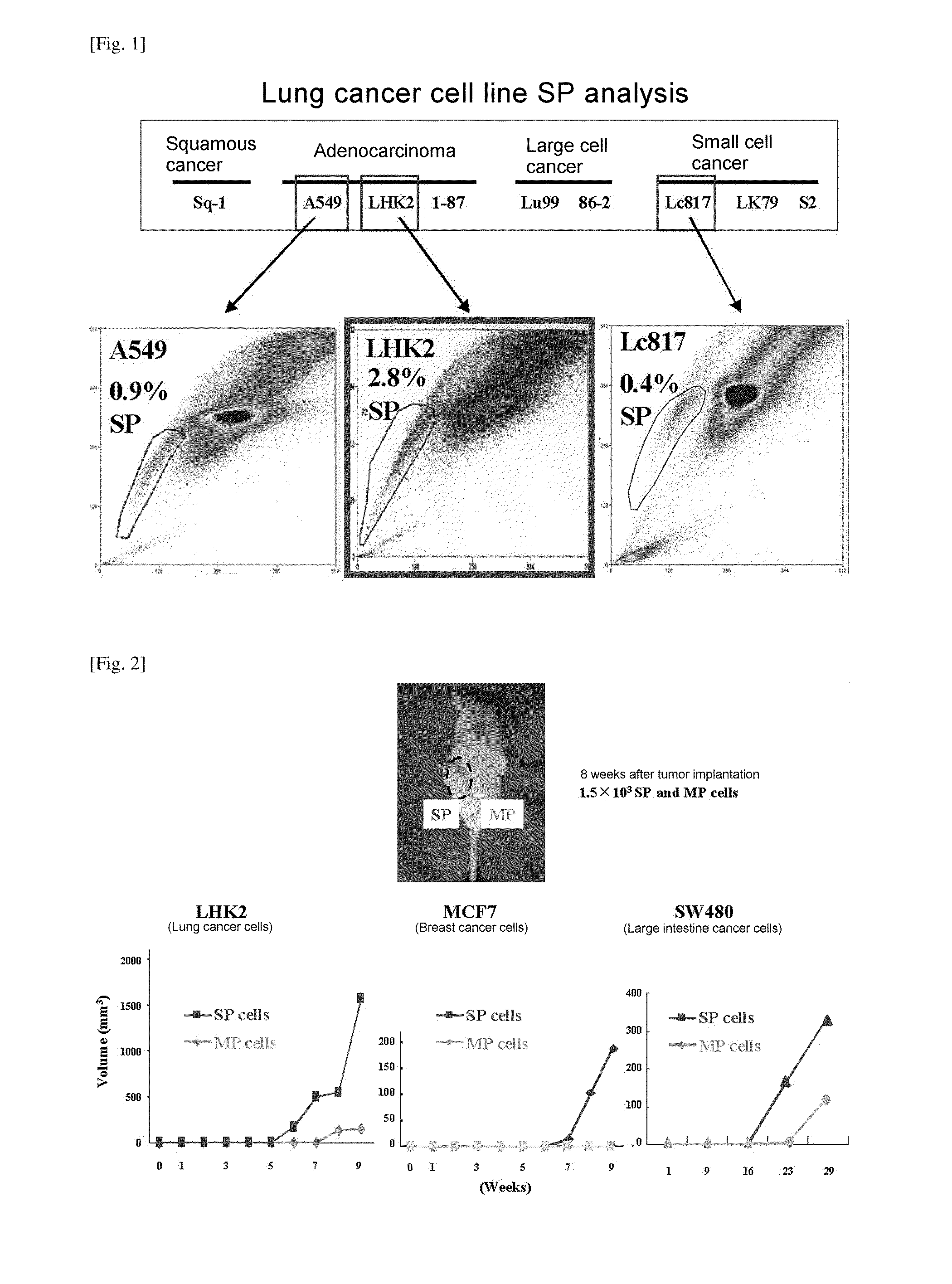
[0130]Tumorigenicity was confirmed by inoculating mice with fractioned SP cells and MP cells using each of LHK2, MCF7, and SW480 cell lines by the same method as in Experimental Example 1. The same number of SP cells and MP cells were suspended in 100 μl of 1×PBS on ice and mixed with 100 μl of Matrigel. NOD / SCID mice (obtained from Oriental Kobo Co., Ltd.) were inoculated subcutaneously to the dorsal skin with 100 μl of a cell-Matrigel mixed liquid at 1500 cells per site of each of SP and MP cells; when a tumor started to form, the lengths of the longest diameter and the shortest diameter were measured, and the volume was calculated as approximated to a spheroid and compared. The results are shown in FIG. 2.

[0131]From the results, it was found that, compared with MP cells, which did not form a tumor in even one mouse when implanted with 150 cells, the fractioned SP cells from LHK2 had much higher tumorigenicity such that 2 out of 5 mic...

example 3

Experimental Example 3

Confirmation of Expression by DNA Microarray

[0132]a) Extraction of mRNA

[0133]SP cells and MP cells isolated by the same method as in Experimental Example 1 were centrifuged at room temperature at 1500 rpm for 5 minutes, and supernatant was removed. Extraction of mRNA was carried out using an RNeasy Kit (Qiagen) in accordance with the kit protocol.

b) Amplification and Labeling of mRNA

[0134]The extracted mRNA was subjected to mRNA amplification using an RNA amplification kit obtained from Sigma Genosys, and the amplified mRNA was subjected to labeling using an mRNA labeling kit obtained from Sigma Genosys so that material extracted from the SP fraction was labeled with Cy5 and material extracted from the MP fraction was labeled with Cy3, and the dyes were exchanged, and that extracted from the SP fraction was labeled with Cy3 and that extracted from the MP fraction was labeled with Cy5.

c) Microarray

[0135]SP fraction-derived mRNA and MP fraction-derived mRNA, whic...


PUM

PropertyMeasurementUnit
Temperatureaaaaaaaaaa

Abstract

A molecular marker for detecting a cancer stem cell in a cell mass which is a subject of interest, wherein the molecular marker can be detected in a cancer stem cell contained in the subject of interest but cannot be detected in a normal cell and a cancer cell that is different from a cancer stem cell; a method for determining the presence or absence of a cancer stem cell in a subject of interest by using the molecular marker as an measure; a kit for determining the presence or absence of a cancer stem cell, which comprises at least a reagent for detecting the molecular marker; a polypeptide encoded by the molecular marker; an antibody capable of recognizing an epitope of an expression product of a gene derived from the molecular marker; a nucleic acid capable of inhibiting the expression of the molecular marker; and a nucleic acid vaccine comprising a gene derived from the molecular marker.

Description

TECHNICAL FIELD[0001]The present invention relates to a molecular marker for detecting cancer stem cells in a cell population as a detection target, the molecular marker being detected in cancer stem cells contained in the detection target but not being detected in normal cells or cancer cells other than cancer stem cells.BACKGROUND ART[0002]It is thought that cancer stem cells are the main cause of recurrence or metastasis of cancer, and the importance of targeting cancer stem cells in cancer therapy has been pointed out. However, the proportion of cancer stem cells in tumor tissue is very low (Non-Patent Document 4), and it is very difficult to specifically recognize and treat cancer stem cells. The development of techniques for detecting cancer stem cells and new therapies targeting cancer stem cells are important issues in cancer care.[0003]Currently, molecular markers such as CD133, CD24, and CD44 are known as a cancer stem cell marker (Non-Patent Documents 1 to 7), but it is s...

Claims


Application Information

Patent Timeline
IPC IPC(8): A61K49/00C12Q1/48C12Q1/68G01N33/566C12Q1/02C12N5/095C07K7/00C07K16/18A61K38/02A61K39/395C07H21/00A61K39/00C07H21/04C07H21/02A61K31/7088A61P35/00A61P43/00C12Q1/04
CPCC12N15/113C12N15/1138C12N2310/14C12Q1/6886C12Q2600/112C12N5/0638C12Q2600/136C12Q2600/158G01N33/5017G01N33/574C07K7/06C12Q2600/118A61K38/177A61P35/00A61P43/00G01N33/57492G01N2333/705
Inventor TORIGOE, TOSHIHIKOHIROHASHI, YOSHIHIKOSATOH, NORIYUKIKAMIGUCHI, KENJIROMORITA, RENANISHIZAWA, SATOSHITAKAHASHI, AKARI
Owner SUMITOMO DAINIPPON PHARMA CO LTD
Learn More