

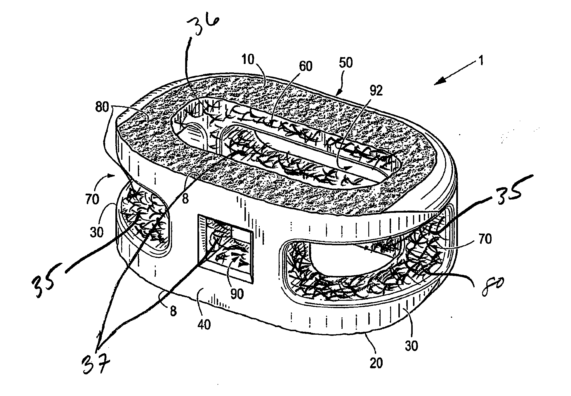
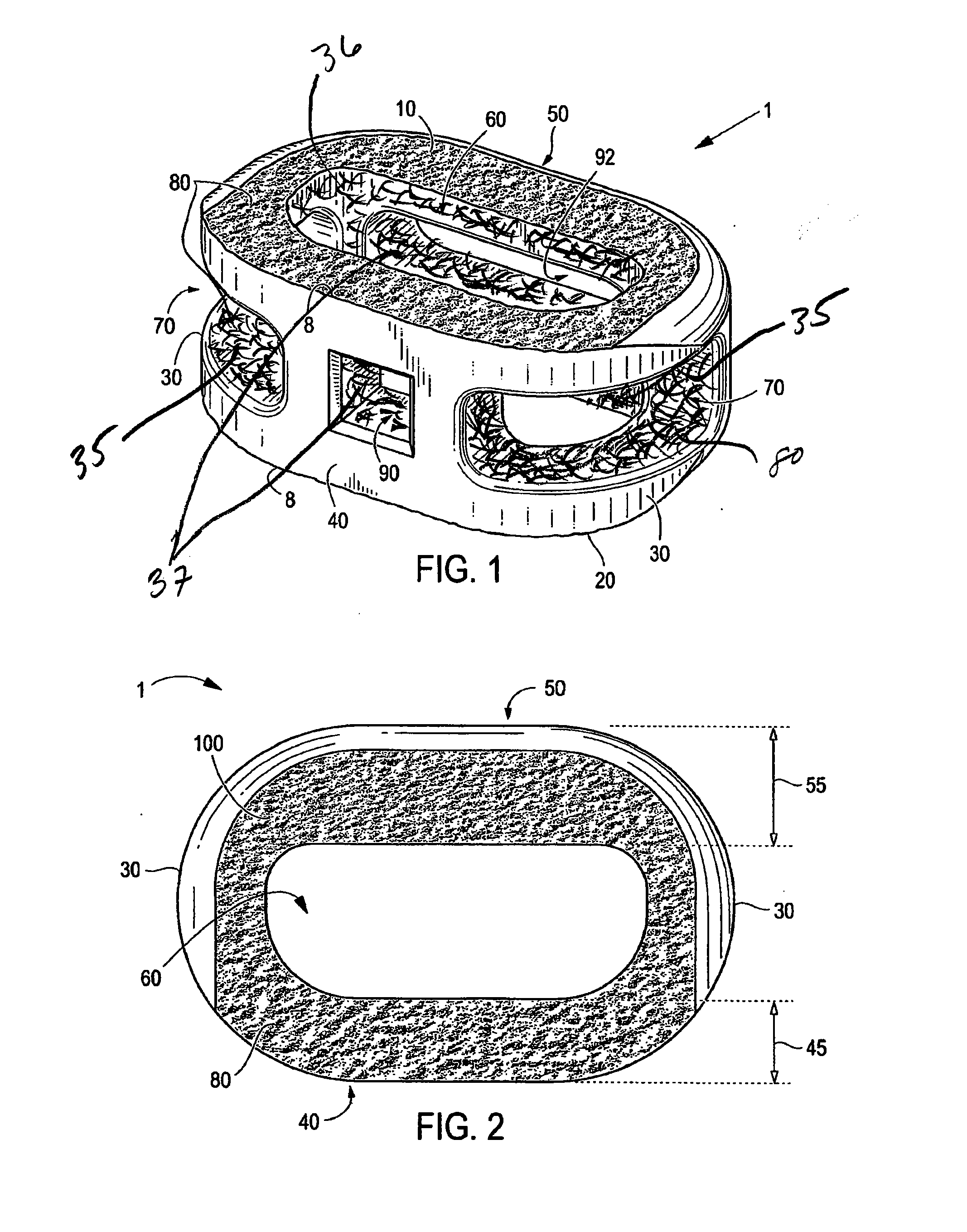
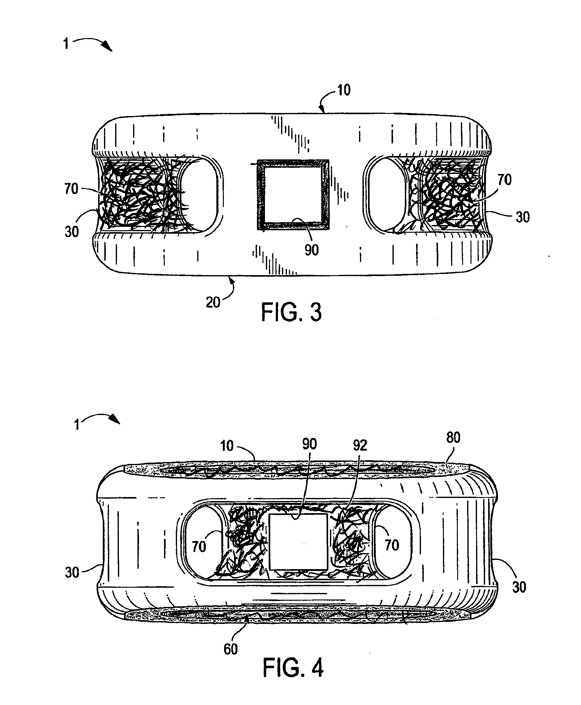
Interbody Spinal Implant Having Internally Textured Surfaces
a spinal implant and surface treatment technology, applied in the field of spinal implants with internal textured surfaces, can solve the problems of impaired discs, affecting the anatomical functions of vertebrae, and affecting the function of discs, so as to maximize the surface area and maximize the effect of radiographic visualization
- Summary
- Abstract
- Description
- Claims
- Application Information
AI Technical Summary
Benefits of technology
Problems solved by technology
Method used
Image

Examples
examples
Test Method
[0063]Human osteoblast-like MG63 cells were cultured on tissue culture polystyrene (TCPS), PEEK, or smooth [sTi6Al4V] and rough [rTi6Al4V] surfaces. FIG. 5 shows a confocal laser miscroscopy image of the PEEK surface; FIG. 6 shows a confocal laser miscroscopy image of the sTiAlV surface; and FIG. 7 shows a confocal laser miscroscopy image of the rTiAlV surface. FIG. 8 shows a SEM image of the PEEK surface at 1K× and 20K× magnification; FIG. 9 shows a SEM image of the sTiAlV surface at 1K× and 20K× magnification; FIG. 10 shows a SEM image of the rTiAlV surface at 1K× magnification Gene expression was measured by qPCR. Osteoblast maturation was assessed by analysis of cell number, alkaline phosphatase activity (ALP), and secreted osteocalcin, osteoprotegerin, TGF-β1, BMP2, BMP4, and BMP7. Data are mean±SEM (n=6 / condition), analyzed by ANOVA with Bonferroni's modification of Student's t-test.
[0064]Human MG63 osteoblast-like cells were harvested 24 hours after confluence on T...
PUM
| Property | Measurement | Unit |
|---|---|---|
| roughness | aaaaa | aaaaa |
| roughness | aaaaa | aaaaa |
| roughness | aaaaa | aaaaa |
Abstract
Description
Claims
Application Information