
Method and Apparatus for an Implantable Inertial-Based Sensing System for Real-Time, In Vivo Detection of Spinal Pseudarthrosis and Adjacent Segment Motion

a biosensor and inertial technology, applied in the field of implantable biosensor systems, can solve the problems of patient pseudarthrosis, screw loosening, weakening of implanted instruments, etc., and achieve the effect of increasing the range of motion in terms of tilt angle between adjacent segments and fused vertebrae, and reducing the risk of adjacent segment diseas

Inactive Publication Date: 2011-12-01
PHARMACO KINESIS
View PDF6 Cites 57 Cited by
  • Summary
  • Abstract
  • Description
  • Claims
  • Application Information

AI Technical Summary

Benefits of technology

[0014]The implanted accelerometer allows information about the instrumentation to be passed on to the treating surgeon and the physical therapist. The goal is to create instrumentation that can be implanted in the bone adjacent to the instrumentation. The accelerometer will notify the physician that there is motion within the fusion. If the bony fusion does not occur, then the implanted instrumentation will weaken, the screws will become loose, and the patient develops a pseudarthrosis. When a complete solid fusion occurs, however, the instrumentation will remain in place. An accelerometer at the segments above and below will let the physician know over time if there is increased motion that may lead to stress and pressure on the adjacent facet segments, accounting for pain.
[0015]The accelerometer is implanted so that it forms a geometric configuration that can be used to determine the position of the patient. This will allow the physical therapist the ability to provide the patient with the best bending motion to strengthen the back without stressing the rest of the fusion. In essence, with this implanted accelerometer, the physical therapist and the physician are now the bio-mechanist of the human skeletal system.
[0021]In one embodiment of the illustrated invention, four motion sensors are placed in the L2-L5 lumbar vertebrae, one within the detector unit on L3 or L4, and the other three on each of the adjacent vertebra. The motion sensors provide information on relative tilt motion between the two vertebrae above and below the fusion joint. The relative tilt motion between these two spinal bone segments, which is sent via the inductive communication link, helps a physician monitor the increased range of motion that might lead to adjacent segment disease. When the two bone segments are totally fused, the range of motion in terms of tilt angle between the adjacent segment and the fused vertebrae increase. The risk of adjacent segment disease could be minimized by providing the patient with a feedback alert when the tilt angle exceeding threshold level is detected.
[0022]It is yet another object of the illustrated embodiments of the invention is to facilitate restoration of health to the patient and assist in physical rehabilitation. The system actively detects the tilt angles and provides warning when the measurement exceeds the threshold. This new sensing / feedback method provides peace of mind when exercising for physical rehabilitation and could lead to a healthier patient.

Problems solved by technology

If the bony fusion does not occur, then the implanted instrumentation will weaken, the screws will become loose, and the patient develops a pseudarthrosis.

Method used


Image

  • Method and Apparatus for an Implantable Inertial-Based Sensing System for Real-Time, In Vivo Detection of Spinal Pseudarthrosis and Adjacent Segment Motion
  • Method and Apparatus for an Implantable Inertial-Based Sensing System for Real-Time, In Vivo Detection of Spinal Pseudarthrosis and Adjacent Segment Motion
  • Method and Apparatus for an Implantable Inertial-Based Sensing System for Real-Time, In Vivo Detection of Spinal Pseudarthrosis and Adjacent Segment Motion

Examples


Embodiment Construction

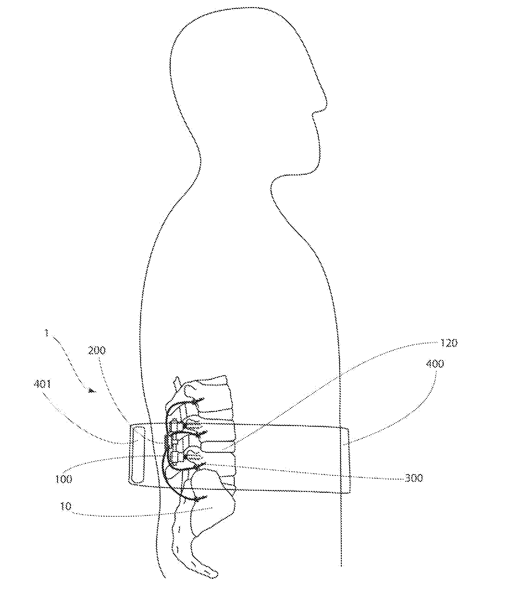
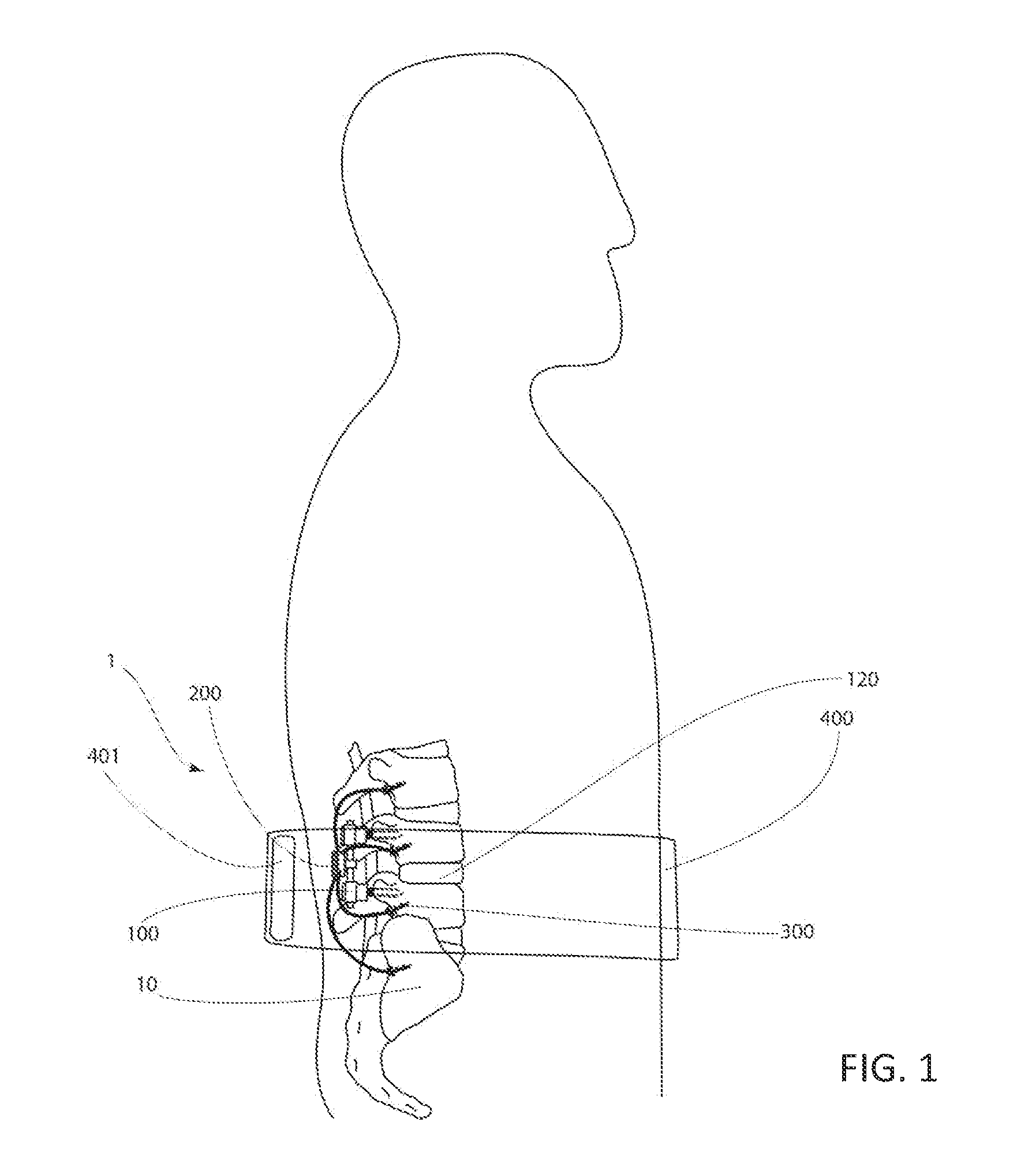
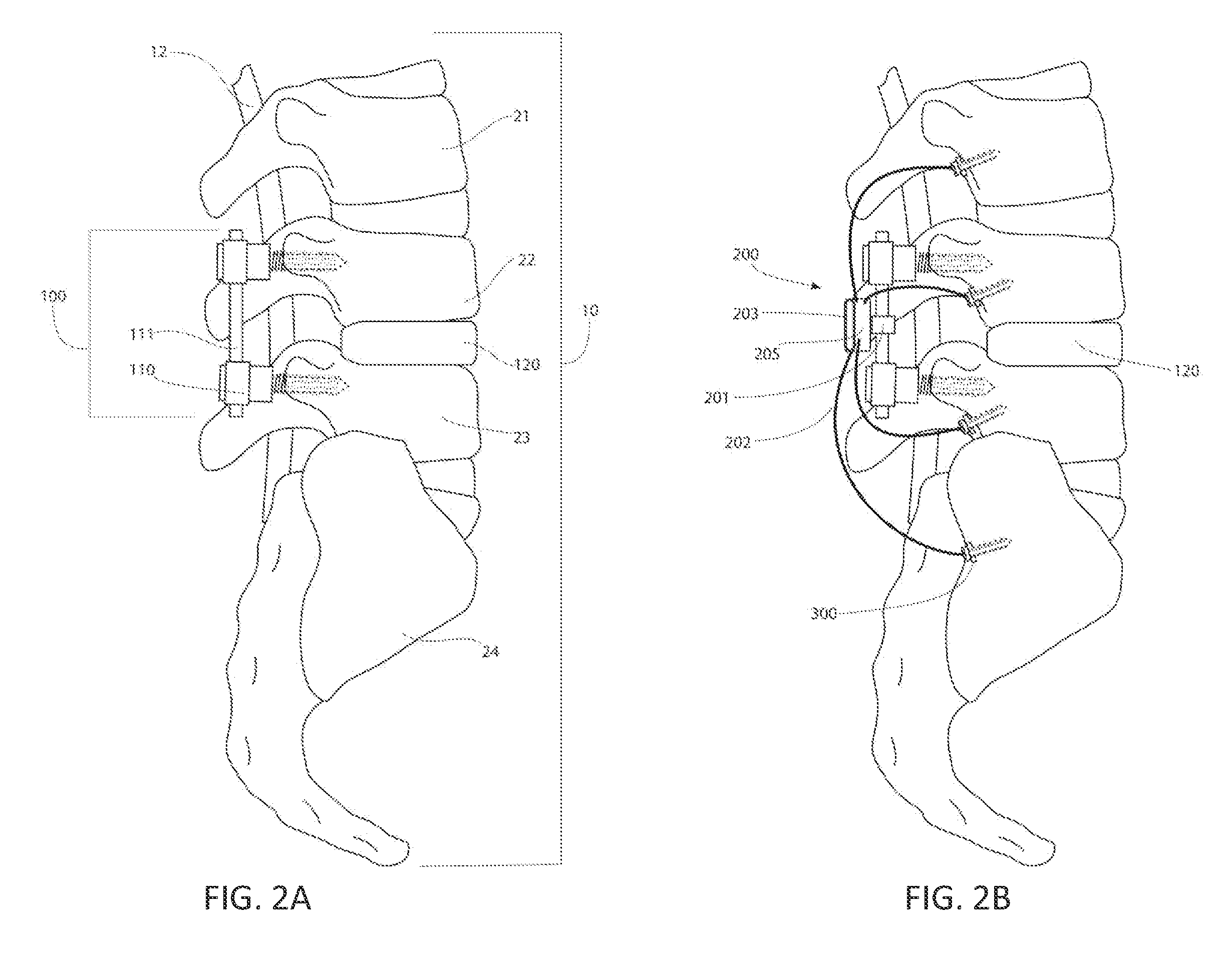
[0095]FIG. 1 is a lateral cross sectional view of a fusion sensing system 1 in its relation to the spine 10 of a patient. In this embodiment, the fusion sensing system 1 comprises an implant electronics assembly, generally denoted by reference numeral 200, coupled to spine stabilization hardware assembly, generally denoted by reference numeral 100, for interbody fusion of L4 and L5 discs of the lumbar spine using an interbody cage 120 and external wearable system 400. The fusion sensing system 1 couples a plurality of motion sensors 300 mounted into the spine 10 as best seen in FIG. 2b. The fusion sensing system 1 is powered via induction coils by a reader 401 coupled to the wearable system 400 that is worn externally by the patient. The reader 401 also comprises means for communicating to the implant electronics assembly 200 via the inductive coupling or link between the induction coils 441, 541 in FIGS. 11 and 12.

[0096]FIG. 2a is a lateral lumbar view of the spinal column 10 of a ...


PUM


Abstract

A vertebral processor designed to collect and interpret data from multiple surgically implanted accelerometers. Each accelerometer is surgically implanted into a vertebra of a patient utilizing a bone screw. Additional accelerometers are implanted in adjacent vertebrae. The data from the accelerometers is compared by an algorithm to determine the relative movement of the accelerometers implanted in adjacent vertebrae. Data is generated via the algorithm and compared against the expected behavior of the surgically implanted accelerometers as if they were connected to a rigid body, thus determining the level of success of a spinal fusion procedure for those adjacent segments. The apparatus may be utilized with or without spinal stabilization hardware, and with or without fusion cages or artificial discs. The vertebral processor is supplemented by an external system worn by the patient, which provides for an inductive charging power source and for data transfer.

Description

BACKGROUND OF THE INVENTION[0001]1. Field of the Invention[0002]The invention relates to the field of implantable biosensor systems, specifically to remotely powered and controlled accelerometer devices that are surgically implanted into adjacent vertebrae for the purpose of determining the success of spinal fusion procedures.[0003]2. Description of the Art[0004]Back pain is one of the most commonly treated medical conditions in the United States today and is second only to the common cold as the most common reason for physician visits. Back pain not only has a profound effect on patients, it has an exceedingly high societal cost. Back pain is the second most common cause of lost productive time for pain disorders and results in the largest amount of total lost time. It has been stated that of all medical conditions, back pain results in the most lost productivity. It is estimated that the direct and indirect costs associated with the disorder are approximately 50 billion dollars pe...

Claims


Application Information

Patent Timeline
IPC IPC(8): A61B5/103A61F2/44
CPCA61B5/1114A61B5/413A61B5/45A61B5/6878A61B17/7074A61B2562/043A61F2/4455A61F2/4657A61B2505/05A61B2560/0219A61F2/442
Inventor SHACHAR, YEHOSHUACHEN, THOMASWUJORDAN, BRETTCHAN, HERWINLUBOFF, PALADINZIMMERMAN, KYLE
Owner PHARMACO KINESIS
Learn More