Method of tendon repair with amnion and chorion constructs

a technology which is applied in the field of amnion and chorion constructs for tendon repair, can solve the problems of poor spontaneous regenerative capacity of tendons, bone healing, and typical time-consuming

Inactive Publication Date: 2011-12-15
LIVENTA BIOSCI
View PDF7 Cites 80 Cited by
  • Summary
  • Abstract
  • Description
  • Claims
  • Application Information

AI Technical Summary

Benefits of technology

[0023]In another general aspect, the present invention relates to a method of preparing a construct for use in surgical repair of tendon, the method comprising drying an allograft comprising at least one layer of human amnion and chorion tissues on a frame of a cylindrical shape, wherein the allograft is generally cylindrical with a C-shaped cross-section to allow for ease of implantation over the tendon.

Problems solved by technology

When tendons are ruptured due to trauma or surgical intervention, they typically take longer than, for example, bone to heal.
Tendons have poor spontaneous regenerative capabilities and complete regeneration is almost never achieved despite intensive remodeling.
Patients whose tendon(s) have been ruptured experience pain, reduced mobility, reduced lubrication between the articulating and adjacent tissues and a heightened risk of post trauma scarring, adhesions and pain.
A primary cause of this is damage to the protective sheathing of the tendon.
That inflammation then causes the tendon to swell, which constricts further the ability of the tendon to glide between adjacent structures and the tendon no longer moves smoothly through its sheath.
However surgery imposes much higher relative risks of perioperative mortality and morbidity, e.g. infection including MRSA, bleeding, deep vein thrombosis, lingering anesthesia effects, stiffness, suture reaction, persistent pain or weakness after the injury and repair, etc.
Peritendonous adhesions are a contributor to poor outcomes in patients undergoing tendon surgery.
The formation of adhesions between the tendon and surrounding tissue reduces the ability of the repaired tendon to glide normally.
This limits post-operative rehabilitation as a result of a reduction in range of motion and an increase in inflammatory pain.
In published clinical studies, none of these approaches have shown to consistently reduce the incidence of adhesions or scar formation following repair of tendon injury.
In 1962, the fetal membrane was used to treat pelvic basins after total exenteration in dogs, however, trials in human proved disappointing.
Despite the clinical and published record regarding the safety and efficacy of amnion in broad surgical use, issues regarding reproducibility, safety and the precise form of amnion for each prospective indication have prevented amnion from achieving broad commercial distribution.

Method used


Image

  • Method of tendon repair with amnion and chorion constructs
  • Method of tendon repair with amnion and chorion constructs
  • Method of tendon repair with amnion and chorion constructs

Examples


example 1

[0080]This example illustrates a surgical procedure for the application and wrapping of a tendon with a construct of the present invention.

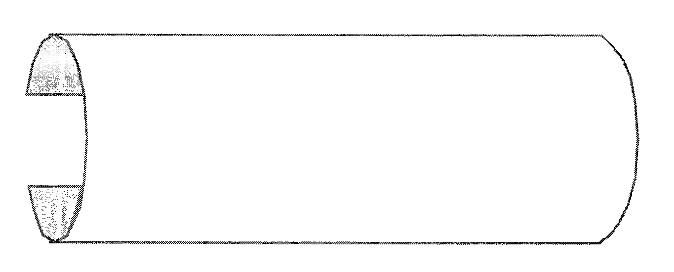
[0081]The application of the allograft construct commences upon completion of repair to the tendon. Repair to the tendon and its structures can include, but are not limited to, synovectomy, tendon tubularization, debridement of the tendon, retinacula repair, sheath repair or end-to-end anastomosis. The cylindrical allograft construct is typically about 8 mm to 10 mm in diameter and has a length of about 60 mm.

[0082]The tendon, being exposed, is now ready for the application of the construct. The sheath is either opened temporarily by the use sutures or with skin hooks. The sutures are placed adjacent to the tendon repair site, on opposing sides of the tendon and another set of sutures proximal to the first set of sutures. Alternatively, this can also be accomplished by an assistant holding the sheath open with skin hooks. The tendon is gently lif...

example 2

[0083]This example illustrates the application of an allograft construct according to an embodiment of the present invention during surgical treatment of posterior tibial tendon dysfunction (PTTD).

[0084]The patient was a 47-year-old woman who presented with a complaint of tenderness in the medial aspect of her right ankle which also occasionally radiated distally into her foot for a period of 6 months. The patient indicated that the pain increased during ambulation and prolonged periods of activity. According to the patient, the pain was not related to any trauma to the foot. The patient noted that she had experienced a progressive flattening of her arch over the past few months. Self-prescribed acetaminophen and ibuprofen did not provide pain relief The patient's medical history revealed hypertension treated with a beta blocker, no previous surgeries and no known drug allergies.

[0085]Upon physical exam the patient had considerable tenderness along the course of the posterior tibial...

example 3

[0093]This example illustrates the application of an allograft construct according to an embodiment of the present invention during surgical treatment of chronic total tendo-Achilles rupture.

[0094]The patient was a 55-year-old man who presented with a five-month history of posterior superior right heel pain. The patient noticed occasional sharp shooting pain in his right heel that began as remitting but eventually progressed to constant tenderness approximately 3-4 weeks after the onset of initial symptoms. Irritating pain, swelling, and tenderness were present with both ambulation and non-weight bearing, but were aggravated with activity. The patient denied any precipitating activity or history of trauma to the area. Self treatment consisted of anti-inflammatory medication.

[0095]Upon examination, the patient's tendo-Achilles was indurated and swollen with an increase in the diameter of the right ankle as compared to the left. The patient experienced pain upon palpation of the poste...


PUM


Abstract

It is described a construct for use in surgical repair of tendon. The construct contains at least one layer of human amnion and chorion tissues and is generally cylindrical with a C-shaped cross-section to allow for ease of implantation over the tendon. Methods for preparing the construct is described. In addition, an improved method for surgical repair of a damaged or diseased tendon using the construct is also described. The improved method reduces adhesions, scar formation, inflammation and risk of post-operative infection.

Description

CROSS-REFERENCE TO RELATED APPLICATIONS[0001]This application is entitled to priority pursuant to 35 U.S.C. § 119(e) to U.S. Provisional Patent Application No. 61 / 353,717, filed Jun. 11, 2010 which is hereby incorporated by reference herein in its entirety.BACKGROUND OF THE INVENTION[0002]1. Field of Invention[0003]This invention relates to methods for reducing inflammation, inhibiting fibrosis, scarring, fibroblast proliferation, post-operative infection while also promoting smooth gliding of the tendon and healing using processed amnion and / or chorion tissues as part of a surgical intervention treatment program to repair damaged or diseased tendons.[0004]2. Background of the Invention[0005]Tendons are a type of regularly arranged dense soft, connective tissue that bridges and attaches muscles to bone. This fibrous and flexible tissue is also called sinew. Tendons are comprised of type I collagen (80% by weight) that are arranged in fibrils. The fibrils form a fascicle, which inclu...

Claims


Application Information

Patent Timeline
Patent Type & Authority Applications(United States)
IPC IPC(8): A61F2/08
CPCA61F2/08A61L2430/10A61L27/3662A61L27/3604
Inventor YOUNG, ROBIN R.JAY, RICHARD M.
Owner LIVENTA BIOSCI
Why Eureka
Try Eureka