Photon counting and energy discriminating detector threshold calibration

a detector and threshold technology, applied in the field of photon counting and energy discrimination detector threshold calibration, can solve the problems of not being able to distinguish energy levels of photons, not providing an energy discrimination capability, and systems with k-edge contrast materials and/or agents that do not fit into the conventional bmd model, and conventional bmd cannot account for the k-edge effect of high z or high atomic number materials such as iodine (i)

Active Publication Date: 2012-04-12
GE MEDICAL SYST ISRAEL
View PDF6 Cites 46 Cited by
  • Summary
  • Abstract
  • Description
  • Claims
  • Application Information

AI Technical Summary

Benefits of technology

[0013]According to one aspect of the invention, an imaging system includes an x-ray source that emits a beam of x-rays toward an object to be imaged, a detector that receives x-rays emitted from the x-ray source, a data acquisition system (DAS) operably coupled to the detector, the DAS configured to count photon hits in the detector that occur at photon energies above at least a low keV threshold, a medium keV threshold, and a high keV threshold, and a computer operably coupled to the DAS. The computer is programmed to vary each of the medium keV threshold and the high keV threshold over a continuous keV range during data acquisition to define low, medium, and high keV bins that are based on the low, medium, and high keV thresholds, obtain photon counts in the low, medium, and high keV bins in a plurality of keV threshold combinations. This acquisition procedure may be repeated while each time different attenuator is placed in the beam. The computer is programmed to calculate a noise variance as a function of at least one of the keV thresholds, and identify a noise minimum and low, medium, and high keV thresholds that correspond thereto.
[0014]In accordance with another aspect of the invention, a method of calibrating an imaging system includes defining photon-counting bins based on low, medium, and high keV thresholds, obtaining photon counts in low, medium, and high photon-counting bins at a plurality of keV threshold combinations, calculating an image noise variance as a function of the keV threshold combinations, determining an image noise minima of the image noise variance, and determining a combination of low, medium, and high keV thresholds that correspond to the determined image noise minima.

Problems solved by technology

However, a scintillator / photodiode typically does not provide an energy discrimination (ED) capability, or an ability to distinguish energy level of photons.
Systems with K-edge contrast materials and / or agents do not fit into the conventional BMD model.
In addition, the conventional BMD cannot account for the K-edge effect of high Z or high atomic number materials such as iodine (I), barium (Ba), tungsten (W), gadolinium (Gd), and xenon (Xe) if the K-edge of the material lies in the active energy region of the incident x-ray spectrum.
A design for resolving K-edge contrast agents has employed monochromatic x-ray beams with which the K-edge material can be resolved by imaging the object at photon energies slightly below and slightly above the K-edge, but prevents integration of monochromatic sources with sufficient x-ray flux into a rotating gantry and so limits the application from use as a practical monochromatic x-ray source in medical CT scanners.
However, as stated, more than two spectra are needed for K-edge detection, which make systems using different x-ray spectra not feasible for K-edge detection.
However, different bin setups result in different image quality, material differentiation, and noise.
More particularly, spectral imaging data obtained having bins defined non-optimally can lead to a needlessly high level of noise in the final images.

Method used


Image

  • Photon counting and energy discriminating detector threshold calibration
  • Photon counting and energy discriminating detector threshold calibration
  • Photon counting and energy discriminating detector threshold calibration

Examples


Embodiment Construction

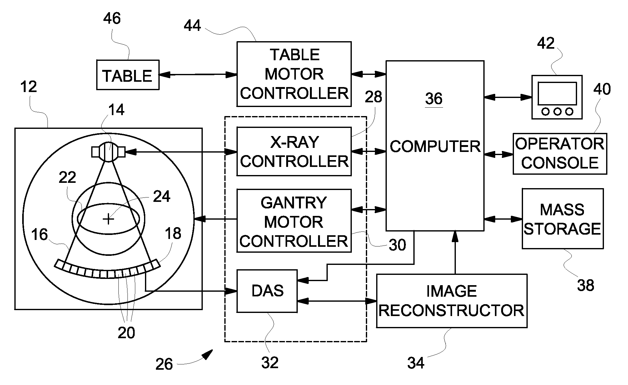
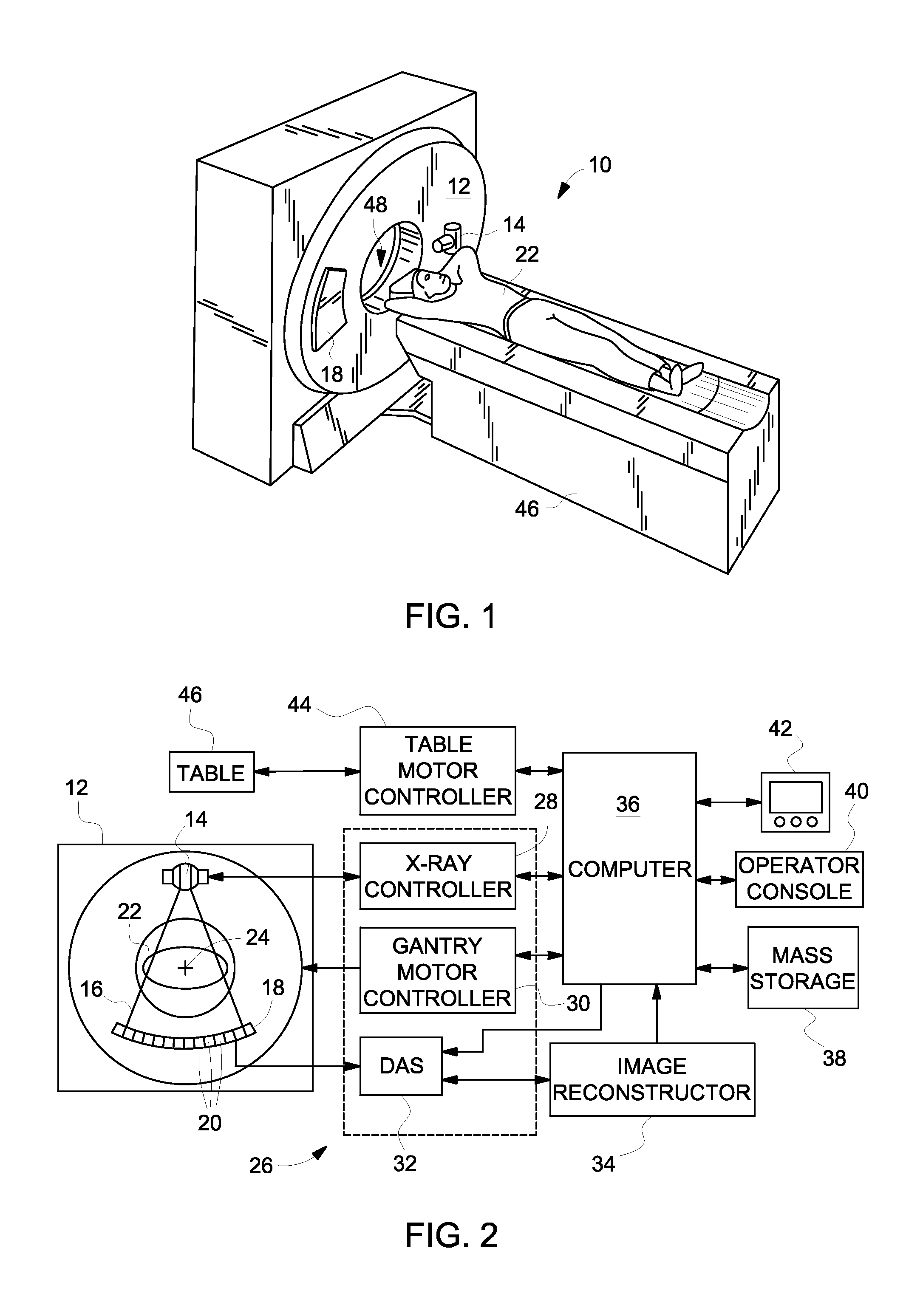
[0027]Diagnostics devices typically comprise x-ray systems, magnetic resonance (MR) systems, ultrasound systems, computed tomography (CT) systems, positron emission tomography (PET) systems, single-photon-emission computed tomography systems (SPECT), and other types of imaging systems. Applications of x-ray sources comprise imaging, medical, security, industrial inspection applications, and portable radiation detectors. The operating environment described herein includes a 64-slice CT system. However, it will be appreciated by those skilled in the art that an implementation of the embodiments described herein is also applicable for use with single-slice or other multi-slice configurations. More generally, an implementation is employable for detection and conversion of x-rays. However, one skilled in the art will further appreciate that an implementation is employable for the detection and conversion of other high frequency electromagnetic energy, high frequency polychromatic electro...


PUM

PropertyMeasurementUnit
photon energiesaaaaaaaaaa
thresholdaaaaaaaaaa
noise varianceaaaaaaaaaa

Abstract

An imaging system includes an x-ray source, a detector that receives x-rays emitted from the x-ray source, a DAS configured to count photon hits in the detector that occur at photon energies above at least a low keV threshold, a medium keV threshold, and a high keV threshold, and a computer operably coupled to the DAS. The computer is programmed to vary each of the medium keV threshold and the high keV threshold over a continuous keV range during data acquisition to define low, medium, and high keV bins that are based on the low, medium, and high keV thresholds, obtain photon counts in the low, medium, and high keV bins in a plurality of keV threshold combinations, calculate a noise variance as a function of at least one of the keV thresholds, and identify a noise minimum and low, medium, and high keV thresholds that correspond thereto.

Description

BACKGROUND[0001]Embodiments of the invention relate generally to an apparatus and method for detector threshold calibration.[0002]Diagnostics devices typically comprise x-ray systems, magnetic resonance (MR) systems, ultrasound systems, computed tomography (CT) systems, positron emission tomography (PET) systems, and other types of imaging systems. Typically, in CT imaging systems, an x-ray source emits a fan-shaped beam toward a subject or object, such as a patient or a piece of luggage. Hereinafter, the terms “subject” and “object” shall include anything capable of being imaged. The beam, after being attenuated by the subject, impinges upon an array of radiation detectors. The intensity of the attenuated beam radiation received at the detector array is typically dependent upon the attenuation of the x-ray beam by the subject. Each detector element of the detector array produces a separate electrical signal indicative of the attenuated beam received by each detector element. The el...

Claims


Application Information

Patent Timeline
Patent Type & Authority Applications(United States)
IPC IPC(8): G01N23/04G01D18/00
CPCG01T7/005G01T1/29
Inventor GREENBERG, LEVRUBIN, DANIELROSNER, BARUCHNAVEH, GALITLEVINSON, REUVENARENSON, JEROME STEPHENROSENBERG, SHALOM
Owner GE MEDICAL SYST ISRAEL
Why Eureka
Try Eureka