X-ray image photographing method and x-ray image photographing apparatus

a technology of x-ray image and x-ray image, which is applied in the direction of material analysis using wave/particle radiation, applications, instruments, etc., can solve the problems of difficult to photograph the structure inside a thick specimen, difficult to freely adjust the ratio of phase contrast and absorption contrast, etc., and achieve high quantitative performance, small artifact, and high resolution

Active Publication Date: 2012-04-12
EXXON RES & ENG CO +1
View PDF1 Cites 5 Cited by
  • Summary
  • Abstract
  • Description
  • Claims
  • Application Information

AI Technical Summary

Benefits of technology

[0020]Accordingly, it becomes possible to photograph an image by a phase contrast in which the outline of the specimen is emphasized by adjusting the arrangement of the specimen and the detector with respect to the X-ray source, to selectively photograph an image of an absorption contrast freely, and to perform CT reconstruction in which quantitative performance is high and artifact is small. Furthe

Problems solved by technology

Accordingly, under high magnification ratio photographing conditions for photographing a fine structure with a high resolution, it is not easy to freely adjust the ratio of phase contrast and absorption contrast while keeping the magnification ratio.
On th

Method used


Image

  • X-ray image photographing method and x-ray image photographing apparatus
  • X-ray image photographing method and x-ray image photographing apparatus
  • X-ray image photographing method and x-ray image photographing apparatus

Examples


first embodiment

Apparatus Configuration

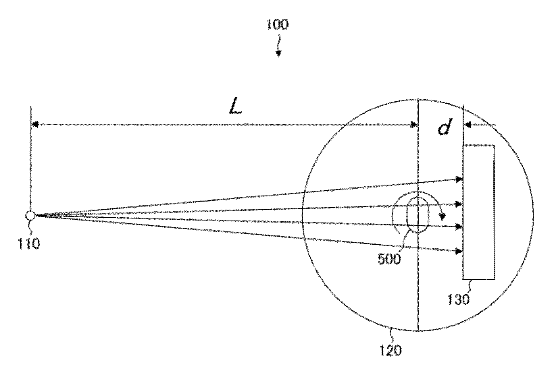
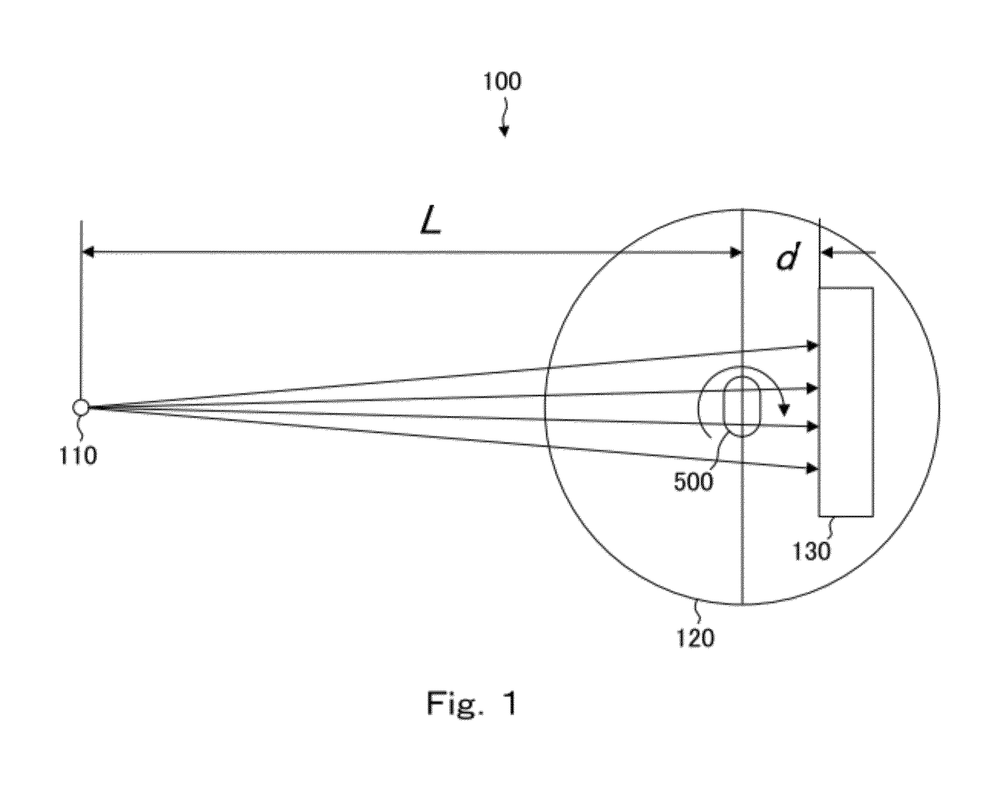
[0032]FIG. 1 is a schematic diagram showing a configuration for an X-ray image photographing method of the invention. Such a configuration is configured by arrangement of each unit of an X-ray image photographing apparatus 100. As shown in FIG. 1, the X-ray image photographing apparatus 100 includes an X-ray source 110, a support table 120, and a detector 130.

[0033]The X-ray source 110 is an X-ray source having a finite focal point size and used in a laboratory. Radiation light by synchrotron radiation is not used in the X-ray source 110. To be more specific, copper, molybdenum, tungsten, etc., is used as an X-ray source target, and the arrangement of each unit is adjusted in accordance with the wavelength of characteristic X-ray, the type of which is not particularly limited. The X-ray source 110 is not necessarily limited to electronic collision type, plasma X-ray, inverse Compton radiation, etc., and a virtual source to be described below is also included.

[...

experimental example

Example 1

[0047]Next, an experiment using the X-ray image photographing apparatus 100 will be described. First, a leaf of a plant was used as the specimen and an X-ray transmission image using a phase contrast was photographed. The X-ray source 110 having a supply power of 875 W, Cr target, and a focal point size of 70 μm, and the detector 130 having a pixel size of 0.65 μm (space resolution of 0.65 μm) were used. The distance L between the X-ray source 110 and the specimen 500 was set to 250 mm. FIGS. 8 and 9 are X-ray transmission images of plant cells photographed when the distances d between the specimen 500 and the detector 130 are set to 1 mm and 5 mm, respectively.

[0048]As shown in FIG. 8, when the distance d is 1 mm, an X-ray transmission image by an absorption contrast in which edge of a cell wall is not emphasized was obtained. At the time, f (λ, d) is 0.5 μm and smaller than 0.65 μm which is the resolution Δ of the detector. Furthermore, as shown in FIG. 9, when the distan...

example 2

[0049]Under the same conditions as the above examples, an X-ray transmission image using an absorption contrast of a carbon fiber reinforced plastic (CRFP, carbon fiber reinforced resin) as a specimen was photographed, and three-dimensional CT reconstruction was performed. The distance L between the X-ray source 110 and the example 500 was set to 250 mm, and the distance d between the specimen 500 and the detector 130 was set 2 to 3 mm which is the middle of the above-mentioned two examples, and adjustment was performed for photographing so that phase contrast is not too emphasized.

[0050]FIGS. 10, 11, and 12 are CT images of two orthogonal cross sections and an enlarged cross section of a carbon fiber reinforced plastic (CRFP, carbon fiber reinforced resin). Carbon fiber has a high strength in fiber direction although the strength is not enough in other directions. Accordingly, fibers whose fiver directions are different are overlapped in a multilayer. As shown in FIGS. 10 to 12, in...


PUM


Abstract

An X-ray image photographing method and apparatus is capable of photographing a high resolution phase contrast image and a high resolution absorption contrast image in a short time by finely adjusting the distance between a specimen and a detector with respect to an X-ray source.
The method enables photographing a fine structure with high space resolution while using an X-ray source having a finite size, and d/L is sufficiently smaller than 1, when L is a distance from an X-ray source to a specimen and d is a distance from the specimen to a detector.

Description

BACKGROUND OF THE INVENTION[0001]1. Field of the Invention[0002]The present invention relates to an X-ray image photographing method and an X-ray image photographing apparatus which enables photographing of a fine structure while using an X-ray source having a finite size.[0003]2. Description of the Related Art[0004]Conventionally, in a field of X-ray transmission image photographing, resolution of a detector has been at most ten and several μm. Accordingly, in order to photograph an image having a high resolution, photographing has been performed by magnifying projection of a specimen by using an X-ray source having a focal point size as small as possible, about 10 μm or less. That is, magnification ratio has been adjusted by adjusting distance relationships by reducing the distance from the X-ray source to the specimen and setting the distance from the specimen to the detector sufficiently large.[0005]On the other hand, a method for performing photographing using a phase contrast ...

Claims


Application Information

Patent Timeline
IPC IPC(8): G01N23/04
CPCA61B6/4035A61B6/484G03B42/02A61B6/589A61B6/588
Inventor OMOTE, KAZUHIKOKAMBE, MAKOTOTAKEDA, YOSHIHIRO
Owner EXXON RES & ENG CO
Why Eureka
Try Eureka