

Feeder cells for target cell induction
a target cell and target cell technology, applied in the field of target cell induction feeder cells, can solve the problems of heterologous cell infection risk, severe visual impairment, donor shortage and allograft rejection, etc., and achieve the effect of simple and easy cultur
- Summary
- Abstract
- Description
- Claims
- Application Information
AI Technical Summary
Benefits of technology
Problems solved by technology
Method used
Image

Examples
example 1
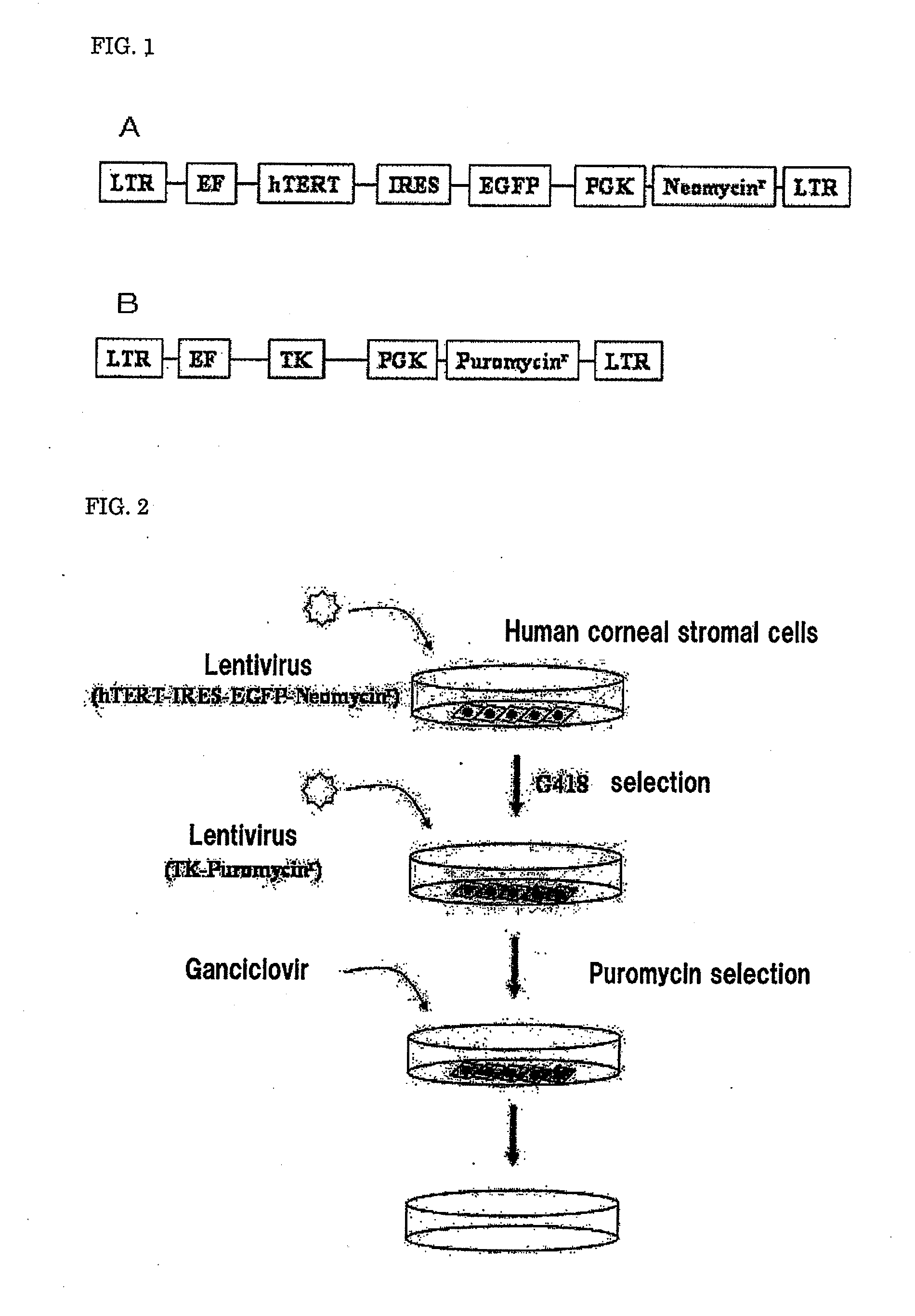
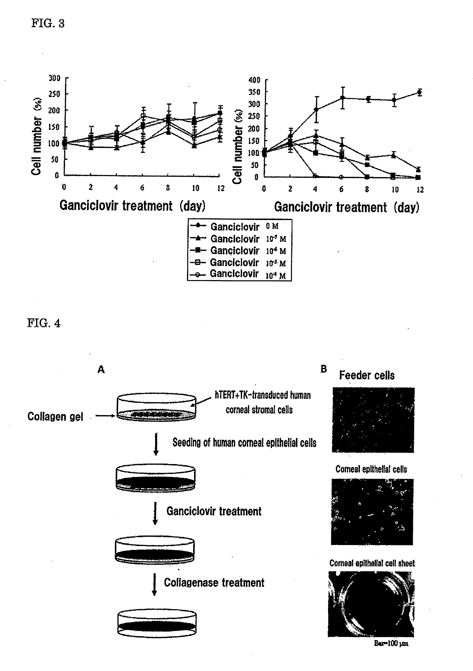
[0093]A human corneal stromal cell line transduced with the hTERT gene and the TK gene (hereinafter referred to as hTERT+TK-transduced human corneal stromal cells) was produced by the following procedure. The schematic view of the production of the hTERT+TK-transduced human corneal stromal cells is shown in FIG. 2.
1. Culture of Human Corneal Stromal Cells
[0094]Epithelial cells and endothelial cells were removed (scraped off with forceps) from a piece of corneoscleral tissue obtained from an imported cornea (USA Eye Bank). The corneal stroma thus obtained was cut into several pieces and cultured with its endothelial side down in a 35-mm dish containing DMEM, 10% FBS, 100 U / ml penicillin and 100 μg / ml streptomycin under conditions of 37° C. and 5% CO2.
2. Preparation of Lentiviral Vectors
[0095]Transduced lentiviral vectors were produced by the following procedure using a packaging plasmid and a gene-of-interest expression vector that is required for the production of the lentivirus sho...
example 2
[0105]A human corneal epithelial cell sheet was produced by the following procedure. The schematic view of the production process for the human corneal epithelial cell sheet is shown in FIG. 4A.
1. Culture of hTERT+TK-Transduced Human Corneal Stromal Cells on Collagen Gel
[0106]A collagen gel (type I collagen) was produced in a 6-well plate using Collagen Gel Culture Kit (Nitta Gelatin). On this collagen gel (Nitta Gelatin), the hTERT+TK-transduced human corneal stromal cells produced in Example 1 were seeded, and cultured under conditions of 37° C. and 5% CO2 (medium: DMEM, 10% FBS, 100 U / ml penicillin and 100 μg / ml streptomycin). The photograph of the hTERT+TK-transduced human corneal stromal cells (feeder cells) obtained by culture is shown in FIG. 4B (top).
2. Preparation of Human Corneal Epithelial Cells
[0107]A piece of corneoscleral tissue obtained from an imported cornea (USA Eye Bank) was incubated in a 2.4 U / ml Dispase solution at 37° C. for 1 hour. Next, the tissue was treate...
example 3
[0110]A corneal epithelial cell sheet was collected in the same manner as in “Culture of hTERT+TK-transduced Human Corneal Stromal Cells on Collagen Gel” of Example 2 except that the hTERT+TK-transduced human corneal stromal cells were treated with mitomycin C (8 μg / ml) at 37° C. for 2 hours before the cells were seeded on the collagen gel.
[0111]From Examples 2 and 3, it was confirmed that a human corneal epithelial cell sheet can be produced using the feeder cells of the present invention. Since the production process of the present invention does not require the use of mouse 3T3 cells, which are commonly used as feeder cells, the risk of infection from heterologous cells is avoidable. In addition, while human corneal stromal cells obtained from primary culture can be maintained only for several passages and thus have limitations on use for transplantation, the feeder cell line of the present invention can be maintained through long-term passage as a result of introduction of the T...
PUM
 Abstract
Description
Claims
Application Information