Detector rotation type radiation therapy and imaging hybrid device

a hybrid device and detection device technology, applied in the field of radiation therapy and imaging hybrid devices, can solve the problems of difficult positioning of tumors, difficult to identify tumors themselves, and change in tumor size, and achieve the effect of reducing the incidence of nuclear fragments

Inactive Publication Date: 2012-06-28
NAT INST OF RADIOLOGICAL SCI
View PDF2 Cites 23 Cited by
  • Summary
  • Abstract
  • Description
  • Claims
  • Application Information

AI Technical Summary

Benefits of technology

[0047]According to the present invention, when performing monitoring for detecting annihilation radiations occurring from an irradiation field due to radiation irradiation in radiation therapy which is conducted by irradiating an affected area with X-rays or a particle beam, it is possible to reduce the incidence of nuclear fragments on detectors without interfering with the treatment beam, thereby enabling measurement of annihilation radiations and three-dimensional imaging of the irradiation field immediately after irradiation or even during irradiation.

Problems solved by technology

In general, X-ray images fail to provide a sufficient contrast between tumor and normal tissues, and it is difficult to identify a tumor itself for positioning.
In addition to such misalignment of the irradiation field at the time of patient setup, other problems have been pointed out such as a change in the size of the tumor from the time of creation of the treatment plan, and respiratory and other variations of the tumor position.
Under the present circumstances, it is difficult to accurately identify whether irradiation is performed according to the treatment plan.
Even if the actual irradiation field deviates from the treatment plan, it is not easy to detect.
This may lead to the omission of annihilation radiations to be measured and the production of errors in position information.
However, lines of coincidence measured are highly uneven in direction, some information necessary for image reconstruction missing.
This significantly reduces the resolution in directions perpendicular to the detector plane, failing to meet the requirement 4.
When combined with a radiotherapy device, such a PET device in turn fails to meet the requirements 1 and 2 because of interference between the PET detectors and the treatment beam.
The requirement 3 is not satisfied, however, since the opposed gamma camera type PET device can be rotated only after beam irradiation, except in some rare cases where beam irradiation is continuously performed from various directions.
If the treatment intensity is extremely high, the detectors may get radioactivated and fail to meet the requirement 2.

Method used


Image

  • Detector rotation type radiation therapy and imaging hybrid device
  • Detector rotation type radiation therapy and imaging hybrid device
  • Detector rotation type radiation therapy and imaging hybrid device

Examples


examples

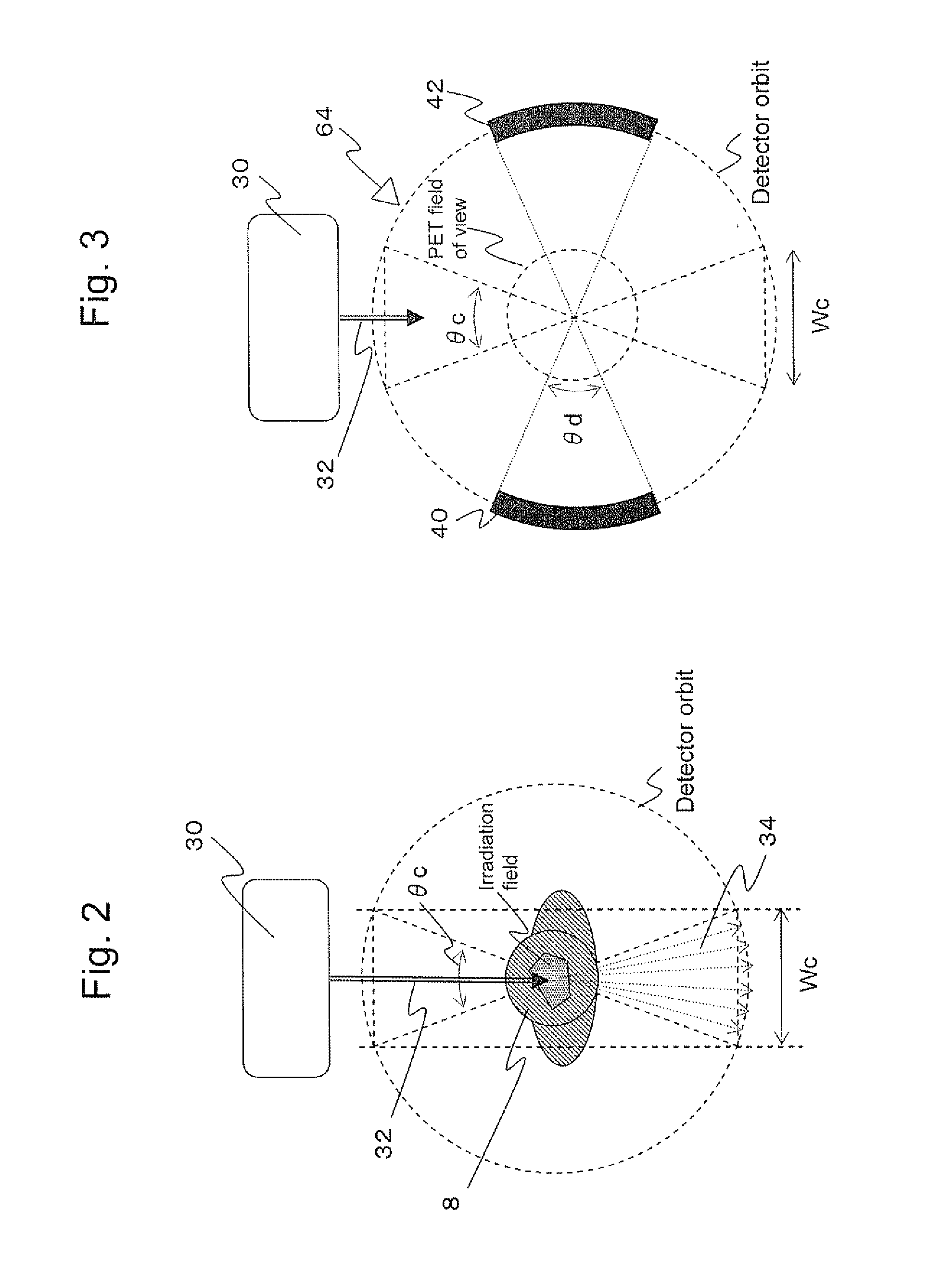
[0079]The Heavy Ion Medical Accelerator in Chiba (HIMAC) of the National Institute of Radiological Sciences in Japan performs treatment beam control with a period of T=3.3 sec. The present invention will be described in terms of application to HIMAC. Assuming that the radius of the orbit of the detectors R=50 cm and the radius of the PET field of view r=20 cm, the lower limit of θd is θd≧47.2°. Table 1 shows upper limits of θd for different irradiation durations ti and different widths We of the critical region. ts=3.3−ti. No device is feasible if the upper limit falls below the lower limit (in the table, denoted as NA). For enhanced sensitivity of the PET device, it is actually desirable to employ the maximum value of θd.

TABLE 1Wc =Wc =Wc =Wc =Wc =20 cm30 cm40 cm50 cm60 cmti = 0.5 sec129.7°117.8°105.6°92.7°79.0°ti = 1.0 sec102.4°90.5°78.3°65.5°51.7°ti = 1.5 sec75.1°63.3°51.0°NANAti = 2.0 sec47.8°NANANANA

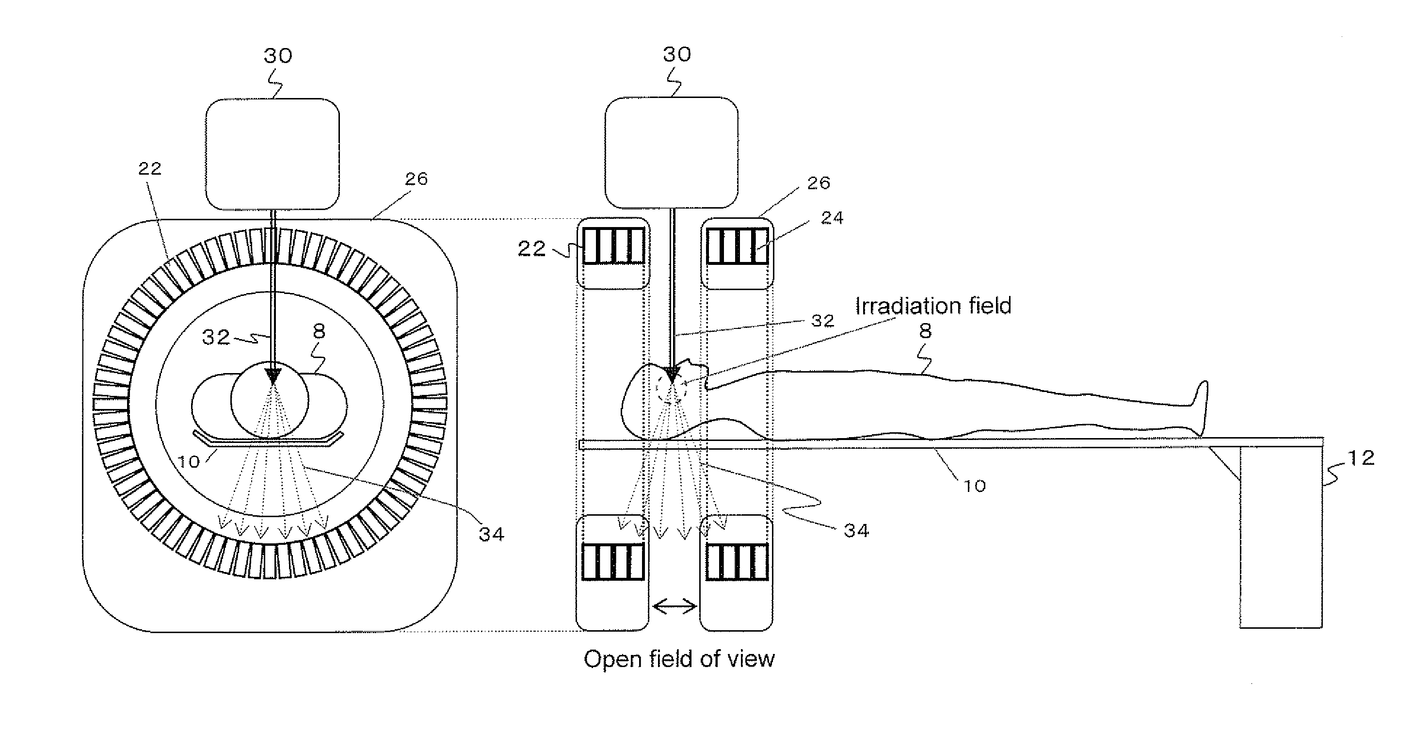
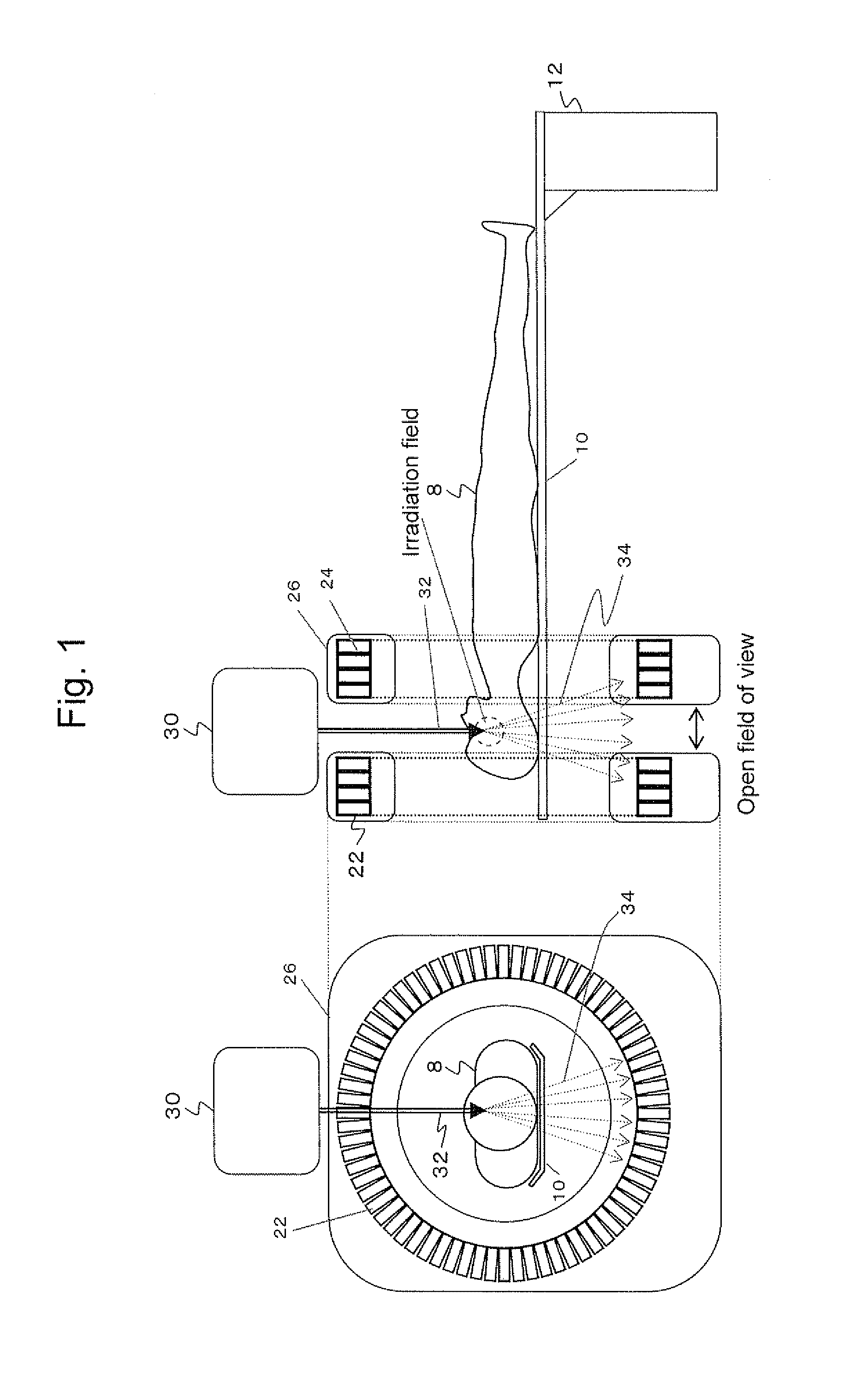
[0080]While the example has dealt with the case with a single irradiation port,...


PUM


Abstract

An imaging device, or a PET device, opposed gamma camera type PET device, or open PET device in particular, that is combined with a radiation therapy device, in which detectors are rotated to reduce incidence of nuclear fragments on the detectors. For example, in an opposed gamma camera type PET device, beam irradiation and detector rotation can be synchronized to prevent the detectors from interfering with the treatment beam and reduce the incidence of nuclear fragments on the detectors. This makes it possible to reduce the incidence of nuclear fragments on the detectors without interfering with a treatment beam, thereby enabling measurement of annihilation radiations and three-dimensional imaging of the irradiation field immediately after irradiation or during irradiation.

Description

TECHNICAL FIELD[0001]The present invention relates to a radiation therapy and imaging hybrid device that can reduce, when performing monitoring for detecting annihilation radiations occurring from an irradiation field due to radiation (also referred to as beam) irradiation in radiation therapy which is conducted by irradiating an affected area with X-rays or a particle beam, the incidence of nuclear fragments on detectors without interfering with the treatment beam, thereby enabling measurement of annihilation radiations and three-dimensional imaging of the irradiation field immediately after irradiation or during irradiation.BACKGROUND ART[0002]Positron emission tomography (PET) is attracting attention as an effective test method for earlier diagnosis of cancer. In PET, a compound labeled with a trace amount of positron emitting nuclei is administered and annihilation radiations emitted from inside the body are detected to create an image of metabolic functions such as sugar metabo...

Claims


Application Information

Patent Timeline
Patent Type & Authority Applications(United States)
IPC IPC(8): A61B6/00
CPCA61N5/1049A61N2005/1052A61B6/037G01T1/1603G01T1/2985A61N2005/1087
Inventor YAMAYA, TAIGAYOSHIDA, EIJINISHIKIDO, FUMIHIKOINANIWA, TAKUMURAYAMA, HIDEO
Owner NAT INST OF RADIOLOGICAL SCI
Why Eureka
Try Eureka