Histological specimen treatment apparatus and method

a technology for treating apparatus and specimens, applied in the field of tissue specimen preservation techniques and instruments, to achieve the effect of reducing the temperature of the container below the melting point of paraffin and less cutting

Active Publication Date: 2012-08-09
WHEELER STEVEN PAUL
View PDF6 Cites 11 Cited by
  • Summary
  • Abstract
  • Description
  • Claims
  • Application Information

AI Technical Summary

Benefits of technology

[0032]In a single pressure and temperature controllable reaction container, a rapid and automatic extraction of cellular solutes that interferes with the impregnation of liquid Paraffin into tissue specimens is combined with the impregnation of the evacuated space with Paraffin. In particular, some embodiments of the invention optimize reaction parameters such as: temperature, pressure, solvent flow rate, reaction container space velocity, and solvent properties, so that specimens are fixed, dehydrated, cleared, and impregnated to produce a Paraffinized tissue section in one hour or less.

Problems solved by technology

Moreover, aeration increases the water concentration.
Although a shortened version of the conventional method is presently practiced, it is feasible only for small biopsies.
Specimens may have to be reprocessed due to sample burning, resulting from poor microwave control, accumulation of extracted cellular solute in the reagent, and / or additional problems inherent to microwave processing.
Microwave processing reagents are toxic, corrosive, chemically unstable, form reactive azeotropes, and are insoluble in Paraffin and thus cross-contamination of microwave reagents.
The process may take 3-hours or more and samples may have to be reprocessed because of sample burring resulting from poor microwave control or the presence of water in the reagent.
In these patents, mineral oil accumulates on the surface of the specimen, causing poor Paraffin adhesion; Oil also contaminates the Paraffin storage vessel results in poor sectioning over the life of the Paraffin.
1) The inventor concluded that ketones, existing in the liquid state can be used as a primary extraction solvent based upon the above stated equation. They are soluble in cellular solutes and are present in low concentrations or are absent in biological systems. Acetone is an effective solvent for small specimens, less than 1.5 mm. Being polar, Acetone will partition itself into the aqueous phase. It has problems extracting large molecular weight lipids that have hydrophobic groups. Paraffin cannot be directly blended with Acetone. It is also insoluble in Acetone.
2) When ketones are mixed with low molecular weight hydrocarbons, a suitable extraction solvent is produced. One such mixture is Acetone and Hexane or mixed Hexanes (A / H). The mixture is excellent for extracting water, lipids, and other cellular solutes that will interfere with Paraffin impregnation. The mixture also has some unique properties. The solvent Dimethyl Sulfoxide (DMSO) is insoluble in Hexane, and Paraffin is insoluble in Acetone. However, an Acetone-Hexane mixture will dissolve both DMSO and Paraffin. That type of dissolving compound mixture has both polar and non-polar regions within the mixture. A blend of about 60% per weight Acetone and 40% per weight Hexane forms an azeotropic mixture with a boiling point of 49.8° C. The azeotrope can be easily separated from the cellular solutes and regenerated for additional use. Accordingly, the preferred solvent is an azeotropic, non-aqueous dehydrant, that is a solvent mixture that maintains a low boiling point and a low viscosity when combined with the solutes found in tissues. Preferably, the dissolving compound mixture should exhibit a viscosity of no more than 0.35 centipoise at 60° C.
3) The A / H compound is an ideal solvent for tissue preservation. It can remove all necessary cellular components that interfere with Paraffin impregnation in a short time, 10 to 30 minutes. The A / H solvent will saturate a tissue specimen, replace the extractable cellular solutes, and can be removed under normal operation conditions as taught in the present invention. It provides a vehicle for transporting Paraffin into the specimen resulting is very short impregnation times.
4) The throughput-limiting factor in tissue processing is the rate of Paraffin impregnation. The experimental value for diffusivity of Paraffin into breast tissue is 5.0×10⁻⁶ cm2 / sec, at 60° C. and −27 inches Hg. Thus, for a 1.0 mm specimen the impregnation time is greater than 60 minutes. By using an A / H solvent and adding 6% per weight of Paraffin, the impregnation time for a 5.0 mm specimen is only 30 minutes. The blending of the Paraffin with the solvent reduces the viscosity of the Paraffin and allows for faster penetration.
Consequently, inconsistent results are obtained.

Method used


Image

  • Histological specimen treatment apparatus and method
  • Histological specimen treatment apparatus and method
  • Histological specimen treatment apparatus and method

Examples


example 1

[0114]

TABLE 1.0Composition of the dissolving compoundComponent Weight-percentAcetone45.8Hexane25.4DMSO7.02-propanol19.8Paraffin2.0

TABLE 1.1Typical processing times and specimen type.Reference Color codeProcessingImpregnationTissueNo.CodeTime (minutes)Time (min)Type101green1030Breast tissue 2 mm102purple1030liver 2 mm103green1030liver 2 mm104green2030liver 5 mm105purple3030Breast tissue 5 mm106purple3030Breast tissue 5 mm107green2030liver 5 mm108purple3030Breast tissue 4 mm109green3030liver 4 mm110purple3030Liver 4 mm111purple3030Breast tissue 2 mm112purple3030Breast tissue 4 mm

[0115]Control samples: Tissue specimens 2 mm to 5 mm thick were used. The reagents and time schedule provided below is used as the control procedure in all examples. After processing, embedding, and sectioning; reference slides are compared with slides processed by the present exemplary embodiment of the invention. Hematoxylin-eosin staining is used for pathological comparison.

TABLE 1.2Control Specimen process...

example 2

[0120]The following is an example of batch solvent blending within the reaction container. Table 2.0 shows wt-% of components before and after batch reactor blending.

TABLE 2.0Solvent Batch blending Composition ofDissolving Compound before and after.Wt-% Before ReactorWt-% After ReactorComponentBlendingBlendingAcetone65.5561.51Hexane16.7615.73DMSO4.594.312-propanol13.1112.30Paraffin—6.15

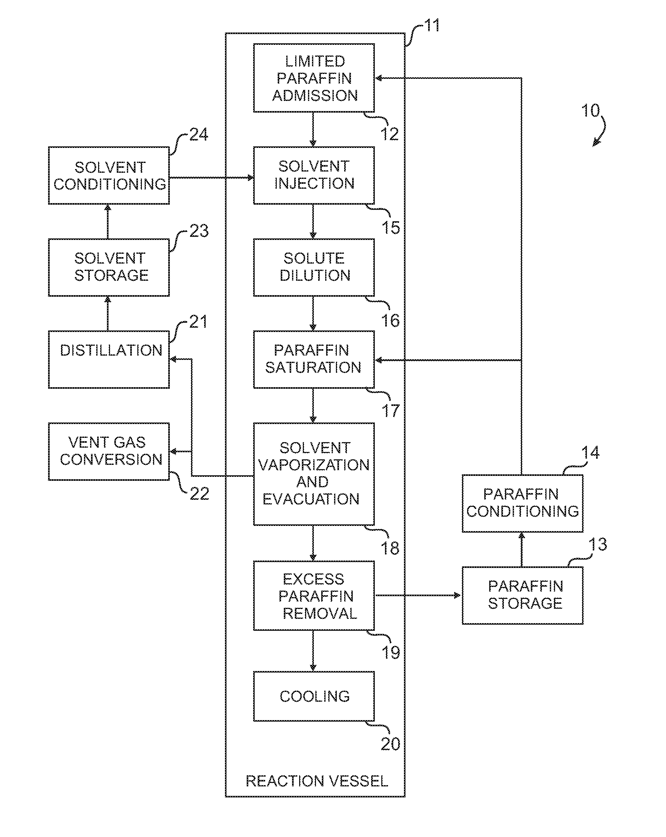
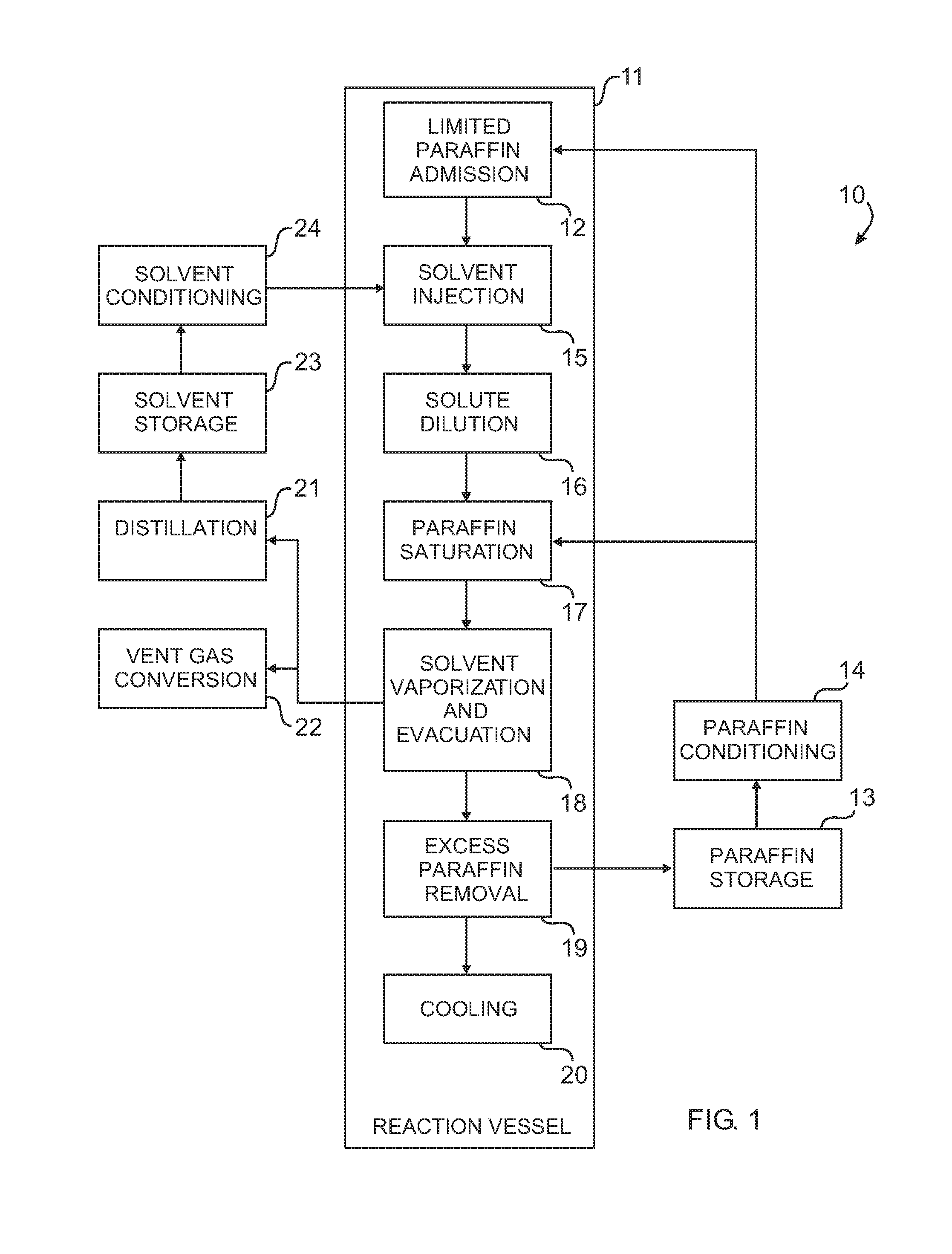
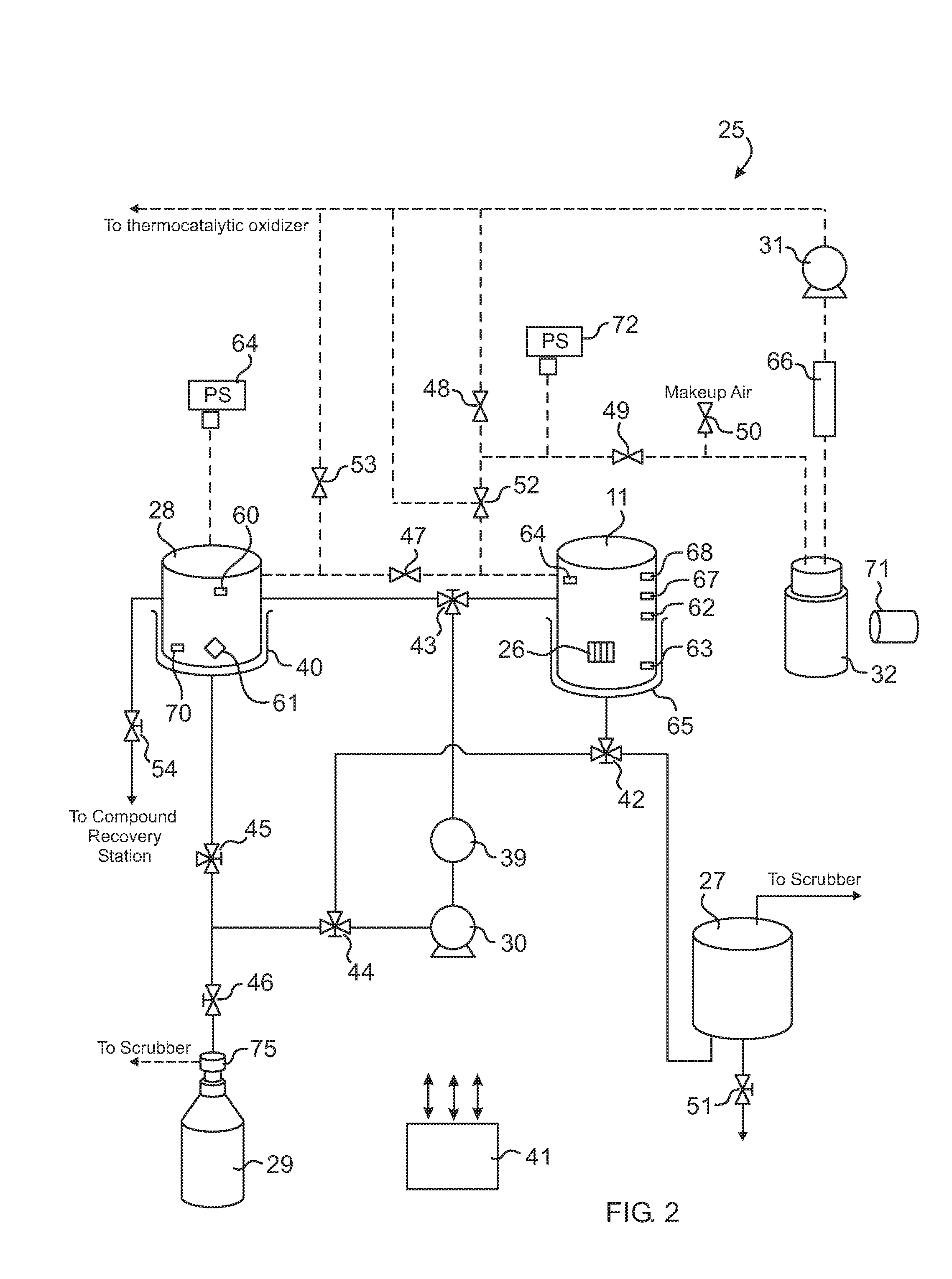
[0121]A measured volume of non-Paraffin based solvent is loaded into the solvent regenerator and heated to 60° C. Liquid Paraffin is transferred into the sealed and heated reaction container until the Paraffin level indicator switch is triggered. Dissolving compound is transferred from the solvent regenerator to the reaction container. The compound and Paraffin are thoroughly blended for 5 minutes. The blend is then transferred to the regenerator. The container is evacuated and loaded with the specimens. The histoprocessing begins.

TABLE 2.1Typical processing times and specimen type.ProcessingImpregnat...

example 3

[0126]The following is another example of batch solvent blending within the reaction container. Table 3.0 shows wt-% of components before and after batch reactor blending.

TABLE 3.0Solvent Batch blending Composition ofDissolving Compound before and after.Wt-% Before ReactorWt-% After ReactorComponentBlendingBlendingAcetone17.4516.68Hexane43.1638.80DMSO5.635.062-propanol33.7610.12Paraffin—30.35

TABLE 3.1Typical processing times and specimen type.ProcessingImpregnationReferenceColor codeTimeTimeTissueNo.Code(minutes)(min)Type301green1030Breast tissue 2 mm302purple1030liver 2 mm303green1030liver 2 mm304green2030liver 5 mm305purple3030Breast tissue 5 mm306purple3030Breast tissue 5 mm307green2030liver 5 mm308purple3030Breast tissue 4 mm309green3030liver 10 mm310purple3030Liver 10 mm311purple3030Breast tissue 2 mm312purple3030Breast tissue 4 mm

[0127]The dissolving compound of Table 3.0 in the solvent regenerator is heated to an equilibrium temperature of 60° C. and a pressure of about 0.8 b...


PUM

PropertyMeasurementUnit
temperatureaaaaaaaaaa
thickaaaaaaaaaa
thickaaaaaaaaaa

Abstract

Method and apparatus for processing tissue specimens against decomposition, putrefaction and autolysis which use a simple three step procedure in a single vessel or container. First, the specimens are saturated with a solvent mixture of a ketone and a hydrocarbon, e.g., an Acetone / Hexane or an Acetone / Xylene mixture, to dissolve lipids and other cellular solutes. The container is then flooded with melted Paraffin. In a last step, the solvent mixture is vaporized and evacuated from the container, allowing the melted Paraffin to replace the vaporized solvent and impregnate the specimens. Raw, i.e., non-processed and non-burred specimens up to 5 mm thick can be processed in about 60 minutes. A solvent regenerator distills the evacuated solvent; and converts vent waste gases into carbon dioxide and water through a thermocatalytic oxidizer.

Description

PRIOR APPLICATION[0001]This is a continuation of U.S. patent application Ser. No. 12 / 128,523, filed 2008 May 28, incorporated herein by reference.FIELD OF THE INVENTION[0002]The present invention relates to techniques and instrumentation for the preservation of tissue specimens. More specifically, the invention teaches the rapid formation of a matrix by the aggregation of a Paraffin medium that impregnates and protects the original tissue structure and prevents decomposition, putrefaction, and autolysis.BACKGROUND[0003]Many of the prior art methods of preparing tissue specimens for histology use incubation in separate solutions of phosphate-buffered 10% formalin for fixation, a series of increasing concentrations of ethanol, and / or isopropanol for dehydration, and xylene for clearing tissue of dehydration agent, prior to impregnation. Because of the time required for this process, usually 8 hours or longer, it is customary to complete these separate steps—fixation, dehydration, clea...

Claims


Application Information

Patent Timeline
Patent Type & Authority Applications(United States)
IPC IPC(8): G01N1/28C12M1/00
CPCG01N1/36G01N1/31
Inventor WHEELER, STEVEN PAUL
Owner WHEELER STEVEN PAUL
Why Eureka
Try Eureka