X-ray tomography method and apparatus used in conjunction with a charged particle cancer therapy system

a cancer therapy system and x-ray tomography technology, applied in the field of solid tumor treatment, can solve the problems of large radiation being delivered outside of the tumor, x-rays not optimal for cancerous tissue treatment, and dangerous tumors in confined places

Active Publication Date: 2012-08-16
BALAKIN ANDREY VLADIMIROVICH +1
View PDF27 Cites 97 Cited by
  • Summary
  • Abstract
  • Description
  • Claims
  • Application Information

AI Technical Summary

Problems solved by technology

Benign tumors cause problems because of their spread, as they press and displace normal tissues.
Benign tumors are dangerous in confined places such as the skull.
X-rays are a form of ionizing radiation and as such can be dangerous.
However, X-rays are not optimal for treatment of cancerous tissue as X-rays deposit their highest dose of radiation near the surface of the targeted tissue and delivery exponentially less radiation as they penetrate into the tissue.
This results in large amounts of radiation being delivered outside of the tumor.
Gamma rays have similar limitations.
These particles damage the DNA of cells, ultimately causing their death.
Cancerous cells, because of their high rate of division and their reduced ability to repair damaged DNA, are particularly vulnerable to attack on their DNA.
Residual particles are used in the accelerator without supplying new particles if sufficient charge is available.

Method used


Image

  • X-ray tomography method and apparatus used in conjunction with a charged particle cancer therapy system
  • X-ray tomography method and apparatus used in conjunction with a charged particle cancer therapy system
  • X-ray tomography method and apparatus used in conjunction with a charged particle cancer therapy system

Examples


example i

[0158]In one example, the initial cross-section distance 810 is about fifteen centimeters and the final cross-section distance 820 is about ten centimeters. Using the provided numbers, the concentration of the magnetic field is about 15 / 10 or 1.5 times at the incident surface 670 of the gap 510, though the relationship is not linear. The taper 860 has a slope, such as about 20, 30, 40, 50, or 60 degrees. The concentration of the magnetic field, such as by 1.5 times, leads to a corresponding decrease in power consumption requirements to the magnets.

[0159]Referring now to FIG. 9, an additional example of geometry of the magnet used to concentrate the magnetic field is illustrated. As illustrated in FIG. 8, the first magnet 610 preferably contains an initial cross-sectional distance 810 of the iron based core. The contours of the magnetic field are shaped by the magnets 610, 620 and the yokes 612, 622. In this example, the core tapers to a second cross-sectional distance 820 with a sma...

example ii

[0163]Referring now to FIG. 11, an example is used to clarify the magnetic field control using a feedback loop 1100 to change delivery times and / or periods of proton pulse delivery. In one case, a respiratory sensor 1110 senses the breathing cycle of the subject. The respiratory sensor sends the information to an algorithm in a magnetic field controller 1120, typically via the patient interface module 150 and / or via the main controller 110 or a subcomponent thereof. The algorithm predicts and / or measures when the subject is at a particular point in the breathing cycle, such as at the bottom of a breath. Magnetic field sensors 1130, such as the high precision magnetic field sensors, are used as input to the magnetic field controller, which controls a magnet power supply 1140 for a given magnetic field 1150, such as within a first turning magnet 410 of a synchrotron 130. The control feedback loop is thus used to dial the synchrotron to a selected energy level and deliver protons with ...

example iii

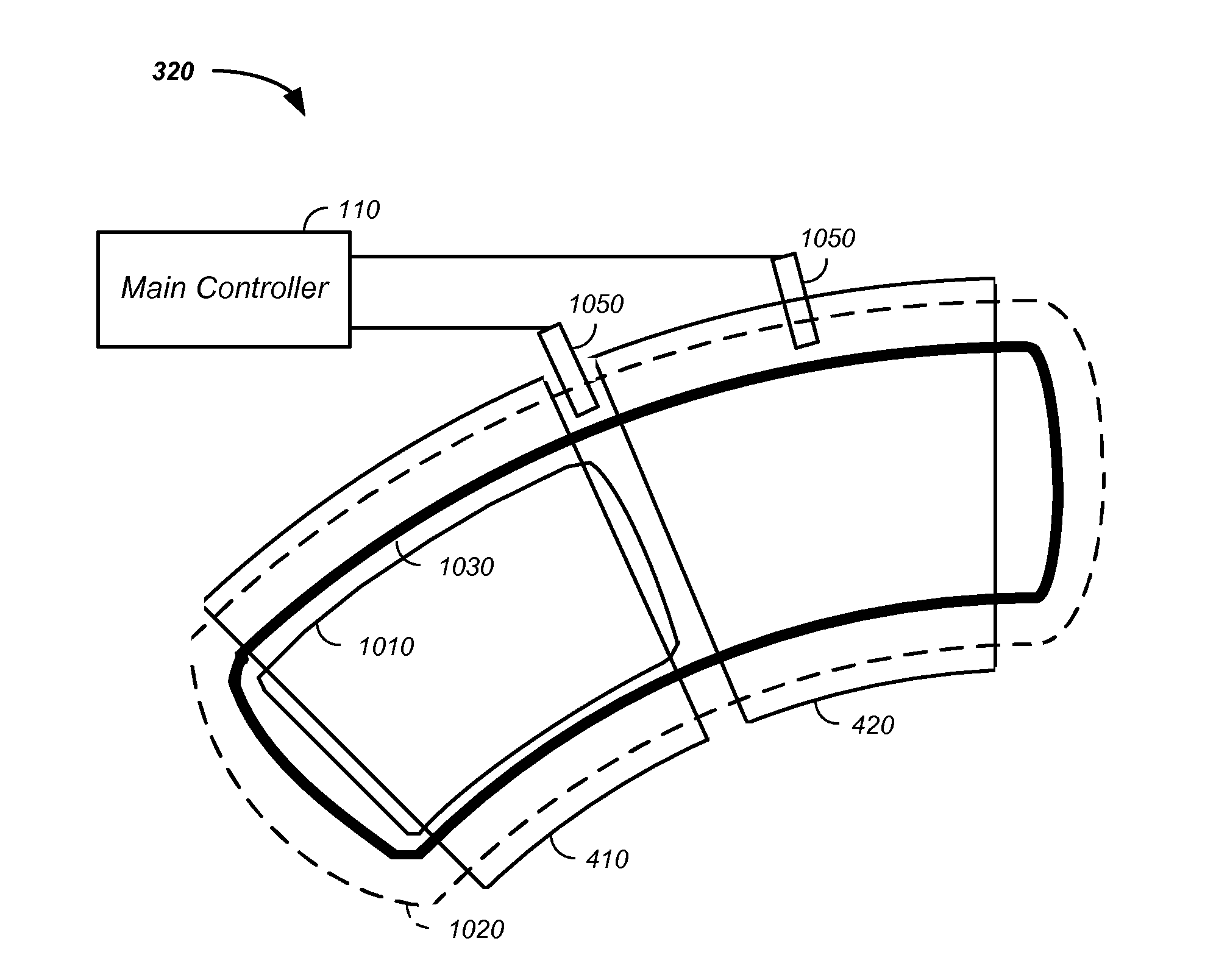
[0166]Referring again to FIG. 10, an example of a winding coil 1030 that covers two turning magnets 410, 420 is provided. Optionally, a first winding coil 1030 covers two magnets 410, 420 and a second winding coil covers another two magnets 430, 440. As described, supra, this system reduces space between turning section allowing more magnetic field to be applied per radian of turn. A first correction coil 1010 is illustrated that is used to correct the magnetic field for the first turning magnet 410. Individual correction coils for each turning magnet are preferred and individual correction coils yield the most precise and / or accurate magnetic field in each turning section. Particularly, the individual correction coil 1010 is used to compensate for imperfections in the individual magnet of a given turning section. Hence, with a series of magnetic field sensors, corresponding magnetic fields are individually adjustable in a series of feedback loops, via a magnetic field monitoring sy...


PUM


Abstract

The invention comprises an X-ray tomography method and apparatus used in conjunction with multi-axis charged particle or proton beam radiation therapy of cancerous tumors. In various embodiments, 3-D images are generated from a series of 2-D X-rays images; the X-ray source and detector are stationary while the patient rotates; the 2-D X-ray images are generated using an X-ray source proximate a charged particle beam in a charged particle cancer therapy system; and the X-ray tomography system uses an electron source having a geometry that enhances an electron source lifetime, where the electron source is used in generation of X-rays. The X-ray tomography system is optionally used in conjunction with systems used to both move and constrain movement of the patient, such as semi-vertical, sitting, or laying positioning systems. The X-ray images are optionally used in control of a charged particle cancer therapy system.

Description

CROSS REFERENCES TO RELATED APPLICATIONS[0001]This application is:[0002]a continuation of U.S. patent application Ser. No. 12 / 606,913 filed Oct. 27, 2009, which is a continuation-in-part of U.S. patent application Ser. No. 12 / 425,683 filed Apr. 17, 2009, which claims the benefit of:[0003]U.S. provisional patent application No. 61 / 055,395 filed May 22, 2008;[0004]U.S. provisional patent application No. 61 / 137,574 filed Aug. 1, 2008;[0005]U.S. provisional patent application No. 61 / 192,245 filed Sep. 17, 2008;[0006]U.S. provisional patent application No. 61 / 055,409 filed May 22, 2008;[0007]U.S. provisional patent application No. 61 / 203,308 filed Dec. 22, 2008;[0008]U.S. provisional patent application No. 61 / 188,407 filed Aug. 11, 2008;[0009]U.S. provisional patent application No. 61 / 188,406 filed Aug. 11, 2008;[0010]U.S. provisional patent application No. 61 / 189,815 filed Aug. 25, 2008;[0011]U.S. provisional patent application No. 61 / 201,731 filed Dec. 15, 2008;[0012]U.S. provisional p...

Claims


Application Information

Patent Timeline
Patent Type & Authority Applications(United States)
IPC IPC(8): A61B6/00A61B6/03
CPCA61B6/032A61B6/0421A61N5/1049A61N2005/1061A61N2005/1097A61N5/1064A61N5/1067A61N5/107A61N2005/1087
Inventor BALAKIN, VLADIMIR
Owner BALAKIN ANDREY VLADIMIROVICH
Why Eureka
Try Eureka