Supercharge Your Innovation With Domain-Expert AI Agents!

Labeled skin lesion biopsy punch and uses thereof

a skin lesion and biopsy technology, applied in medical science, surgery, vaccination/ovulation diagnostics, etc., can solve the problems of imposing the requirement for additional biopsies, the possibility of performing the inability to perform surgery on the wrong area, so as to facilitate non-invasive and continued monitoring of the biopsy site, and achieve effective treatment strategies.

Inactive Publication Date: 2012-09-20
GILCHREST BARBARA +1
View PDF4 Cites 10 Cited by
  • Summary
  • Abstract
  • Description
  • Claims
  • Application Information

AI Technical Summary

Benefits of technology

[0012]Accordingly, embodiments of the present invention include one or more of the following advantages. As the biopsy device and method and systems enable concurrent delivery of the dye, e.g., fluorescent dye marker at the biopsy location site at the time of the biopsy procedure, it allows for a physician to definitively identify and determine the exact site or location of the biopsy at a later timepoint, e.g., for a diagnostic and / or follow-up assessment by a physician. Additionally, concurrently delivery of the dye, e.g., fluorescent dye marker can be delivered to the biopsy site through the sampling portion of the biopsy device so that the device does not have to be removed before depositing the dye, e.g., fluorescent dye at the biopsy site. Accordingly, another advantage of the present invention relates to being able to mark the location or margins of such a lesion immediately after removing the biopsy tissue mass or sample. In some embodiments, where the biopsy device removes the entire lesion or skin abnormality is removed in its entirety, marking the biopsy site immediately at the same time as the biopsy procedure ensures precise reestablishment of the biopsy location for future identification. The skin marking system and biopsy device allows concurrent marking of the biopsy site at the same time as the tissue sample is removed from the subject, therefore prevents repeated removal of the biopsy device and insertion of a biopsy marking device which may cause unneeded additional discomfort to the patient undergoing the procedure; as well as avoids removal of the biopsy device to be replaced with a marking device, which may introduce an error in placement of the biopsy marker into the desired location; as well as repeated removal and insertion of each of the devices may prolong the duration of the procedure or spread cancer cells etc.
[0013]In some embodiments, the dye, e.g., fluorescent dye can be deposited to indicate the circumference of the biopsy site (e.g., at the biopsy cavity walls), and in some embodiments, at opposite the ends of the biopsy site. Additionally, marking a biopsy site with a fluorescent tattoo facilitates non-invasive and continued monitoring of the biopsy site, which enables effective treatment strategies to be devised. For example, a therapeutic device or therapeutic treatment or agent can be guided by and located to a specific biopsy site at the location of the biopsy fluorescent tattoo.
[0014]Another advantage is the sociological and psychological advantages to the subject, including the fact that the tattoo is substantially invisible in normal light (white light) avoids having a visible mark which could be an unpleasant or unwanted constant reminder of the biopsy procedure and / or could exasperate a fear of reoccurrence of a cancer in the subject at any time in the future.
[0015]In some embodiments, the skin marking biopsy device of the present invention is used to mark and localize a body area of medical interest. In some embodiment, the fluorescent dye is a biocompatible fluorescent dye that is visible under UV illumination, such as, for example, Wood's light and is minimally visible under visible ambient light.
[0016]In some embodiments, the dye, e.g., fluorescent dye can be at least partially, or fully biodegradable. When several biopsy samples (e.g., 3-12) are taken, the biopsy device can deposit different colors of dye, e.g., different colors of fluorescent dye to different biopsy locations, so they can be distinguished from one another by the type of fluorescent color used in the dye. For example, in some embodiments, a fluorescent dye can be white, or yellow or orange, or other colors under a defined wavelength, e.g., UV or blacklight, yet substantially invisible under normal (white) light. In some embodiments, a subject can undergo multiple different biopsy, each marked with a different color fluorescent dye, e.g., white, or yellow or orange, or another color, enabling subsequent distinction of each biopsy sites from one another when they are identified and visualized at a later timepoint under the defined wavelength, e.g., e.g., UV or blacklight, e.g., Wood's light.
[0017]One aspect of the present invention relates to a tissue marking system comprising combining a biopsy device with a biocompatible dye, wherein the biopsy device applies the biocompatible dye to the biopsy cavity walls or surface of a subject's tissue during a biopsy procedure, and wherein the biocompatible dye is visible under a predetermined wavelength. In some embodiments, the biocompatible dye is a fluorescent dye, for example, a biocompatible dye is not visible or minimally visible under normal (white) light, and is visible under UV light or black light. In some embodiments, the biocompatible dye, e.g., fluorescent dye is visible at under 400 nm wavelength, for example at about 365 nm wavelength.

Problems solved by technology

In the U.S., there are more than 1.3 million non-melanoma skin cancers (NMSC) occur each year, which cause significant morbidity and incur high cost to society.
This creates the possibility of performing surgery on the wrong area or imposing the requirement for additional biopsies.
However, these tattoos are visible under ordinary light and thus may be distasteful to many patients.
Also, the physicians performing the biopsy is often not the physician who performs the definited surgery.
In locations like scalp, back, arms, where visible and unequivocal anatomical landmarks are sparse, digital photography becomes difficult to carry out.
Pictorial documentation has traditionally been used, but the advent of electronic medical record system has made pictorial documentation difficult to do.
As of yet, Logician, the computerized charts at the Boston Medical Center, for example, cannot accommodate digital photographs.
When the biopsy occurs in area that is difficult to reach, such as back of the ear, top of the scalp and back, the patient may have a hard time identifying the biopsy site.
The difficulty is especially more concerning in the elderly population with the highest incidence of NMSC who have difficulty caring for and remembering the location of the biopsy.
The downside to the carbon graphite and India ink tattoo is that these tattoos are permanent and readily visible.

Method used


Image

  • Labeled skin lesion biopsy punch and uses thereof
  • Labeled skin lesion biopsy punch and uses thereof
  • Labeled skin lesion biopsy punch and uses thereof

Examples


example 1

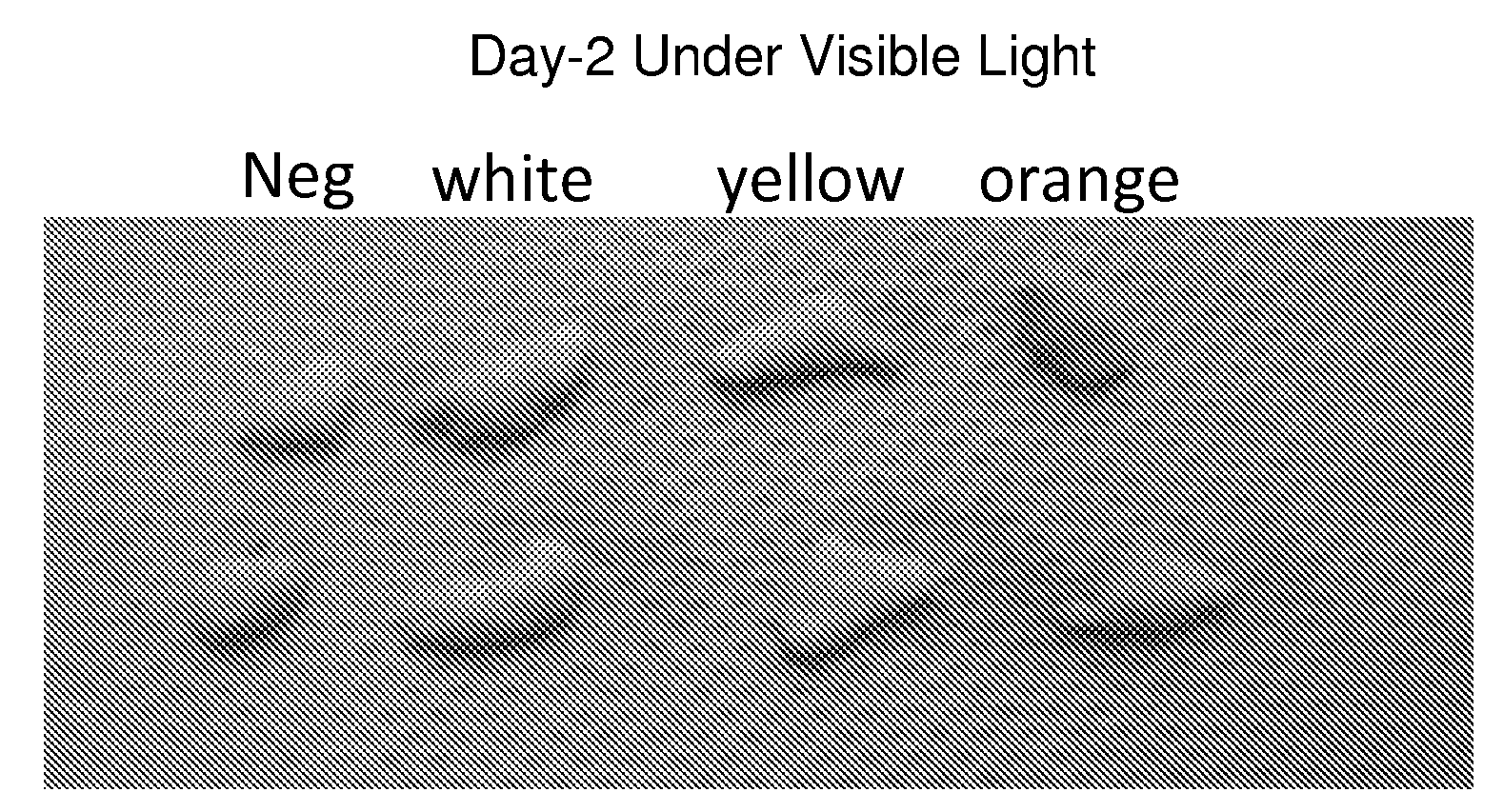
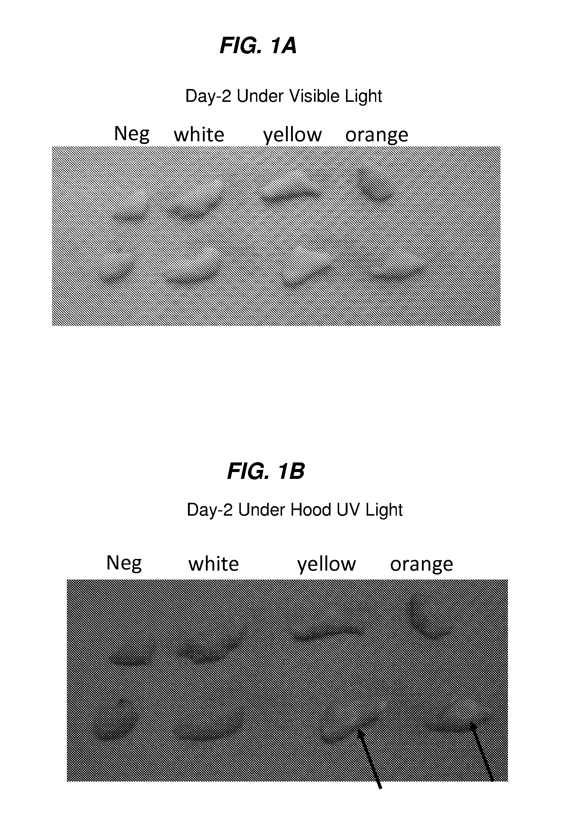
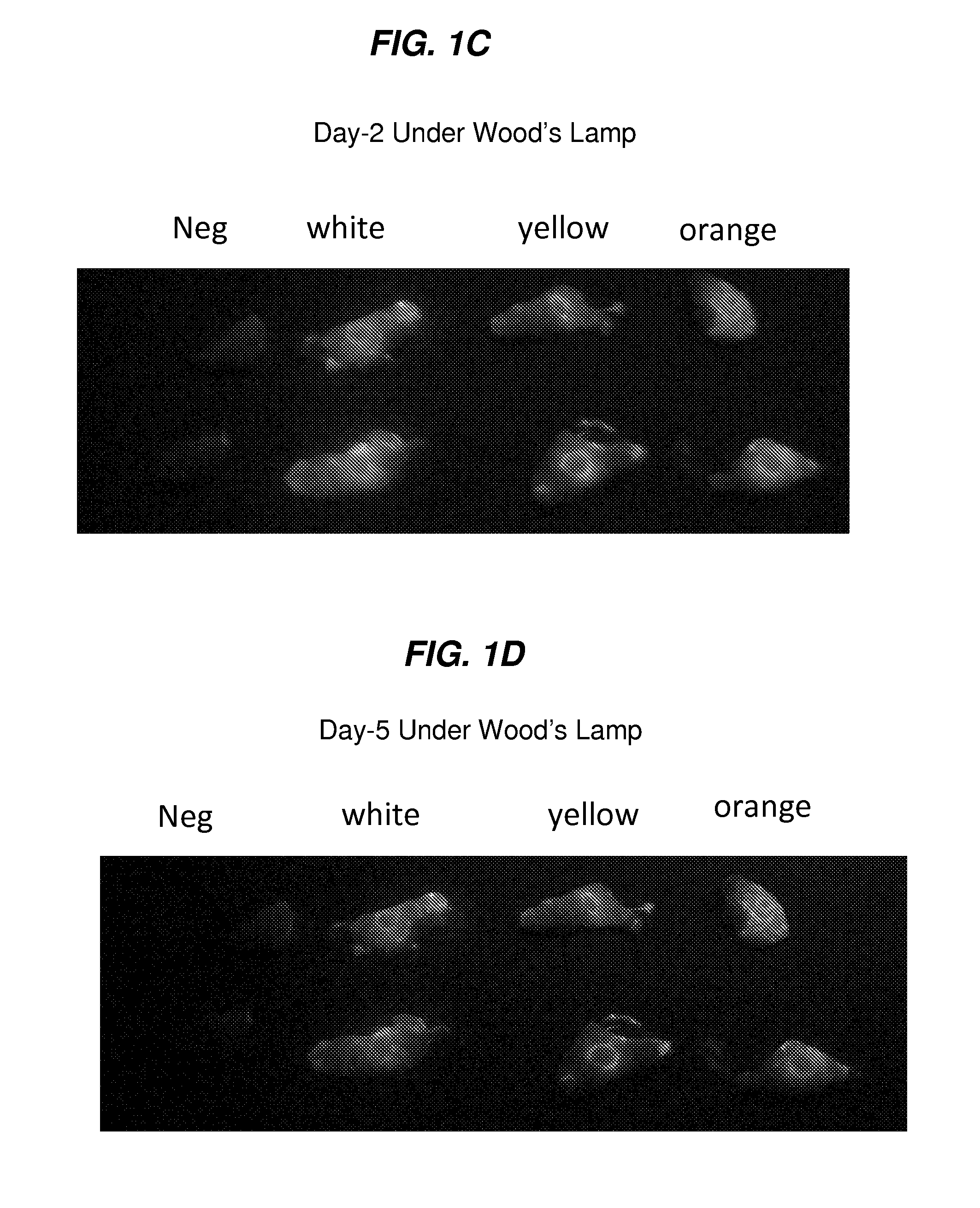
[0218]In vitro study demonstrating fluorescence tattoo is invisible under normal light and visible under UV light after multiple washes.

[0219]Methods:

[0220]Human fetal foreskin is harvested, and the biocompatible dye, e.g., fluorescent tattoo deposited to each sample using a biopsy device, e.g., needle biopsy device, and the human fetal foreskin tissue was washed in a combination of media and alcohol and cultured in Transwell microplate. Each day the human fetal foreskin tissue samples were removed under the ventilated hood and was washed with both media and alcohol (to mimics daily skin care). The human fetal foreskin tissue samples were harvested on Day 5 following application of the dye and analyzed using histological staining.

[0221]The inventors used neonatal foreskin as an example of human skin. The inventors assessed the following: (a) ease of application and the feasibility of applying the tattoo, (b) the optimal protocol for the application of the fluorescent tattoo, (c) the...

example 2

[0226]In vivo study demonstrating fluorescence tattoo is invisible under normal light and visible under UV light for at least 6 months following.

[0227]The inventors demonstrated the use of the biopsy device and the system and methods as disclosed herein on a subject (23 year male) with basal cell nervus syndrome with a lesion in the middle of his back, which was suspected to be a recurrent basal cell carcinoma (BCC) with multiple LN2 and ED&C by FP (see FIG. 6A). The biopsy procedure was performed with a punch biopsy with the tissue cutting edge coated with a biocompatible dye, e.g., fluorescent dye. The dye was deposited at the exact location of the biopsy when the biopsy was performed, and was visible under UV light using Wood's lamp on the day of the biopsy (see FIG. 6B). The dye was not visible under normal white light. On the 3 month follow-up timepoint after the biopsy procedure, the subject was assessed and the location of the biopsy procedure was not visible under normal (wh...


PUM


Abstract

The present invention relates to a biopsy device, methods, kits and systems for marking the location of a biopsy site in order to later identify the location of a biopsy or surgery in a subject, where the marker is a dye or tattoo which is not visible to the naked eye under normal (white) light, but is visible under UV light. Some embodiments relate to marking a biopsy site for later visualization of the marker.

Description

CROSS REFERENCE TO RELATED APPLICATIONS[0001]This application claims benefit under 35 U.S.C. 119(e) of U.S. Provisional Patent Application Ser. No: 61 / 271,003 filed Jul. 16, 2009, the contents of which are incorporated herein in their entirety by reference.FIELD OF THE INVENTION[0002]The present invention generally relates to the field of skin marking dyes and systems and methods for their use in the identification prior to, during and after medical procedures, such as the identification of a specific location of a biopsy site. The present invention relates to markers to be employed at biopsy sites to permanently or temporarily mark the site, and to methods and apparatus for applying the permanent or temporary marker. Other aspects of the present invention also relate to methods and devices for marking and defining particular locations in body tissue, particularly human tissue, and more particularly relates to methods and devices for permanently or temporarily defining the location ...

Claims


Application Information

Patent Timeline
IPC IPC(8): A61B10/02A61B10/06
CPCA61B10/0266G01N1/30A61B2019/545A61B2019/5408A61B2090/3908A61B2090/395
Inventor GILCHREST, BARBARA ANNCHUANG, GARY
Owner GILCHREST BARBARA
Features
  • R&D
  • Intellectual Property
  • Life Sciences
  • Materials
  • Tech Scout
  • Unparalleled Data Quality
  • Higher Quality Content
  • 60% Fewer Hallucinations
Learn More