
System and method for detection of acoustic shadows and automatic assessment of image usability in 3D ultrasound images

a technology of ultrasound image and detection method, applied in the field of detection of acoustic shadows and evaluation of image quality in 3d ultrasound image, can solve the problems of image usability, image degradation, and often unusable images, and achieve the effects of eliminating unusable data, improving free-hand compounding, and accurate separation of feature spa

Active Publication Date: 2012-09-27
SIEMENS MEDICAL SOLUTIONS USA INC
View PDF10 Cites 59 Cited by
  • Summary
  • Abstract
  • Description
  • Claims
  • Application Information

AI Technical Summary

Benefits of technology

[0023]Classification results for both human and phantom images are presented and compared to manual classifications. An algorithm according to an embodiment of the invention achieved 95% sensitivity in some cases, 91% sensitivity on average, and 91% specificity for usable regions of human scans. All results were obtained using a linear SVM kernel, which is the simplest one, but they should be even better with complex kernels since they allow a more accurate separation of the feature space.
[0024]Example applications of this algorithm could include improved compounding of free-hand 3D ultrasound volumes by eliminating unusable data and improved automatic feature detection by limiting detection to only usable areas.

Problems solved by technology

Image acquisition is sensitive to skin contact and transducer orientation and requires both time and technical skill to be done properly.
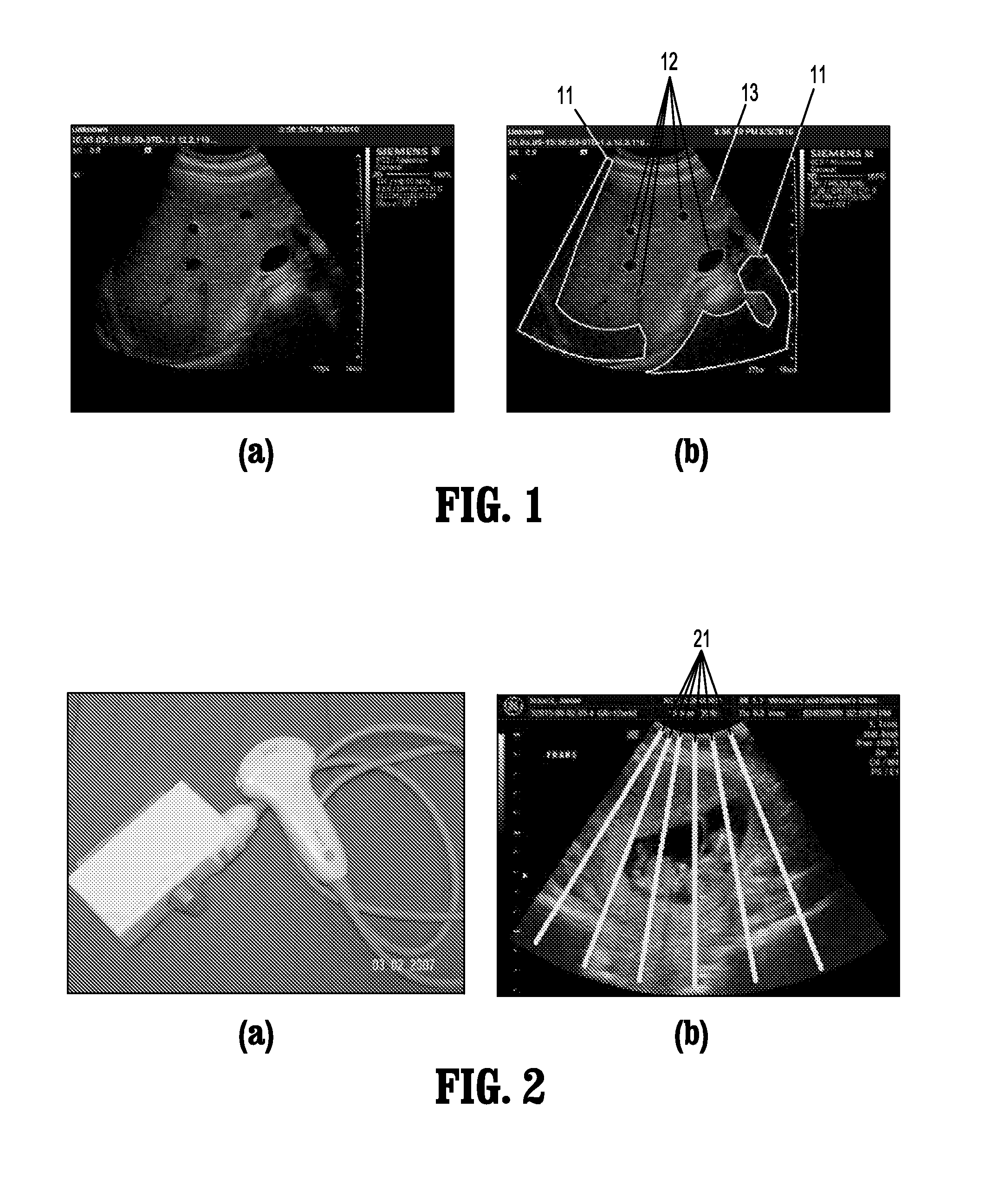
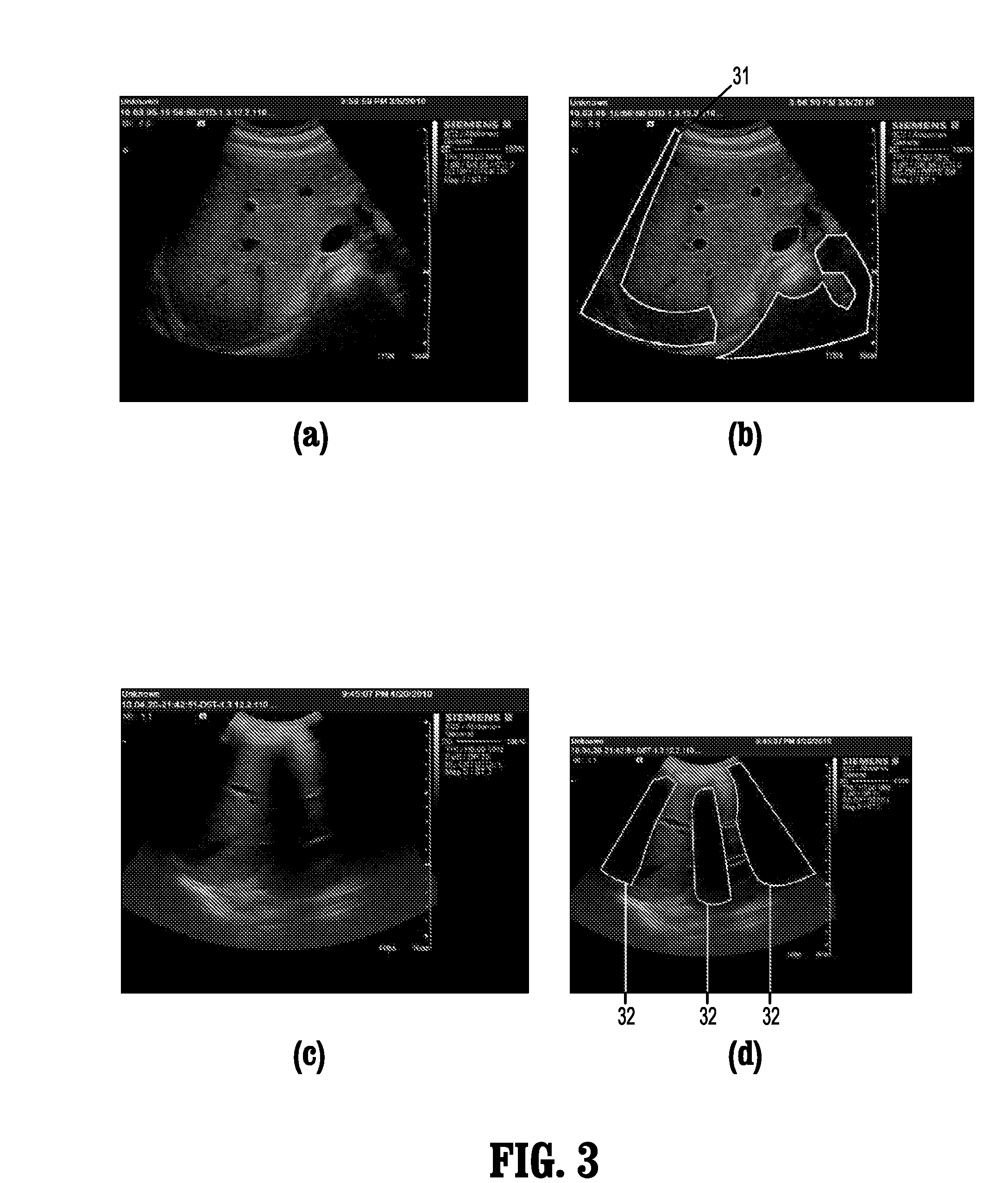
Images commonly suffer degradation from acoustic shadows and signal attenuation, which present as regions of low signal intensity masking anatomical details and making the images partly or totally unusable.
A variety of factors, including occluding anatomy, poor contact, and signal degradation can obscure anatomical details, leading to regions of images that are clinically useless.
But it becomes a very different challenge when an image contains anatomical structures that appear dark in B-scans.
The vessel / shadow distinction is more challenging given that they are not clearly separated.
However, it also creates challenges for other applications, such as ultrasound image registration and segmentation of lesions or structures.
A challenge in applying these methods to acoustic shadow detection is that these methods generally ignore the basic geometric property of acoustic shadow: that the acoustic shadow exists as regions with constraint configurations.
However, geometric based methods do not take full advantage of the texture properties and the configuration information of the regions.

Method used


Image

  • System and method for detection of acoustic shadows and automatic assessment of image usability in 3D ultrasound images
  • System and method for detection of acoustic shadows and automatic assessment of image usability in 3D ultrasound images
  • System and method for detection of acoustic shadows and automatic assessment of image usability in 3D ultrasound images

Examples


Embodiment Construction

[0051]Exemplary embodiments of the invention as described herein generally include systems and methods for detecting acoustic shadows and evaluating image quality in 3D ultrasound images. Accordingly, while the invention is susceptible to various modifications and alternative forms, specific embodiments thereof are shown by way of example in the drawings and will herein be described in detail. It should be understood, however, that there is no intent to limit the invention to the particular forms disclosed, but on the contrary, the invention is to cover all modifications, equivalents, and alternatives falling within the spirit and scope of the invention.

[0052]As used herein, the term “image” refers to multi-dimensional data composed of discrete image elements (e.g., pixels for 2-D images and voxels for 3-D images). The image may be, for example, a medical image of a subject collected by computer tomography, magnetic resonance imaging, ultrasound, or any other medical imaging system ...


PUM


Abstract

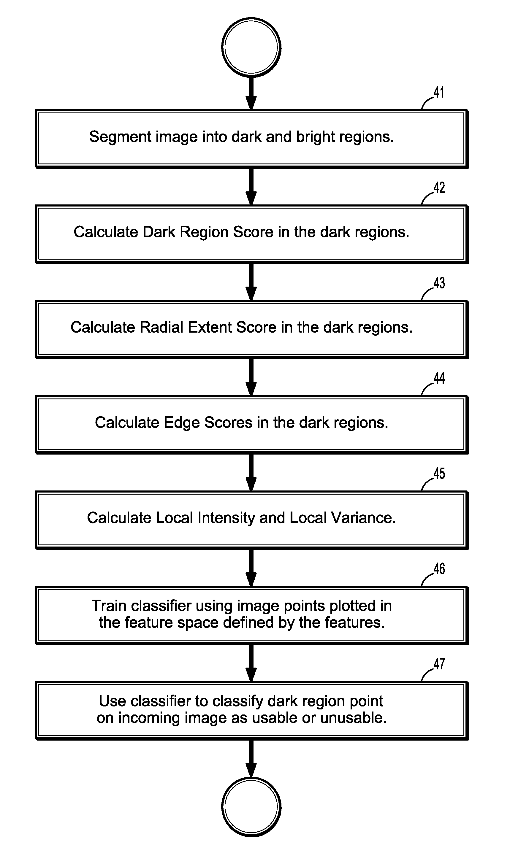
A method for automatically assessing medical ultrasound (US) image usability, includes extracting one or more features from at least one part of a medical ultrasound image, calculating for each feature a feature score for each pixel of the at least one part of the ultrasound image, and classifying one or more image pixels of the at least one part as either usable or unusable, based on a combination of feature scores for each pixel, where usable pixels have intensity values substantially representative of one or more anatomical structures.

Description

CROSS REFERENCE TO RELATED UNITED STATES APPLICATIONS[0001]This application claims priority from “Detection of Acoustic Shadows in 3D Ultrasound Images For Registration”, U.S. Provisional Application No. 61 / 250,061 of Hong, et al., filed Oct. 9, 2009, and “Automatic assessment of ultrasound image usability” of Stoll, et al., filed Jul. 30, 2010, the contents of both of which are herein incorporated by reference in their entireties.TECHNICAL FIELD[0002]This disclosure is directed to detecting acoustic shadows and evaluating image quality in 3D ultrasound images.DISCUSSION OF THE RELATED ART[0003]Ultrasound is the most commonly used form of medical imaging. It offers clinicians the ability to view body structures quickly, easily, relatively in expensively and without radiation. Among its many uses are evaluating trauma, monitoring fetal development, identification and characterization of lesions, and guiding interventions. Nevertheless, skill and training are required to acquire high-...

Claims


Application Information

Patent Timeline
IPC IPC(8): G06K9/46G06K9/48G06K9/62
CPCG06T7/0002G06T2207/30168G06T2207/30004G06T2207/10132
Inventor FUNKA-LEA, GARETHSTOLL, JEFFREYVALENTE, LUCA
Owner SIEMENS MEDICAL SOLUTIONS USA INC
Learn More