

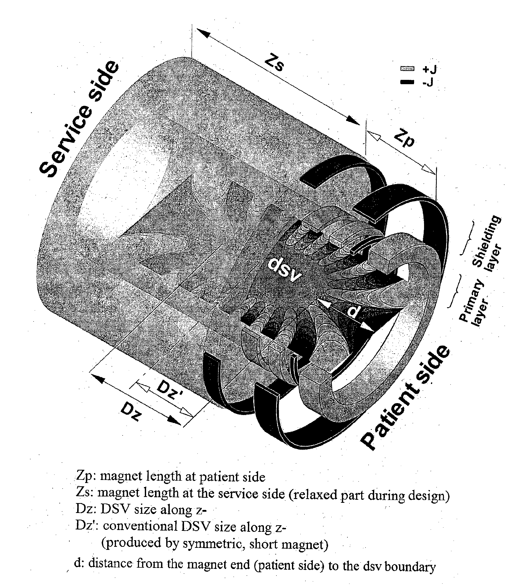
Open-bore magnet for use in magnetic resonance imaging
a magnetic resonance imaging and open-bore technology, applied in the field of magnets, can solve the problems of difficult design and construction of short or compact magnets, many people suffer from claustrophobia, and the axial dsv size of a short magnet can be enlarged, and the axial dsv size can be enlarged. , the effect of reducing the for
- Summary
- Abstract
- Description
- Claims
- Application Information
AI Technical Summary
Benefits of technology
Problems solved by technology
Method used
Image

Examples
example 1 (
1.5 T Whole-Body Magnet)
[0078]This example, shown schematically in FIG. 4, illustrates a superconducting magnet according to one embodiment of the present invention. In broad overview, the magnet employs thirteen coils and has a cold bore length and a cold bore inner radius of approximately 1.34 and 0.49 meters, respectively. More importantly, the shortest distance between the cold-bore magnet end and the dsv edge is only 0.36 meters, which is difficult to achieve using other coil configurations. In this example, the axial distance between the magnet centre and the imaging centre is 1.2 cm. On the primary windings of the magnet, all of the coils are wound in the same direction (i.e. have the same polarity) apart from the coils second from the ends. These coils are wound in the opposite direction to all others on the primary (i.e. have reverse polarity).
[0079]Relative to the imaging centre, the coil blocks on the primary winding have asymmetric electromagnetic topology. The total cur...
example 2 (
3 T Extremity Magnet (Versions a, b))
[0085]This example, shown schematically in FIGS. 10 and 15, illustrates a 3 T superconducting magnet design using a structure according to second and third embodiments of the invention.
[0086]As shown in FIG. 10, in the design version ‘a’, the coil structure is less than 55 cm in total length while a homogeneous dsv is generated: 23.5 cm along the axial direction and 7.5 cm in the radial direction, wherein the homogeneity of the dsv varies by less than 5 ppm over that volume. On the patient side of the primary layer, the coil next to the end coil is of opposite polarity to all others in the primary coil set. Six middle coils in this example are positioned in the centre region of the magnet. There is no negative coil next to the end coil on the service side. In this example, the axial distance between the magnet centre and the imaging centre is 1.2 cm. The coil structure again provides the advantage when combined with the other features of producin...
PUM
 Abstract
Description
Claims
Application Information