Method for the quantitative and qualitative characterization of antigen-specific t cells recognizing a specific antigen

a t cell and quantitative characterization technology, applied in the field of qualitative and quantitative characterization of antigen-specific t cells recognizing a specific antigen, can solve the problems of severe affecting the sensitivity of the method and the loss severely affects the accuracy of quantitative and qualitative analysis, so as to improve the handling of small cell numbers and associated cell loss, the effect of optimal detection

Inactive Publication Date: 2012-11-01
MILTENYI BIOTEC
View PDF4 Cites 3 Cited by
  • Summary
  • Abstract
  • Description
  • Claims
  • Application Information

AI Technical Summary

Benefits of technology

[0022]The present invention solves the above technical problem by a method using magnetic cell separation using activation markers of antigen-specific T cells to increase the frequency of target T cells before further analysis of these cells, e.g. flow-cytometric analysis and the handling of the small cell numbers and associated cell loss was improved by manipulation of the cells directly on the columns used for enrichment.
[0023]It was surprising that we could establish a method for the specific enrichment of target T cells based on activation marker expression, e.g. CD154 expression, which allowed to separate rare antigen-specifically activated T cells from unspecific background. First, this was achieved by adjusting the conditions in a way that mainly brightly labelled T cells, which resemble those T cells which have been activated by antigen, are enriched versus low expressing “background” cells, resembling weakly activated, e.g. low affinity, T cells or T cells activated already in vivo at an earlier timepoint, i.e. residual activation marker expression or T cells activated by non TCR signals, i.e. bystander activation. Second, we have developed a new strategy to allow simultaneous detection of activation markers on the surface and accumulation of intracellular cytokines for their optimal detection by intracellular cytokine staining. This was achieved by subsequent stimulation with and without secretion inhibitors for optimized time periods. Another restriction of rare cell analysis is the cell loss during several processing steps, i.e. surface staining, fixation, intracellular staining. This cell loss severely affects the sensitivity of the method as well as the accuracy of quantitative and qualitative analysis. Therefore the invention includes staining of the cells following enrichment directly on the enrichment column, e.g. a magnetic column, especially MACS columns, which dramatically reduce cell loss, i.e. recovery of more target cells allowed us to accurately determine the target cell numbers as well as the analysis of phenotypic and functional subpopulations.

Problems solved by technology

This cell loss severely affects the sensitivity of the method as well as the accuracy of quantitative and qualitative analysis.

Method used


Image

  • Method for the quantitative and qualitative characterization of antigen-specific t cells recognizing a specific antigen
  • Method for the quantitative and qualitative characterization of antigen-specific t cells recognizing a specific antigen
  • Method for the quantitative and qualitative characterization of antigen-specific t cells recognizing a specific antigen

Examples


example 1

CD154+ Enrichment Increases the Sensitivity of Detection of Antigen-Specific CD4+ T Cells

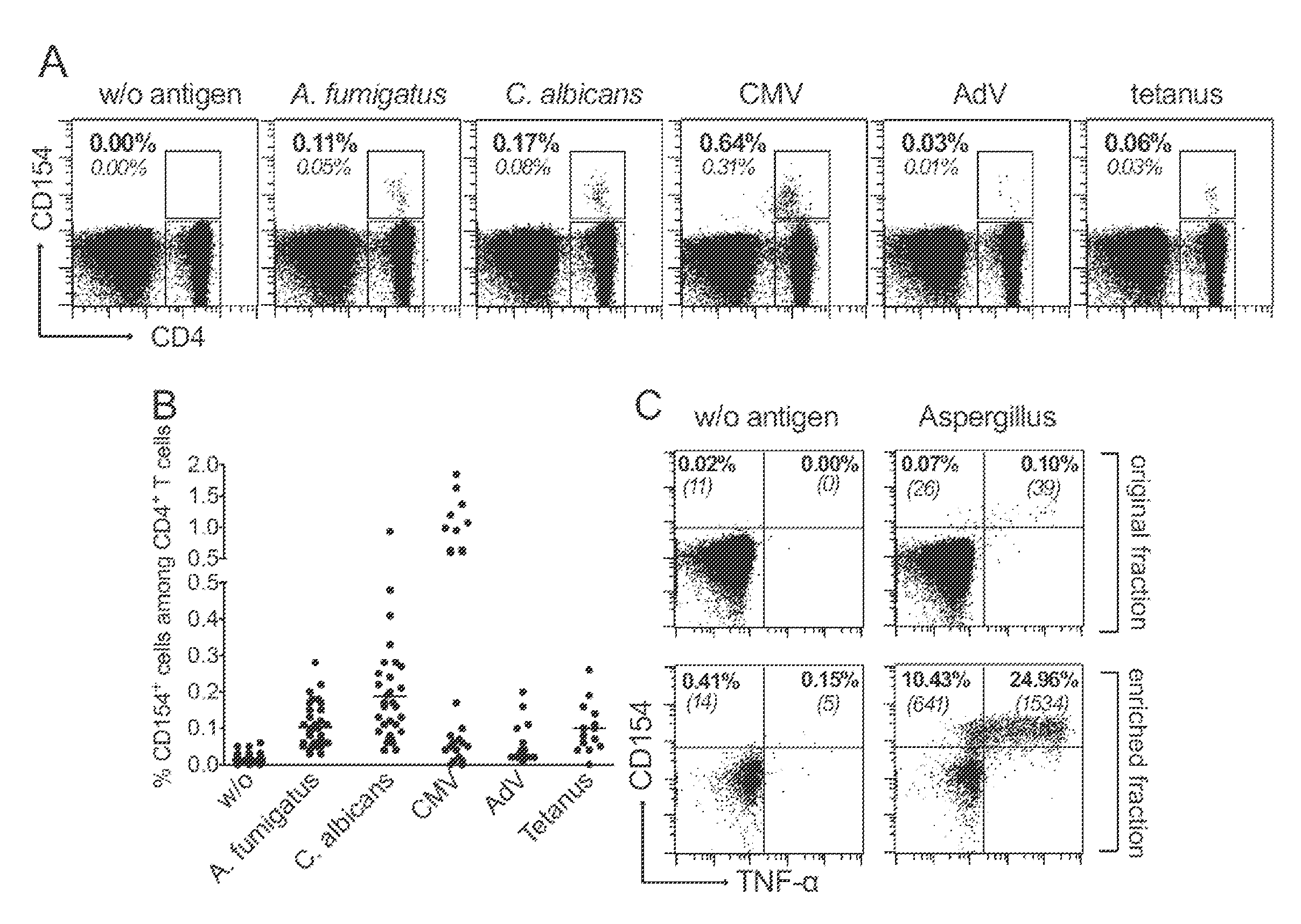
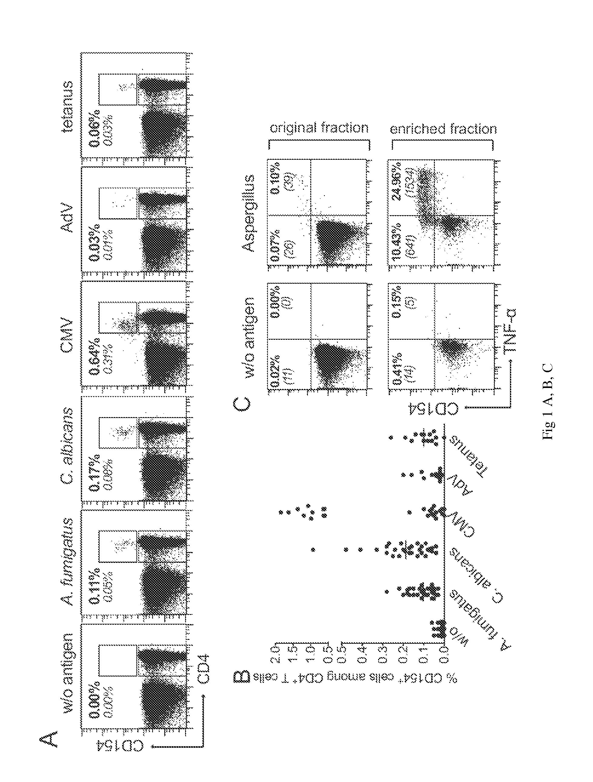
[0144]CD154 expression allows the detection of the entire pool of antigen reactive CD4+ T cells, without any restriction to a certain subset producing particular cytokines. We therefore used this marker to perform a comprehensive characterization of T cells specific for various viral (Cytomegalovirus, Adenovirus), bacterial (Tetanus toxoid), or fungal (Aspergillus fumigatus, Candida albicans) recall antigens.

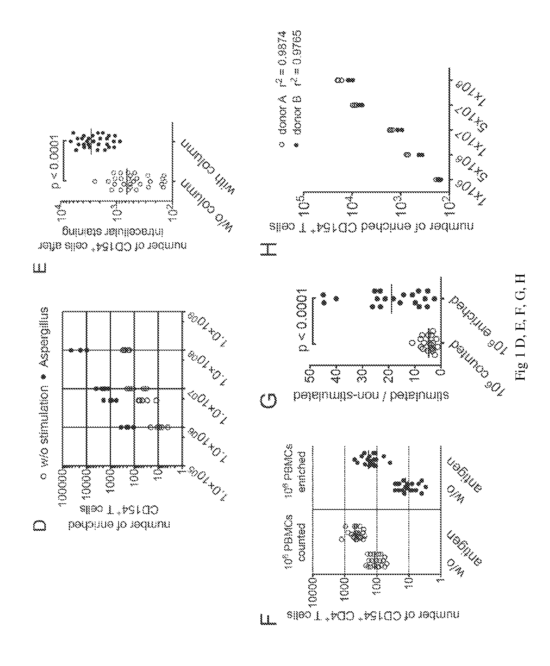
[0145]Human peripheral blood mononuclear cells (PBMCs) were stimulated in vitro with lysates of the respective antigens. As shown in FIGS. 1A and B, CD154 expressing CD4+ T cells can readily be detected following seven hours stimulation of PBMCs. However, it is also evident that for certain antigens and / or donors the frequencies of antigen-specific CD4+ T cells are often below 0.1%, which is in general the limit of detection for standard flow-cytometry. In particular, at frequencies below 0....

example 2

CD154+ Enrichment Specifically Identifies Antigen-Reactive T Cells

[0155]To exclude non-specific stimulation by fungal components potentially present in the total antigen lysates we blocked the T cell activation by addition of an HLA-DR-neutralising antibody. This antibody blocked the CD4+ T cell responses against Aspergillus-, Candida- and CMV-lysates as well as against a peptide pool of CMV pp65 but neither T cell activation by SEB and antiCD3 / CD28 (FIG. 2A) nor the activation of CD8+ T cells (not depicted). Similarly, the CD4+ T cell responses against the lysates but not against the pp65 peptide pool were prevented when APCs were fixed with formaldehyde before incubation with the antigens (FIG. 2B). These data clearly show that T cell activation against native antigens occurs through an MHC class II- and antigen processing-dependent mechanism.

[0156]To further confirm the specificity of the antigen activated CD154+ cells, we used the CD154+ enrichment for the rapid generation of po...

example 3

Phenotypic and Functional Characterization of Pathogen-Reactive CD4+ T Cells

[0164]Until now, the cytometric analysis of antigen-reactive T cells using cytokines or activation markers as a read-out was mainly restricted to the analysis of the memory T cell pool, because the frequency of naïve T cells was below the level of detection. For this reason, we also analyzed the phenotype of antigen-reactive T cells following CD154k enrichment. As expected, C. albicans, CMV, AdV and tetanus stimulated T cells mainly belonged to the CD45RO+ memory T cells (FIG. 3A, B) and were distributed between the CD45RO+CCR7+ central memory and CD45RO+CCR7− effector T cell subset, depending on the pathogen. C. albicans-reactive T cells were found to be mainly within the central memory compartment, whereas CMV generates a strong effector memory phenotype. For A. fumigatus, AdV and tetanus the distribution between the two subsets was less uniform but displayed strong donor dependent variability. Interesting...


PUM

PropertyMeasurementUnit
timeaaaaaaaaaa
sizeaaaaaaaaaa
sizeaaaaaaaaaa

Abstract

The invention relates to a method for sensitive quantitative and/or qualitative analysis of target T cells comprising the steps a) enrichment of said cells from a mixture of said cells and other cells in a sample by the use of one or more activation markers expressed on antigen-activated T cells in a parallel cell sorting process and b) analysis of the cells of step a).
The invention relates also to a method of diagnosing diseases based on the analysis of the target T cells.

Description

CROSS-REFERENCE TO RELATED APPLICATIONS[0001]This application claims the priority benefit of European Patent Application No. 11164382.1, filed Apr. 29, 2011, which is incorporated herein by reference in its entirety.FIELD OF THE INVENTION[0002]The invention relates to a method for the qualitative and quantitative characterization of antigen-specific T cells recognizing a specific antigen in body fluids by the use of antigen-specific T cell activation markers.BACKGROUND OF THE INVENTION[0003]T cells are the central organizers and effectors of the immune system and are responsible for effective immunity against pathogens and tumors as well as for keeping unresponsiveness (tolerance) against autoantigens and harmless non-self antigens, such as food. To achieve this goal T cells are educated either during their early development in the thymus or later on in the periphery to acquire distinct effector functions, which can be stably inherited from a single cell to its progeny and in this w...

Claims


Application Information

Patent Timeline
Patent Type & Authority Applications(United States)
IPC IPC(8): G01N33/569
CPCG01N33/54326G01N33/56972G01N2800/24G01N2333/38G01N2333/40G01N2333/70575
Inventor SCHEFFOLD, ALEXANDERBACHER, PETRA
Owner MILTENYI BIOTEC
Why Eureka
Try Eureka