

Phasor Method to Fluorescence Lifetime Microscopy to Discriminate Metabolic State of Cells in Living Tissue
a lifetime microscopy and phasor technology, applied in the field of phasor and phasor method to discriminate the metabolic state of cells in living tissue, can solve the problems of limited success in ad hoc assigning autofluorescence to specific tissue components, and it is difficult to associate specific tissue components with exponential decay
- Summary
- Abstract
- Description
- Claims
- Application Information
AI Technical Summary
Benefits of technology
Problems solved by technology
Method used
Image

Examples
Embodiment Construction
[0057]As a preliminary matter, it should be noted that numerous modifications we made in the phasor method and the analysis software as disclosed herein, with respect to the 2008 phasor method published (reference 36). Such modifications, include, but are not limited to:
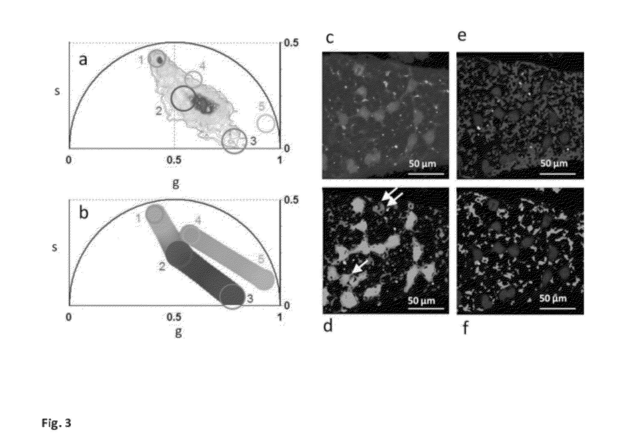
[0058]a) modification of the phasor method to perform image segmentation to measure the average phasor value of regions of interest in the tissues. The region of interest of cells is selected by using a circular of custom diameter or an arbitrary shape Different regions of the image, such as cells, can be attributed statistically to different average phasor values. (FIG. 4).
[0059]b) modification of the phasor method to measure the relative concentrations of fluorophores and map their spatial distribution in living tissues. (FIG. 3 and FIG. SM2).
[0060]c) modification of the phasor method to perform analysis of the FLIM data with higher harmonics (ω=nωσ with n=2, 3) of the laser repetition rate (ωσ=2πf), where f is the...
PUM
| Property | Measurement | Unit |
|---|---|---|
| pH | aaaaa | aaaaa |
| pH | aaaaa | aaaaa |
| pH | aaaaa | aaaaa |
Abstract
Description
Claims
Application Information