

Methods of enriching and detecting fetal nucleic acids
a technology of fetal nucleic acids and enrichment methods, which is applied in the field of methods of enriching and detecting fetal nucleic acids, can solve the problem of exposing both the mother and the fetus to a certain amount of risk
- Summary
- Abstract
- Description
- Claims
- Application Information
AI Technical Summary
Benefits of technology
Problems solved by technology
Method used
Image

Examples
example 1
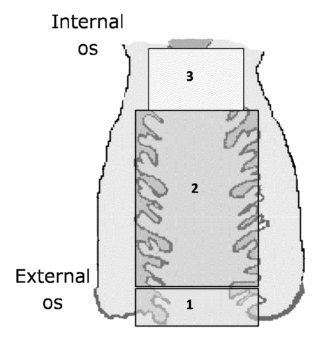
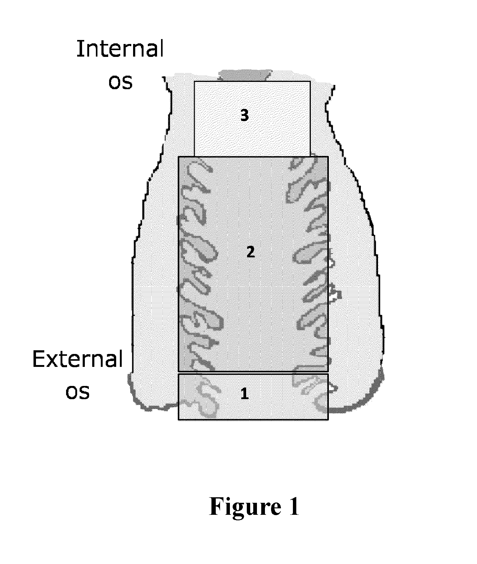
[0106]Prior to sampling, the length of the patient's cervical canal was determined by ultrasound or using the following published estimates:
GestationalEstimated cervicalage (wks)length (cm)62-37383-49410412 4-4.5
[0107]A speculum was used to aid in the collection of the sample. The speculum was carefully inserted into the vagina, letting the speculum follow the path of least resistance. The speculum was opened thereby making the cervix visible to the operator.
[0108]Cervical mucus samples (transcervical samples) were collected with one of the three different devices listed below:
[0109](i) Pap smear cytology brush (Medico EndoScan plus) (n=24),
[0110](ii) Rare Cellect™ biological sampling device described in WO 2010 / 085841 (Genetic Technologies Ltd, Australia) (n=47), and
[0111](iii) Aspiracath (Cook Australia) (n=32).
[0112]A total of one hundred and three samples were collected from patients undergoing elective termination of pregnancy (TOP) during first trimester of p...
example 2
Preparation of Cell Free DNA from Supernatant Samples
[0117]The sampling device was removed from the transport container using sterile forces and transferred into an organ culture dish containing 1 ml D-PBS.
[0118]The mucus was carefully removed from the brush using sterile forceps. The sample was manually tweezed apart using sterile forceps into small pieces. Gentle pipetting using a 1 ml pipette further disaggregates the sample. The entire sample was then passed through a cell strainer (100 μm Nylon strainer, Becton Dickinson, USA) into a sterile 50 ml FALCON™ tube. The brush was then transferred back into the 15 ml FALCON™ tube where it was washed with the original 4 ml of transport media or D-PBS present in the transport container. The brush was washed by gently agitation, swirling the 15 ml FALCON™ tube up and down a few times.
[0119]The entire sample was then passed through the same cell strainer into the sterile 50 ml FALCON™ tube. A further 4 ml D-PBS wa...
example 3
DNA Extraction of Cell Free from Supernatant Samples
[0123]The cell free DNA from the supernatant collected in Example 2 was extracted using the Qiagen DNA Blood Maxi kit (Qiagen, USA). The DNA extraction process involved equilibrating the samples to room temperature (RT). Once the samples reached RT, 500 μl of Qiagen protease per 10 ml sample, 12 ml of buffer AL is also added to the sample and mixed vigorously. The sample is incubated at 70° C. for 10 minutes.
[0124]Prior to transferring the lysate to the column 10 ml 100% ethanol was added to the lysate. The entire lysate is transferred to the maxi column and centrifuged at 3000 rpm for 3 minutes (note: this may require multiple lysate to column spins). The maxi column was then washed with 5 mL of Buffer AW1 and centrifuged at 5000 rpm for 1 min. Followed by a further 5 mL wash with buffer AW2 and centrifuged at 5000 rpm for 15 min. AE buffer was then used to elute the sample off the column. Approximately, 600 μL Buffer AE was added...
PUM
| Property | Measurement | Unit |
|---|---|---|
| Time | aaaaa | aaaaa |
| Mass | aaaaa | aaaaa |
| Mass | aaaaa | aaaaa |
Abstract
Description
Claims
Application Information