

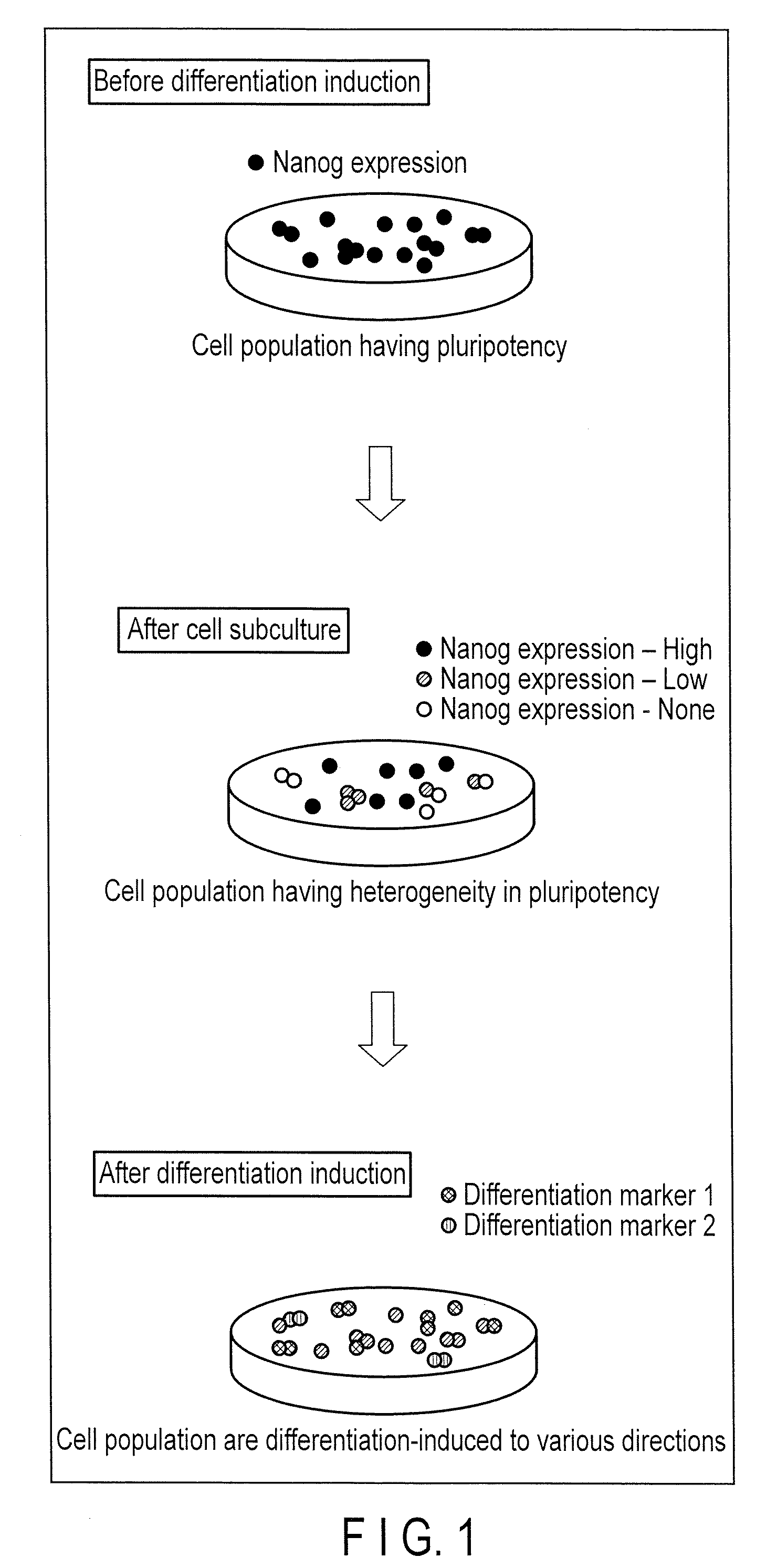
Method for monitoring state of differentiation in stem cell
a stem cell and differentiation state technology, applied in the field of stem cell differentiation state monitoring, can solve the problems of inability to continuously investigate the cell state and the way the stem cell actually works
- Summary
- Abstract
- Description
- Claims
- Application Information
AI Technical Summary
Benefits of technology
Problems solved by technology
Method used
Image

Examples
example 1
Examples Using Undifferentiation Marker
Example 1-1
Observation and Analysis of Transient Expression Cells
[0132](1) Preparation of ES Cells into which a Fusion Gene of a Promoter Region of an Undifferentiation Marker Gene and a Luciferase Gene is Introduced
[0133]For cloning of Nanog gene promoter region, the information of promoter sequence was obtained with reference to Non-Patent Literature “T. Kuroda et al., Molecular and Cellular Biology, 2005, Vol. 25, No. 6, pp. 2475-2485”.
[0134]Nanog gene promoter region sequence was obtained using a mouse genomic DNA as a template. As a primer for amplifying Nanog gene promoter region, the following primers were used.
forward primer:(SEQ ID NO: 1)CTACTCGAGATCGCCAGGGTCTGGAreverse primer:(SEQ ID NO: 2)CTACTCGAGCGCAGCCTTCCCACAGAAA
[0135]The obtained Nanog gene promoter sequence was inserted into pGL4-basic vector (Promega) to prepare “Nanog gene expression-specific luminescent vector pNanog-GL4”.
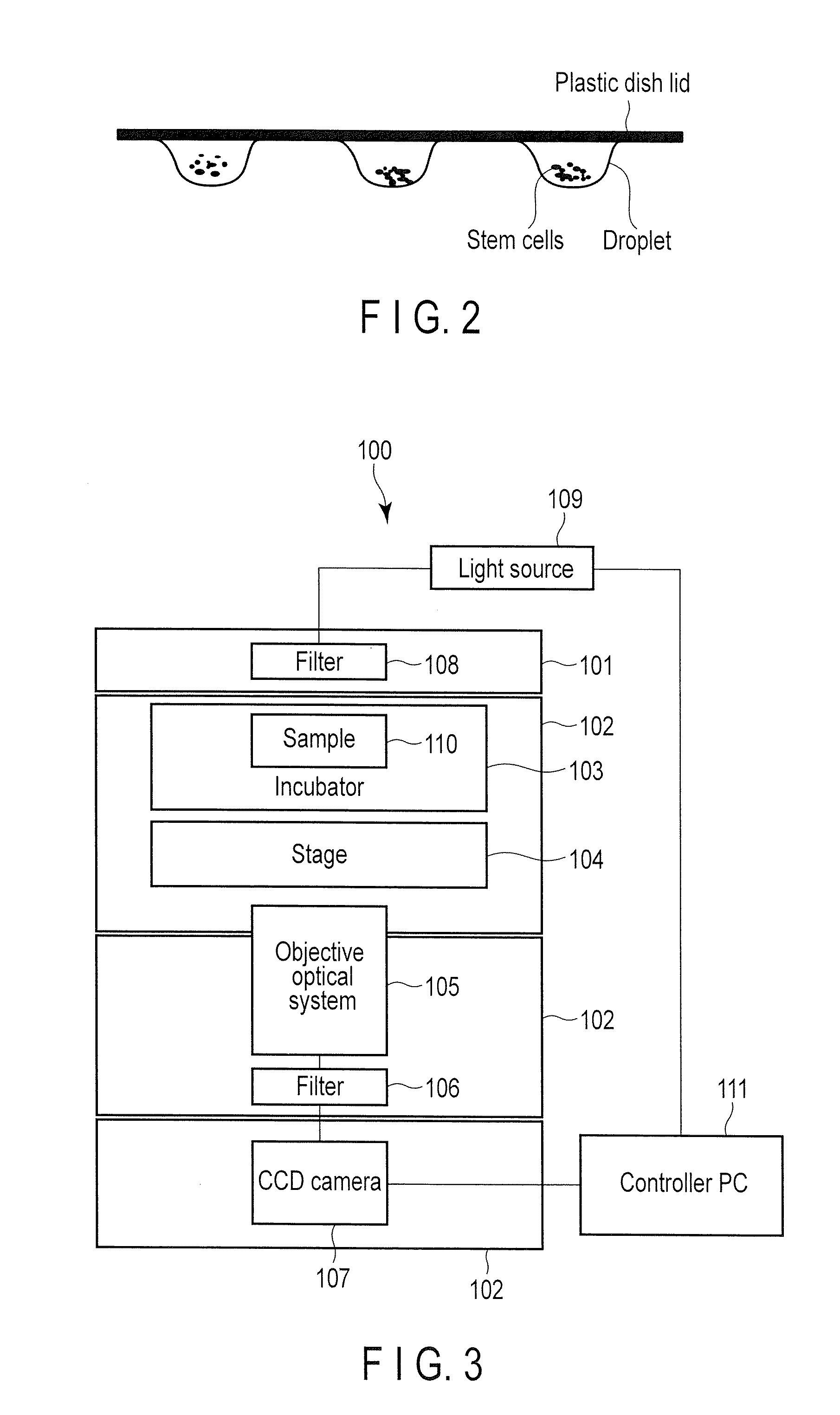
[0136]Feeder cells were prepared to culture ES cells....
example 1-2
Long Term Observation and Analysis of Stable Expression Cells
[0148]In the present example, a vector containing a drug resistance gene is used as a gene transfer vector, cells into which gene transfer was performed (stable expression cells) were selected by drug selection, and a long term observation and analysis were performed. The detail of the present example is set forth below.
[0149](1) Preparation of ES Cells into which a Fusion Gene of a Promoter Region of an Undifferentiation Marker Gene and a Luciferase Gene is Introduced
[0150]For cloning of Nanog gene promoter region, the promoter sequence was obtained with reference to Non-Patent Literature “T. Kuroda et al., Molecular and Cellular Biology, 2005, Vol. 25, No. 6, pp. 2475-2485”.
[0151]Nanog gene promoter region sequence was obtained using a mouse genomic DNA as a template. As a primer for amplifying Nanog gene promoter region, the following primers were used.
forward primer:(SEQ ID NO: 1)CTACTCGAGATCGCCAGGGTCTGGAreverse primer...
example 1-3
Long Term Observation and Analysis of Stable Expression Cells Under Drug Stimulus
[0164]Mouse ES cells used in Example 1-2 were used, which is constitutively expressing Nanog-Eluc. The cells were differentiation-induced with FGF (Fibroblast Growth Factor), and then observed under condition with signal transduction inhibitor. FGF signal is a signal transduction which induces stem cell differentiation, and it has been reported that the stem cell returns from the differentiated state to the undifferentiated state by the action of an inhibitor (T. Kunath et al., Development 134, pp. 2895-2902, 2007). The preparation method of ES cells is in the same manner as in Example 1-2.
[0165](1) Preparation of ES Cells
[0166]After seeding in a dish and culturing overnight, the mouse ES cells constitutively expressing Nanog-Eluc were cultured overnight with DMEM culture medium containing 15% KSR (Knockout Serum [Gibco]) and LIF (leukemia inhibitory factor). After the culture, the culture medium was re...
PUM
| Property | Measurement | Unit |
|---|---|---|
| concentration | aaaaa | aaaaa |
| luminescent | aaaaa | aaaaa |
| luminescence | aaaaa | aaaaa |
Abstract
Description
Claims
Application Information