Apparatus and method for quantitative noncontact in vivo fluorescence tomography using a priori information

Inactive Publication Date: 2013-01-24
RGT UNIV OF CALIFORNIA
View PDF3 Cites 17 Cited by
  • Summary
  • Abstract
  • Description
  • Claims
  • Application Information

AI Technical Summary

Benefits of technology

[0012]In this disclosure, a first-of-its-kind fully integrated tri-modality system is described that combines fluorescence, diffuse optical and x-ray tomography (FT/DOT/XCT) into one instrument. This system performs quantitative fluorescence tomography using multimodality imaging. XCT anatomical information is used as structural a priori data while optical background heterogeneity information obtained by DOT measurements is used as functional a priori data. The performance of the hybrid system is evaluated using

Problems solved by technology

However, the improvement is limited.
The inverse problem of fluorescence tomography is severely ill-posed, so the recovered fluorescence par

Method used


Image

  • Apparatus and method for quantitative noncontact in vivo fluorescence tomography using a priori information
  • Apparatus and method for quantitative noncontact in vivo fluorescence tomography using a priori information
  • Apparatus and method for quantitative noncontact in vivo fluorescence tomography using a priori information

Examples


Embodiment Construction

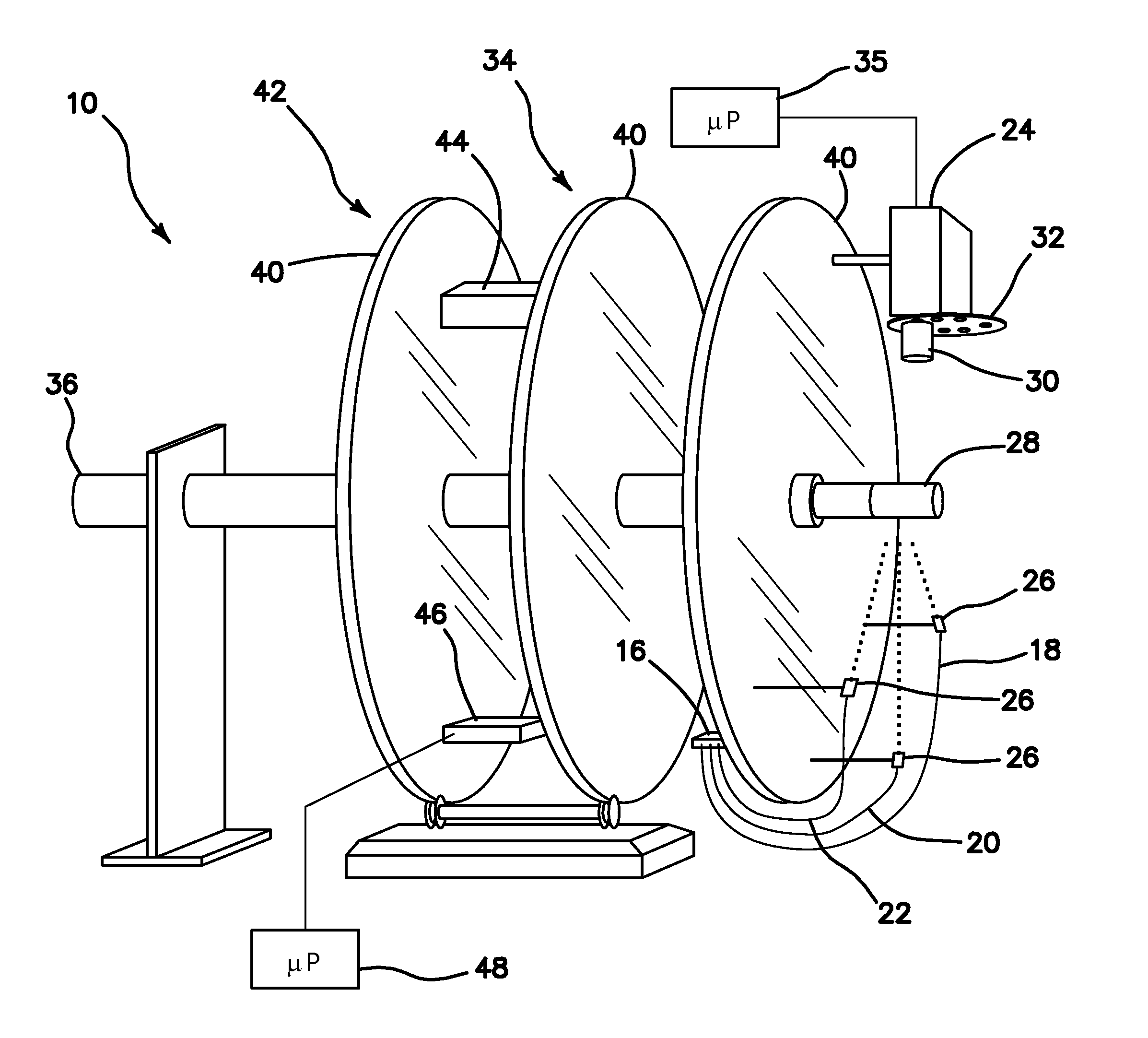
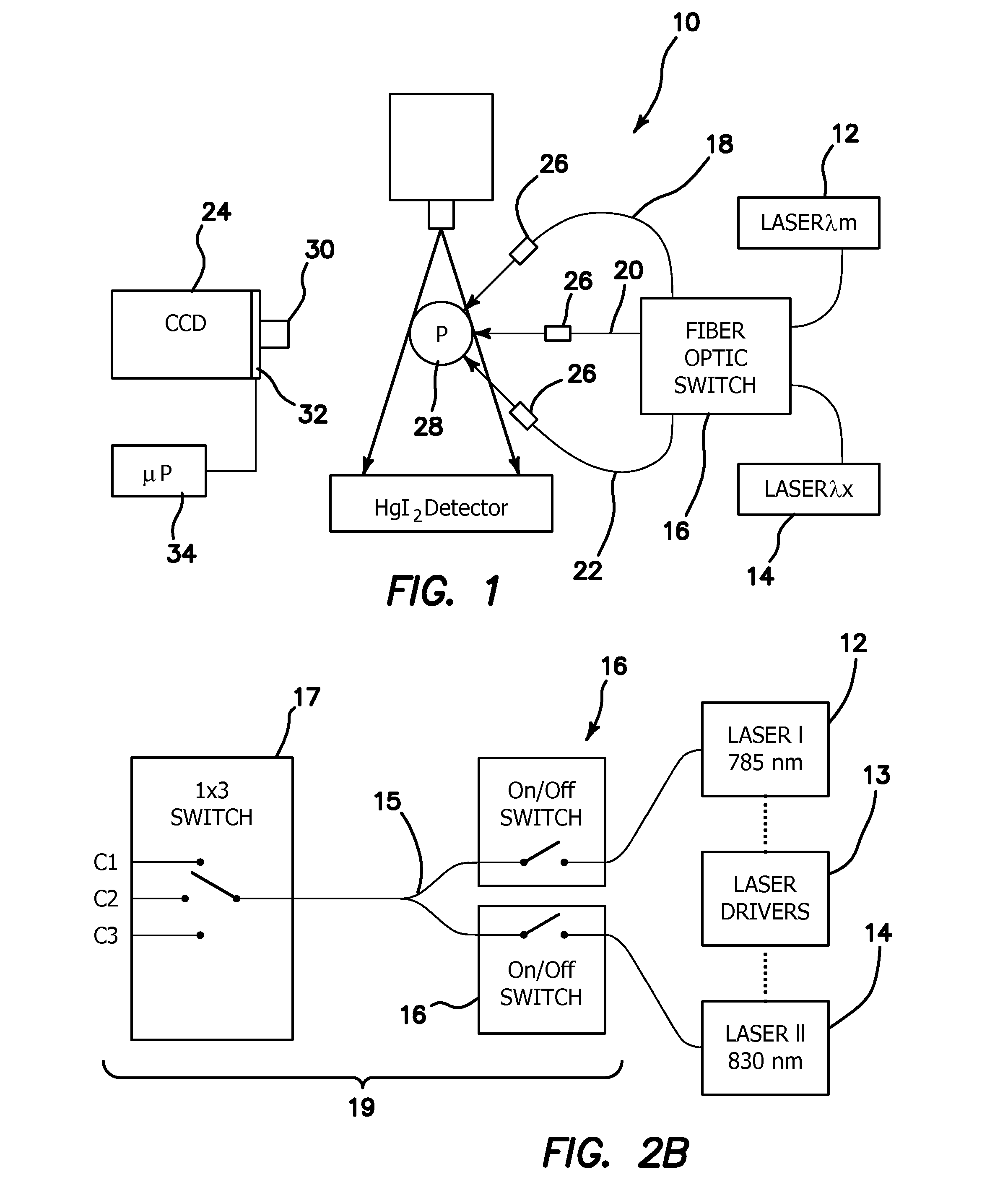
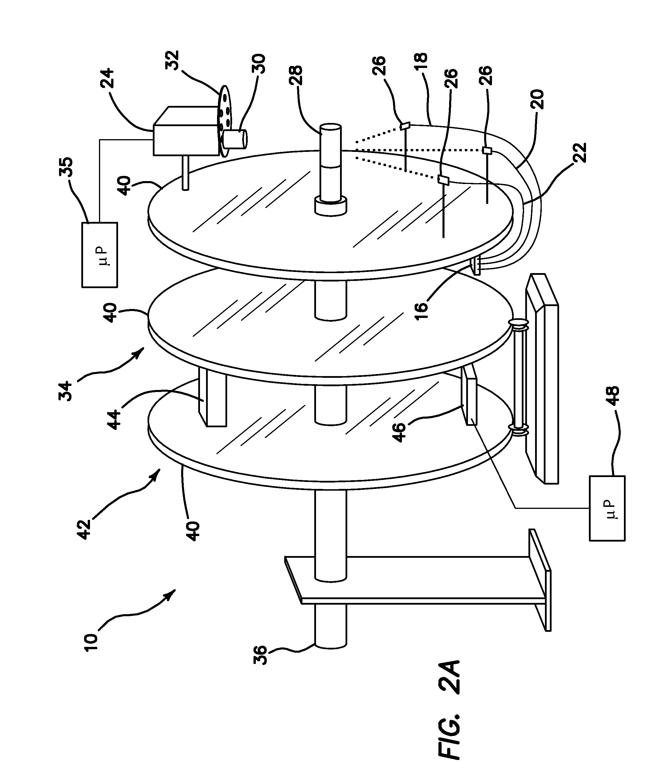
[0064]We have built a first-of-its-kind gantry-based multi-modality system 10 that combines FT, DOT and XCT in an integrated platform. The XCT offers anatomical information while the DOT provides optical background heterogeneity to improve the FT images further. The performance of the system 10 was evaluated using multi-modality phantoms. We first assessed the linearity of the system response using fluorescence inclusions with various concentrations located in a homogeneous, tissue-mimicking phantom. Next, size and location dependence of the recovered fluorophore concentration was investigated. For both studies, the fluorophore concentration maps reconstructed with and without XCT structural a priori information were compared. Finally, we investigated the recovery of the fluorophore concentration of the inclusion in the presence of background heterogeneity. We demonstrated that fluorophore concentration could be accurately recovered only when both functional (DOT) and structural (XC...


PUM


Abstract

An apparatus for providing an integrated tri-modality system includes a fluorescence tomography subsystem (FT), a diffuse optical tomography subsystem (DOT), and an x-ray tomography subsystem (XCT), where each subsystem is combined in the integrated tri-modality system to perform quantitative fluorescence tomography with the fluorescence tomography subsystem (FT) using multimodality imaging with the x-ray tomography subsystem (XCT) providing XCT anatomical information as structural a priori data to the integrated tri-modality system, while the diffuse optical tomography subsystem (DOT) provides optical background heterogeneity information from DOT measurements to the integrated tri-modality system as functional a priori data. A method includes using FT, DOT, and XCT in an integrated fashion wherein DOT data is acquired to recover the optical property of the whole medium to accurately describe photon propagation in tissue, where structural limitations are derived from XCT, and accurate fluorescence concentration and lifetime parameters are recovered to form an accurate image.

Description

RELATED APPLICATIONS[0001]The present application is related to U.S. Provisional Patent Application Ser. No. 61 / 430,036, filed on Jan. 5, 2011, which is incorporated herein by reference and to which priority is claimed pursuant to 35 USC 119.GOVERNMENT RIGHTS[0002]This invention was made with government support under EB007873 awarded by The National Institutes of Health. The government has certain rights in the invention.BACKGROUND[0003]1. Field of the Technology[0004]The disclosure relates to the field of apparatus and methods used for imaging biological specimens and in general small animal imaging, preclinical imaging systems, x-ray imaging, molecular imaging and optical imaging.[0005]2. Description of the Prior Art[0006]Fluorescence imaging has become an essential in vivo small animal imaging tool in recent years. Both commercial availability and relative ease of use has enabled widespread adoption of this technique. Most commercial fluorescence imaging systems utilize a planar ...

Claims


Application Information

Patent Timeline
IPC IPC(8): A61B6/02
CPCA61B6/02A61B6/583A61B5/0073A61B2503/40A61B5/0035A61B6/032A61B6/4233A61B6/4417A61B6/508A61B6/5205A61B6/5247A61B5/0059G01N21/4795G01N21/6456A61B5/0071
Inventor NALCIOGLU, ORHANGULSEN, GULTEKINLIN, YUTING
Owner RGT UNIV OF CALIFORNIA
Why Eureka
Try Eureka