Antibody binding to lysophosphatidylglucoside, and composition comprising the same

Inactive Publication Date: 2013-06-06
RIKEN
View PDF0 Cites 5 Cited by
  • Summary
  • Abstract
  • Description
  • Claims
  • Application Information

AI Technical Summary

Benefits of technology

[0054]The present invention provides: an antibody capable of suppressing a repellent effect of Lyso-PtdGlc on axon extension of a neuron; and a composition for suppressing a repellent effect of Lyso-PtdGlc on axon extension of a neuron, the composition comprising the antibody as

Problems solved by technology

Nevertheless, such a research outcome suggests that unlike a proposed differentiation mechanism at a late stage of development, defects in the early pattern formation cause premature inward extension of TrkA and TrkC afferent nerves

Method used


Image

  • Antibody binding to lysophosphatidylglucoside, and composition comprising the same
  • Antibody binding to lysophosphatidylglucoside, and composition comprising the same
  • Antibody binding to lysophosphatidylglucoside, and composition comprising the same

Examples


example 1

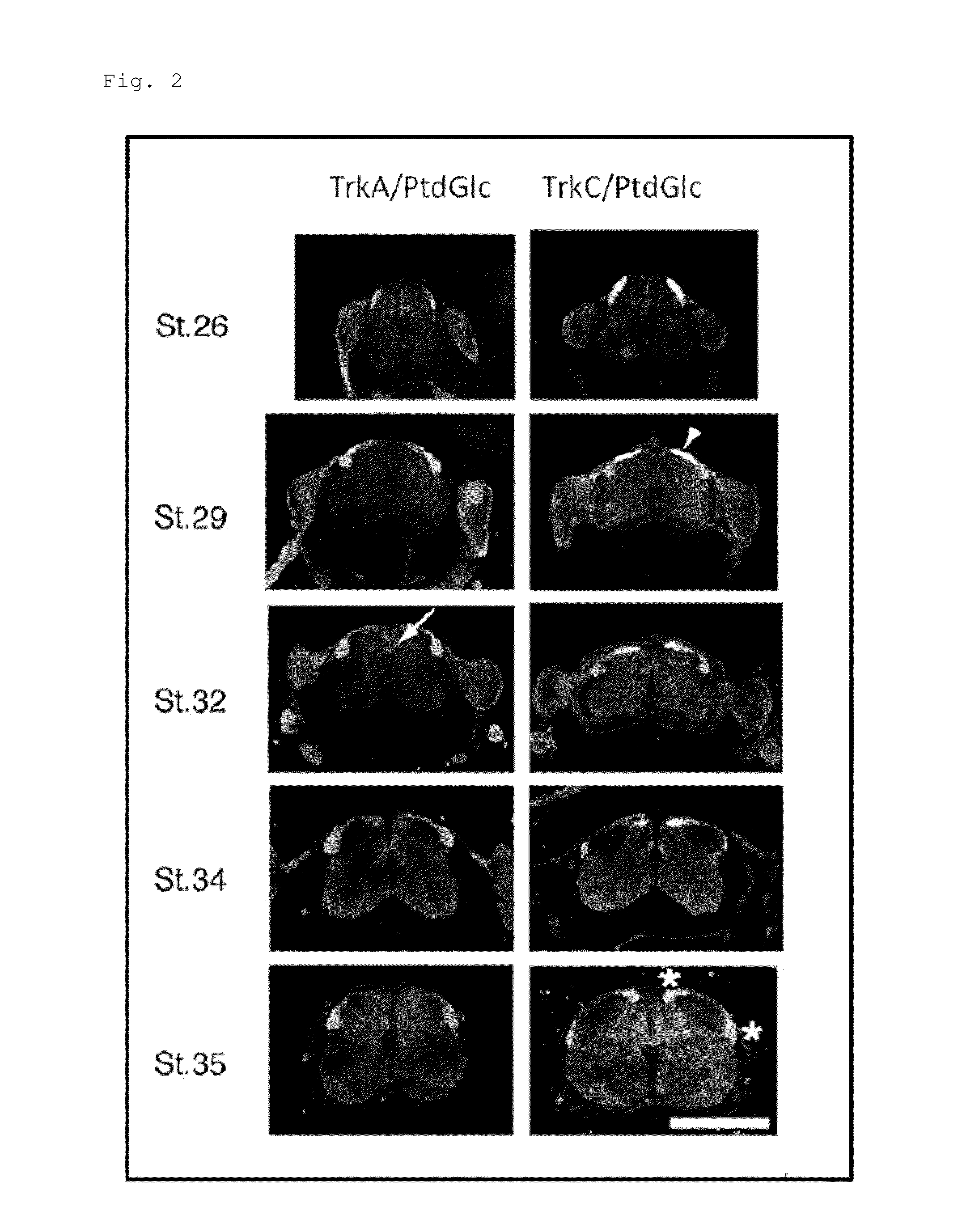
[0147]To observe spatial and temporal relations between phosphatidyl-β-D-glucoside (PtdGlc) and DRG sensory neuronal cells in the spinal cord, co-immunostaining (labelling) was performed on embryos at HH Sts. 26, 29, 32, 34, and 35 (corresponding to embryos between approximately E5 (embryonic day 5) to E9 (embryonic day 9)) using the anti-TrkA antibody or the anti-TrkC antibody, and the DIM21 antibody. FIG. 2 shows the obtained result. Note that the white line in FIG. 2 is a scale bar, representing a length of 500 μm. Moreover, in FIG. 2, a portion emitting magenta fluorescence indicates a site stained with the DIM21 antibody, a portion emitting green fluorescence indicates a site stained with the anti-TrkA antibody (left lanes in FIG. 2) or the anti-TrkC antibody (right lanes in FIG. 2).

[0148]As apparent from the result shown in FIG. 2, in the embryonic spinal cord at HH St. 26, a site having a strong immunoreactivity to DIM21 is restricted to the dorsal white matter, while a weak ...

example 2

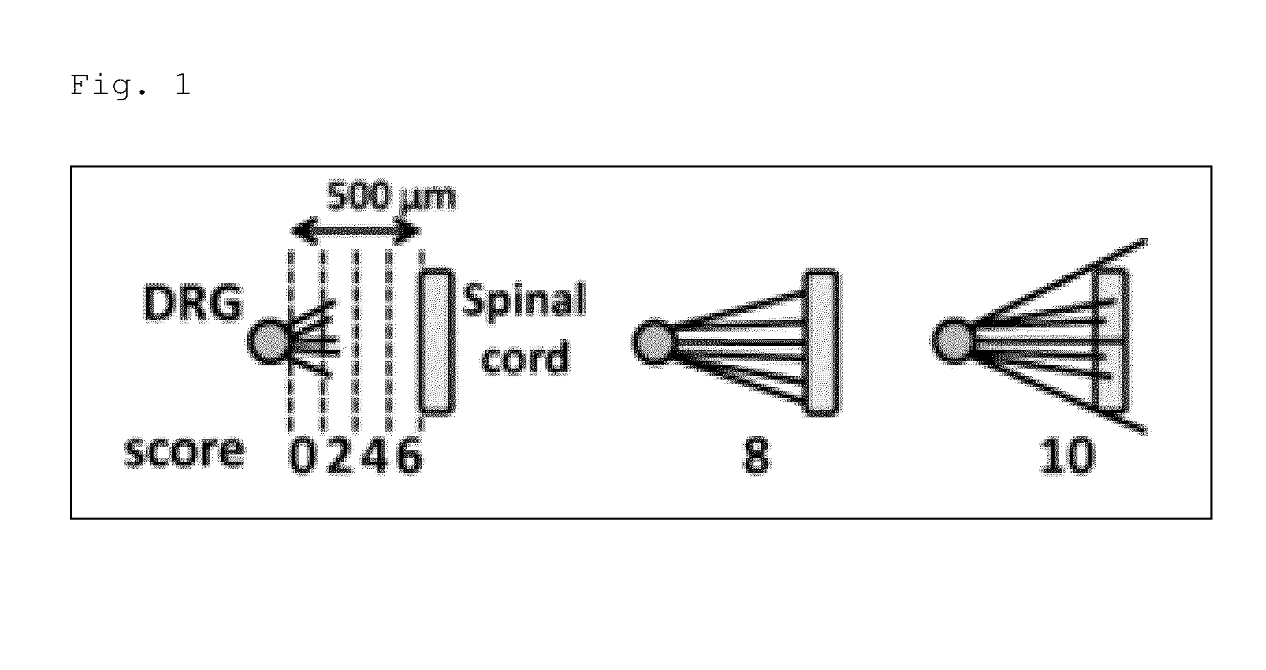
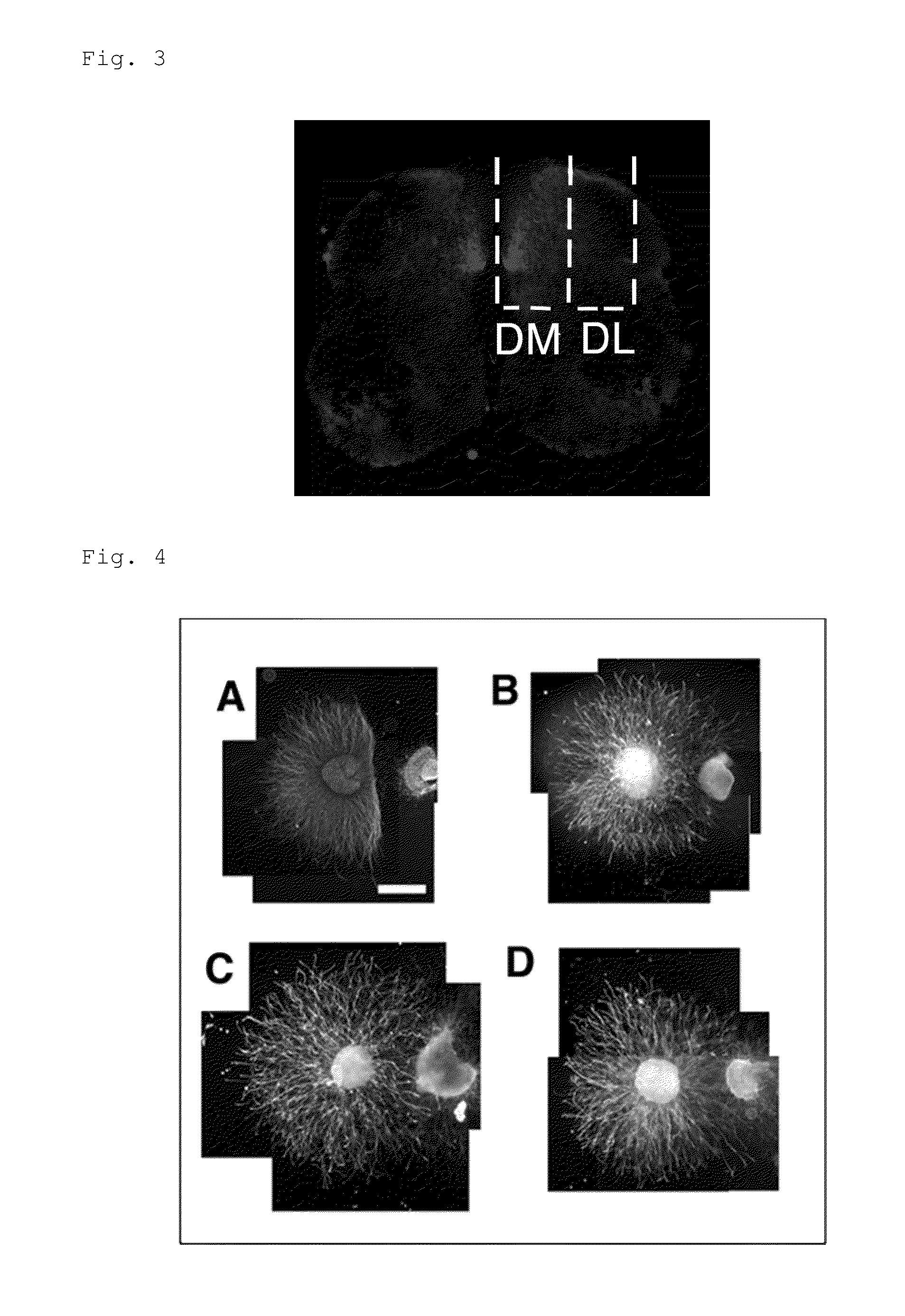
[0155]In order to verify that PtdGlc or a derivative thereof is a signaling molecule for DRG sensory nerves and acts as a chemorepellent particularly on a TrkA-expressing neuron as described above, organotypic spinal cord / DRG explant culture assay was conducted. A dorsal root ganglion (DRG) explant having a diameter of approximately 250 μm was cultured on a 3-D collagen matrix together with an explant obtained by dissecting the spinal cord. Then, 48 hours after the co-culturing, DRG axon extension was observed. Note that in the present Example, the explant from the spinal cord was dissected and separated into ones derived from dorsal (Dorsolateral, DL) or dorsomedial (DM) side, in other words, sites where the PtdGlc content is low (DL) or high (DM), for use (see FIGS. 2 and 3). FIGS. 4 and 5 show the obtained result. Note that the white line in FIG. 4 is a scale bar representing a length of 500 μm, the numerical values in parentheses in FIG. 5 indicate the number of DRG explants ass...

example 3

[0158]As described in Example 1, PtdGlc is widely expressed in the white matter (see FIG. 2). Thus, it is predicted that the PtdGlc supply source in the spinal cord is not neurons. For this reason, in order to confirm that such a prediction is reasonable, glia in the spinal cord were analyzed by mass spectrometry. Specifically, proliferative glial cells were isolated from the spinal cord at HH St. 35, and cultured for 7 days in vitro. After the culturing was finished, the glial cells and the culture supernatant were analyzed with a nano-liquid chromatography-tandem mass spectrometer (nano-LC / MS / MS). FIG. 6 shows the obtained result. Moreover, using such a glial cell culture, immunostaining was performed instead of the mass spectrometry. FIGS. 8 and 9 show the obtained result. Note that in FIG. 8, a portion emitting green fluorescence indicates a region labelled with an anti-trans it in antibody, and the white line is a scale bar, representing a length of 10 μm. Moreover, in FIG. 9, ...


PUM

PropertyMeasurementUnit
Compositionaaaaaaaaaa

Abstract

In order to identify a factor demonstrating a repellent effect on axon extension of a neuron, lysophosphatidylglucoside (LPG) has been found to have an activity as a chemorepellent effect on axon guidance of a DRG sensory neuron specific to a neuron expressing TrkA, an NGF receptor.
In addition, it has been found out that suppressing the function of LPG makes it possible to suppress a repellent effect of LPG on axon extension of a neuron expressing TrkA. A molecule capable of suppressing the function of LPG can be a composition promoting repairing of a neural circuit in neuronal damages and the like.

Description

TECHNICAL FIELD[0001]The present invention relates to: an antibody being capable of binding to lysophosphatidylglucoside and having an activity of suppressing a repellent effect of lysophosphatidylglucoside on axon extension of a neuron expressing TrkA; and a composition comprising the antibody as an active ingredient.BACKGROUND ART[0002]In the development stage of a nerve, a dynamic structure, a so-called growth cone, is formed at a tip of an axon of the nerve as the axon extends to a target tissue. The growth cone detects signaling molecules therearound, and changes its extension direction in response to diffusible or contact-dependent guidance cues. For the detection and so forth, complex systems including organelles and receptors are utilized (NPL 1). Most of axon guidance factors identified until today are proteins and derivatives thereof. However, the researches on the lipid-based mechanism have revealed the presence of lysophosphatidic acids (NPLs 2, 3), sphingosine-1-phospha...

Claims


Application Information

Patent Timeline
IPC IPC(8): C07K16/18
CPCA61K2039/505C07K2317/92C07K2317/76C07K16/18A61P25/00A61P25/28
Inventor HIRABAYASHI, YOSHIOKAMIGUCHI, HIROYUKIOHTA, KUNIHIRONAKAMURA, AKIHO
Owner RIKEN
Why Eureka
Try Eureka