Abscription based molecular detection of DNA methylation

a molecular detection and molecular detection technology, applied in the field of abscription based molecular detection of dna methylation, can solve the problems of not being able to easily score partially methylated targets in methylated- or unmethylated-specific reactions, and not being able to identify abnormally methylated dna by itself, so as to facilitate the incorporation of oligonucleotide primers, and improve the efficiency of the method

Inactive Publication Date: 2013-06-20
RIBOMED BIOTECHNOLOGIES INC
View PDF2 Cites 3 Cited by
  • Summary
  • Abstract
  • Description
  • Claims
  • Application Information

AI Technical Summary

Benefits of technology

[0028]Capture will typically be via an affinity reagent or binding pair bound or capable of being bound to a solid support. For example, the 5′ capture tag can be biotin, which can be readily incorporated into oligonucleotide primers, and the molecule that binds to the 5′ capture tag can be streptavidin immobilized on a solid support. A wide variety of solid supports are suitable for use in the methods of the present invention, such as beads, tubes, and microtiter plates. Conveniently, steptavidin and other binding pair molecules can be bound to magnetic beads which permit rapid separation of the solid phase from unbound reagents in solution. In certain embodiments of the invention, unbound reagents, primers, and polynucleotides can be washed from immobilized and captured polynucleotides prior to the subsequent steps in the procedure, which may increase the efficiency of the method. However, this is not necessary as the entire method can be performed in a single pot or tube without separation steps.

Problems solved by technology

The appearance of abnormally methylated DNA in bodily fluids by itself does not help to pinpoint which organ is affected by a tumor.
While this is usually true for heavily methylated or completely unmethylated islands, partially methylated targets are probably not readily scored in methylated- or unmethylated-specific reactions.
Due to its complexity and expense, however, bisulfite sequencing is better suited for marker discovery than clinical diagnostics.
Bisulfite treatment destroys a large percentage of the input DNA, resulting in limited sensitivity and a requirement for large amounts of DNA.
There is a potential of false-positive results for MSP-based assays due to incomplete cytosine deamination during bisulfite treatment.
While this problem can usually be corrected by optimizing primer annealing conditions, it may complicate primer design and testing.
Although bisulfite modification is a widely used, the extensive DNA degradation it causes can introduce sampling errors when few molecules are long enough to be amplified (Ehrich et al.
Furthermore, the assays are time-consuming, require a harsh base denaturation step, and have a high-probability of false-positive results due to incomplete cytosine deamination during bisulfite treatment.
Some disadvantages with this procedure are that it is lengthy and is dependent on the presence of MSRE / MDRE recognition sequences within a target DNA.
Furthermore, this approach is relatively inefficient, which can reduce the reliability of the results.
Incomplete digestion leads to frequent false positives, especially when cleavage reactions are subjected to a subsequent amplification step.
Restriction endonuclease cleavage assays have poor sensitivity compared to bisulfite methods, such as MSP, allowing detection of not less than 10% methylated DNA in a sample (Singer-Sam et al.
However, the ChIP procedure is very time-consuming, involves several steps and requires expensive reagents.
However this strategy could suffer the disadvantage associated with the use of restriction endonucleases in that some partially methylated islands will be scored as unmethylated in clinical samples (Yegnasubramanian, et al. supra).

Method used


Image

  • Abscription based molecular detection of DNA methylation
  • Abscription based molecular detection of DNA methylation
  • Abscription based molecular detection of DNA methylation

Examples


example 1

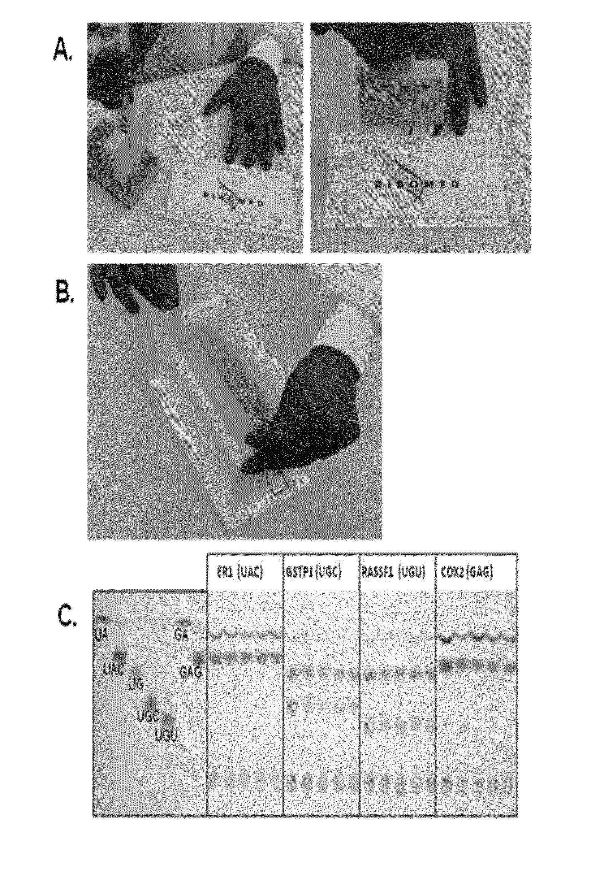
Abscription® Methods

[0118]Abscription® has been previously described; see e.g. U.S. patent application Ser. No. 09 / 984,664 (filed Oct. 30, 2001) now U.S. Pat. No. 7,045,319; Ser. No. 10 / 425,037 (filed Apr. 29, 2003); Ser. No. 10 / 600,581 (filed Jun. 23, 2003); Ser. No. 10 / 602,045 (filed Jun. 24, 2003); Ser. No. 10 / 607,136 (filed Jun. 27, 2003), now U.S. Pat. No. 7,226,738; Ser. No. 10 / 686,713 (filed Oct. 17, 2003); Ser. No. 10 / 976,240 (filed Oct. 29, 2004); Ser. No. 10 / 790,766 (filed Mar. 3, 2004); Ser. No. 10 / 488,971 (filed Oct. 18, 2004); and Ser. No. 10 / 551,775 (filed Sep. 14, 2006) the contents of each of which are incorporated by reference herein in their entirety.

example 2

Mass Spectrometry Detection of Abscripts

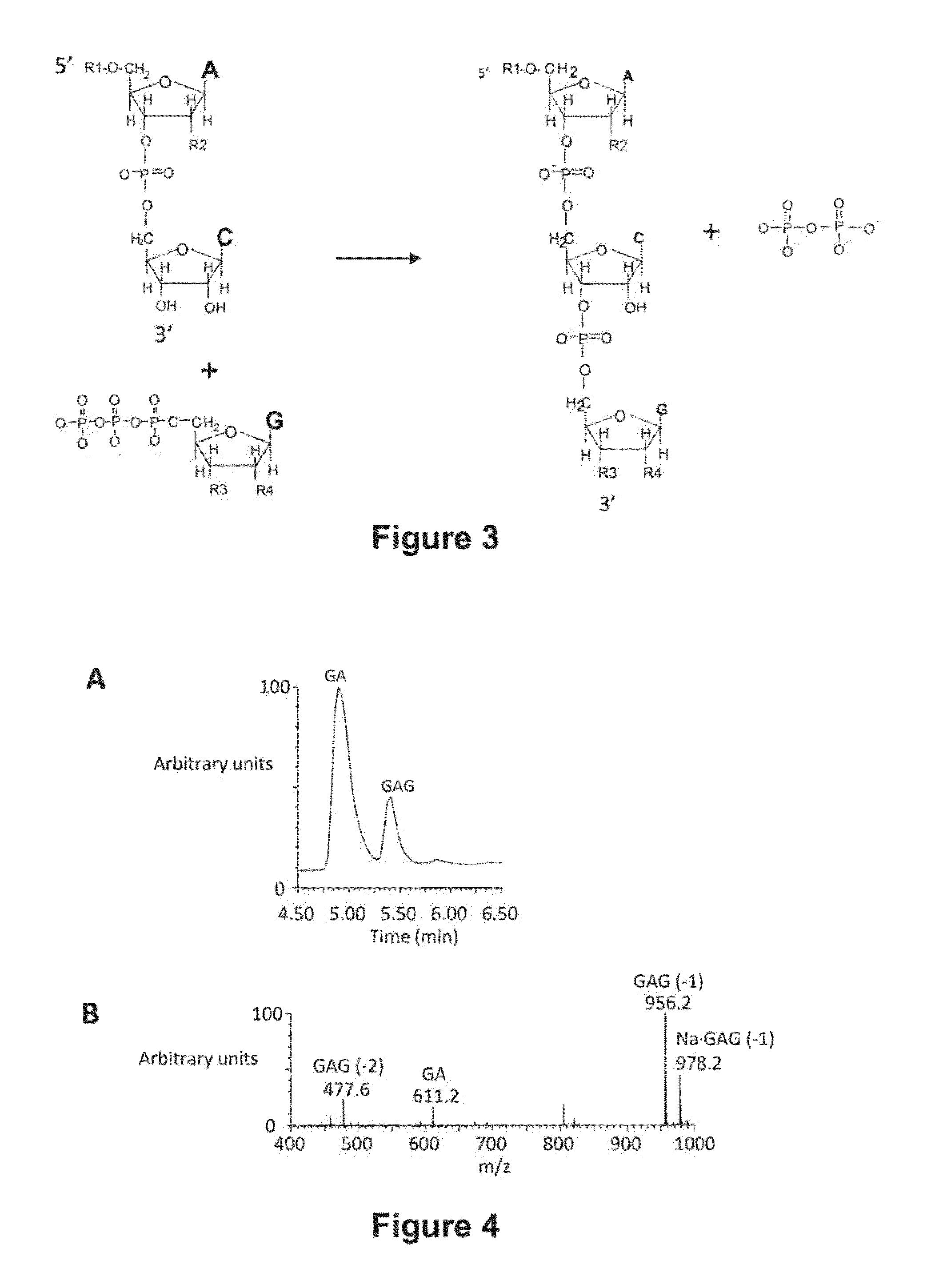
[0119]Trinucleotide Abscripts are detected by mass spectrometry following their fractionation from dinucleotide initiators by HPLC. The output of the fractionation is plotted as total ion count verses chromatographic retention time as illustrated in FIG. 4A. The chromatographic profile for any ion can be similarly plotted. The contributions of particular m / z species at a specific retention time can be summed to give the amount of Abscript as the area under the chromatographic peak. FIG. 4B shows the ion spectrum associated with the Abscript GAG (retention time of 5.4 min). The yield of GAG would be the sum of species with m / z values of 477.6, 956.1 and 978.2. These species account for doubly charged, singly charged and the sodium adduct respectively.

example 3

Preparation of GST-MBD Protein

[0120]A GST fusion protein that contains the methyl binding domain (MBD) from mouse MBD2 was constructed as illustrated in FIG. 5. The codons for the MBD domain were optimized for expression in E. coli. The construct contains a thrombin cleavage site between the GST and MBD domains. The GST protein also contains four surface cysteine residues that were used for attachment of APCs or Biotin. The details of the GST-MBD protein are provided in U.S. Patent Application No. 61 / 053,648, filed May 15, 2008 the contents of which are incorporated by reference herein, and in particular, Examples 2-11 describing the preparation and use of MBD fusion proteins.

[0121]The GST domain allows the fusion protein, or its complexes with methylated DNA, to be isolated on Glutathione resins or beads and eluted with glutathione, or to be captured or detected with antibodies that recognize GST.

[0122]MBD from the MBD2b protein was chosen for final constructs because MBD2b has the...


PUM

PropertyMeasurementUnit
retention timeaaaaaaaaaa
temperaturesaaaaaaaaaa
thicknessaaaaaaaaaa

Abstract

The present invention provides methods for detecting biomarkers based on Abscription®, abortive transcription technology. Particularly, the present invention provides bisulfate free methods for detecting methylation of CpG islands from small samples containing DNA, including formalin fixed, paraffin embedded samples. The methods are suitable for multiplexing and can be used to analyze multiple CpG islands from a single sample in a short time.

Description

RELATED APPLICATION[0001]This application is a continuation in part of U.S. patent application Ser. No. 12 / 724,416, filed Mar. 15, 2010, now U.S. Pat. No. 8,263,339, issued Sep. 11, 2012, which in turn claims the benefit of priority under 35 USC §119 of U.S. Provisional Application Ser. No. 61 / 160,335 filed Mar. 15, 2009, the entire disclosures of which are incorporated herein by reference.STATEMENT OF GOVERNMENT INTEREST[0002]This invention was made with government support under from the National Institutes of Health through National Cancer Institute Small Business Innovative Grant number 1R43CA132851-1 and NCl Contract number HHSN261200900047C. The Government may have certain rights in the invention.BACKGROUND OF THE INVENTION[0003]Cancer is actively avoided through the expression of numerous tumor-suppressor genes that regulate the cell division cycle and mediate interactions among cells. Studies of benign and malignant tumors have shown that cancer develops through a multi-step ...

Claims


Application Information

Patent Timeline
Patent Type & Authority Applications(United States)
IPC IPC(8): C12Q1/68
CPCC12Q1/6886C12Q1/6883C12Q1/686C12Q2600/118C12Q2600/154C12Q2565/627C12Q2537/143C12Q2531/113C12Q2521/331C12Q2565/125C12Q2565/137
Inventor HANNA, MICHELLE M.MCCARTHY, DAVID
Owner RIBOMED BIOTECHNOLOGIES INC
Why Eureka
Try Eureka