Methods and Systems for Detection of Microorganisms

a microorganism and detection method technology, applied in the field of methods and systems for the detection of microorganisms, can solve the problems of requiring several days of procedures, affecting the detection efficiency of microorganisms, so as to facilitate the detection of ribosomes

Active Publication Date: 2013-08-22
LAB OF AMERICA HLDG
View PDF4 Cites 21 Cited by
  • Summary
  • Abstract
  • Description
  • Claims
  • Application Information

AI Technical Summary

Benefits of technology

[0012]Thus, embodiments of the present invention rely on bacteriophage-based and ribosome-based methods for amplification of the detection of bacteria. The principles applied herein can be applied to the detection of other microorganisms. Because of the sheer number of the ribosomes present in a microorganism or the rapid increase in the number of infectious agents present in a cell after amplification, it can be easier to detect ribosomes and / or such progeny infectious agents than to detect the microorganisms themselves. In this way, embodiments of the present invention can achieve total amplification of at least 10,000 from a single infected cell.

Problems solved by technology

Bacterial pathogens can cause substantial morbidity among humans and domestic animals, as well as immense economic loss.
Such procedures can require several days.
However, these methods have drawbacks.
Polymerase chain reaction (PCR) tests also include an amplification step and therefore are capable of both very high sensitivity and selectivity, however, the sample size that can be economically subjected to PCR testing is limited.
In practice, most high sensitivity methods employ an overnight incubation and take about 24 hours overall.
Due to the time required for cultivation, these methods can take up to three days, depending upon the organism to be identified and the source of the sample.
This lag time is generally unsuitable as the contaminated food, water (or other product) may have made its way into livestock or humans.

Method used


Image

  • Methods and Systems for Detection of Microorganisms
  • Methods and Systems for Detection of Microorganisms
  • Methods and Systems for Detection of Microorganisms

Examples


example 1

Purified Ribosome Isolation

[0195]E. coli was obtained from ATCC (#11303) and cultured in LB Medium (Difco: 10 g Tryptone, 5 g yeast extract, 10 g NaCl / L) overnight at 37° C. A colony was placed in 5 mL of LB Medium and incubated at 37° C. overnight. The following day the culture was diluted 1:100 in fresh LB and the cells grown to a concentration of Klett 68 (OD at 600 nm=0.8) (20 Klett units on a Summerson Photoelectric Colorimeter=0.1 OD at 540 nm on a spectrophotometer), which corresponds to approximately 4×10⁸ cells / mL after growth for about 3.5 hours. The cells were then pelleted in a Beckman Coulter XL 80K Type 19 rotor (Cat. No. 325632) at 5,000 rpm for 15 minutes. The pellet was resuspended in 1 mL TMS (50 mM Tris-HCl, pH 7.8, 10 mM MgCl2, 100 mM NaCl) with 1 / 10 volume of 10× Bugbuster (Novagen, PN 70921-3), 0.5 mg / ml lysozyme (Sigma L6876), and 10 μg / mL high quality DNaseI (RNase free) (USB 14365). The lysate was clarified in a 1.5 mL microfuge tube in a Beckman Coulter All...

example 2

Polyclonal Antibody Production in Rabbits

[0196]Purified ribosomes, isolated as described in Example 1, were combined with an appropriate adjuvant. The ribosomes and adjuvant were injected beneath the skin of young rabbits (2.5-3.0 kg; 10-16 weeks of age). Blood was collected from the central ear artery with a 19-gauge needle and allowed to clot and retract at 37° C. overnight. The clotted blood was then refrigerated for 24 hours before the serum was decanted and clarified by centrifugation at 2500 rpm for 20 minutes. The rabbits were injected and bled according to the following schedule:

Day −4: Pre Bleed

[0197]Day 0: Immunize via an intradermal (ID) route using CFA (Complete Freund's Adjuvant)

Day 7: Booster injection via an intradermal (ID) route using IFA (Incomplete Freund's Adjuvant)

Day 14: Booster injection via a subcutaneous (SC) route using IFA

Day 28: Booster injection via a subcutaneous (SC) route using IFA

Day 38: Test Bleed

[0198]Day 40: Ship pre-immune bleeds and test bleeds ...

example 3

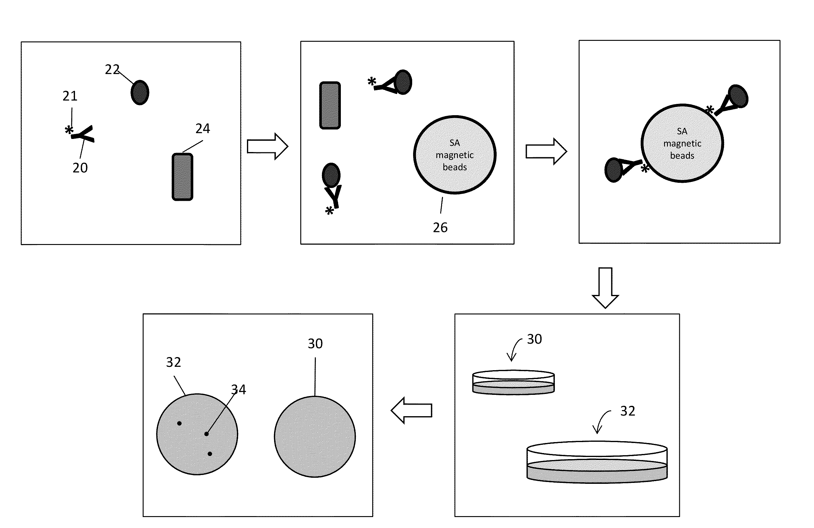
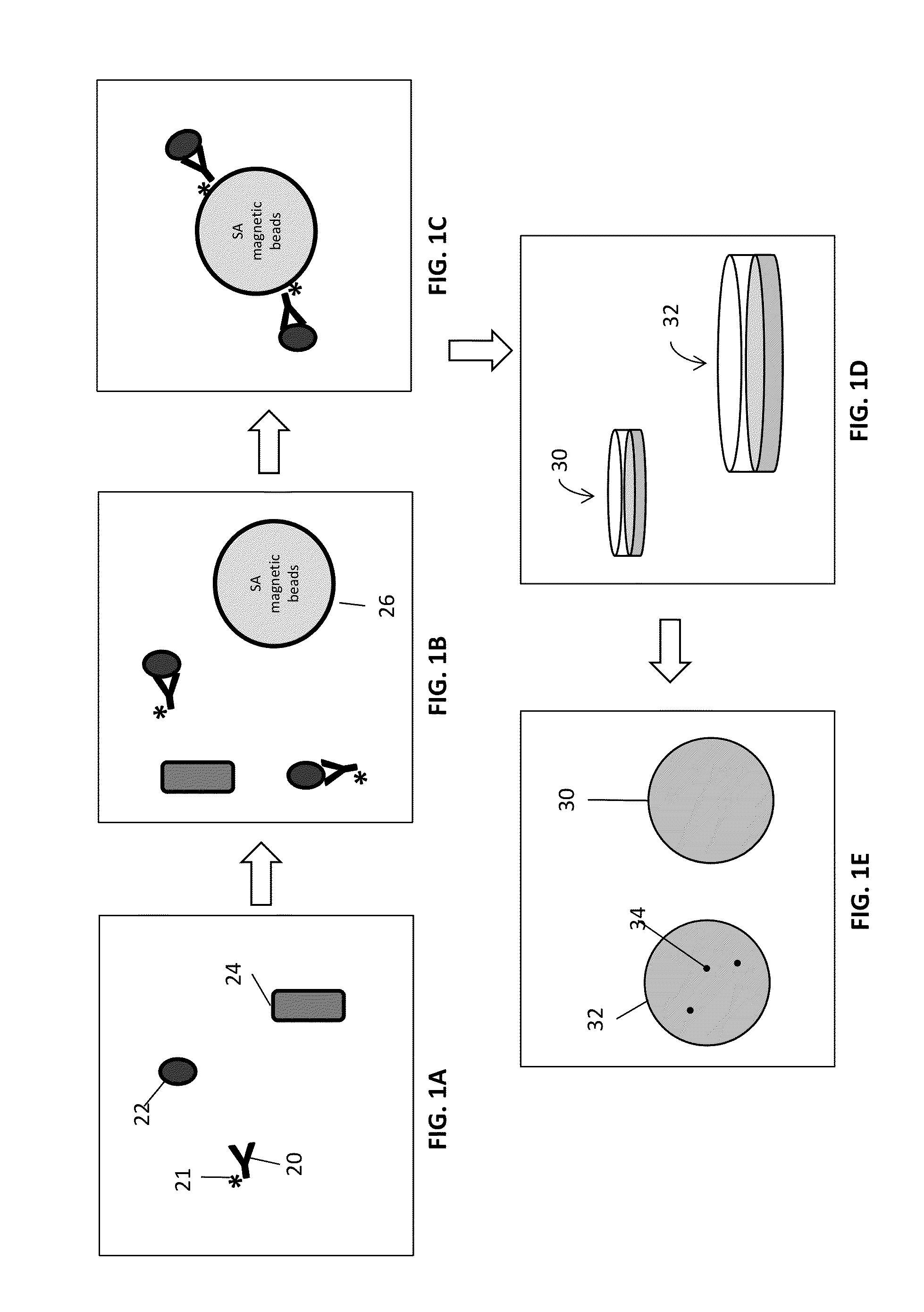
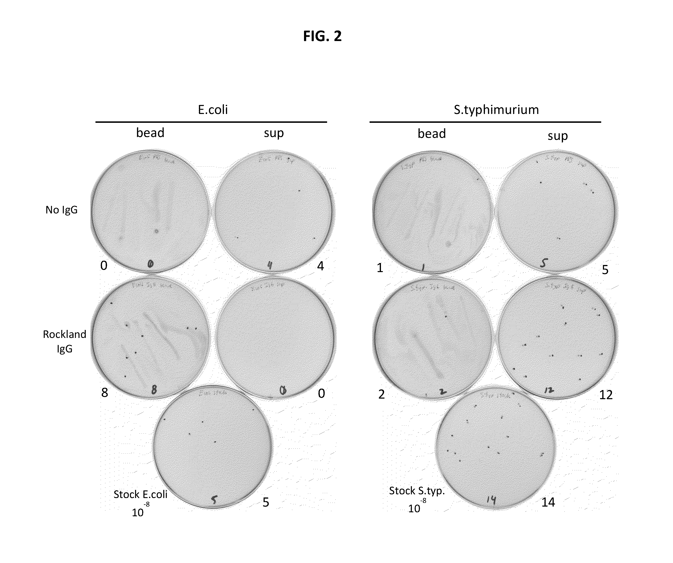
Bacteria Capture from Solution Using Cell-Specific Antibodies and Magnetic Microparticles

[0199]In order to demonstrate capturing intact, viable bacterial cells from solution rabbit polyclonal antibodies to surface epitopes of various bacterial species (e.g., E. coli and S. typhimurium) were generated.

[0200]Cultures of both E. coli and S. typhimurium were grown in liquid media before harvesting and were washed with phosphate buffer (1.1 mM KH2PO4, 5.6 mM Na2HPO4, 154 mM NaCl, pH 7.4). The washed cells were then diluted to a concentration between 5-20 cells per ml. Approximately 250 ng biotinylated, polyclonal anti-E. coli antibody (equivalent to about 1×10¹² antibody molecules) produced by Rockland Immunochemicals Inc., Gilbertsville, Pa. was added to the cell suspension and allowed to incubate for 45 min. A control experiment, where antibody is not added to the cell suspension, was also done.

[0201]Following antibody incubation, 4×10⁸ streptavidin-coated magnetic microparticles (Solu...


PUM

PropertyMeasurementUnit
timeaaaaaaaaaa
diameteraaaaaaaaaa
diameteraaaaaaaaaa

Abstract

Disclosed are methods and systems for the isolation and detection of microbes from a sample. The use of binding agents for isolation of a microbe of interest from a sample are described. In certain embodiments, the methods use ribosome-based and/or bacteriophage-based amplification of the signal in detection of bacteria and other microorganisms. For example, embodiments of the present invention can achieve total amplification of at least 10,000 from a single infected cell.

Description

[0001]This application claims priority to U.S. Provisional Patent Application 61 / 601,231 filed Feb. 21, 2012. The disclosure of U.S. Provisional Patent Application 61 / 601,231 is incorporated by reference herein in its entirety.FIELD OF THE INVENTION[0002]This invention relates to methods and systems for the detection of microorgansims.BACKGROUND[0003]There is a strong interest in the detection of bacteria and other microorganisms in both biological and food based samples. Bacterial pathogens can cause substantial morbidity among humans and domestic animals, as well as immense economic loss. Also, detection of microorganisms is a high priority within the Food and Drug Administration (FDA) given outbreaks of life-threatening or fatal illness caused by ingestion of food contaminated with certain microorganisms, e.g., Escherichia coli or Salmonella spp.[0004]Traditional microbiological tests for the detection of bacteria rely on non-selective and selective enrichment cultures followed b...

Claims


Application Information

Patent Timeline
Patent Type & Authority Applications(United States)
IPC IPC(8): G01N33/569G01N33/58
CPCG01N33/56911G01N33/569G01N33/581G01N33/54326G01N2333/90241G01N2469/10C12Q1/70
Inventor ANDERSON, DWIGHT LYMANCONRAD, ANDREW J.ERICKSON, STEPHENGIL, JOSE S.HOPKINS, BEN BARRETT
Owner LAB OF AMERICA HLDG
Why Eureka
Try Eureka