Method for a rapid determination of spatially resolved magnetic resonance relaxation parameters in an area of examination

a technology of spatial resolution and relaxation parameter, applied in the field of rapid determination of spatial resolution magnetic resonance relaxation parameter, can solve the problems of motion artifacts, difficult long-term relaxation, and major challenge in determining the constant of tb>1/b> of heart tissue,

Active Publication Date: 2013-10-24
SIEMENS HEALTHCARE GMBH
View PDF6 Cites 12 Cited by
  • Summary
  • Abstract
  • Description
  • Claims
  • Application Information

AI Technical Summary

Benefits of technology

[0012]An object of the invention is to provide a rapid method for spatially resolved determination of a relaxation parameter in an area of examination.
[0016]In an alternative preferred embodiment, a saturation pulse is irradiated into the area of examination as a preparation pulse. In this manner, the same initial value for the relaxation is present after every preparation pulse. This allows to avoid image artifacts or incorrect values at the quantification due to different waiting times after the previous preparation.
[0018]Also advantageous is the use of accelerated imaging techniques, such as an iterative reconstruction, parallel imaging, radial scanning, etc. in the so-called single-shot imaging techniques. This technology makes also possible to compensate for motion artifacts due to a variable heart rate during the imaging.
[0021]A further advantage is that the starting point of the data acquisition window within the cardiac cycle may be placed in the phase of only a slight cardiac motion. Thus, especially the important magnetic resonance data of short inversion times end up in the diastole, and are thus affected by the heart motion as little as possible. In addition, magnetic resonance data that are considered imperfect due to a defective movement correction, can be ignored in the further processing. The criterion for this can result both from the heart phase information and from the motion correction process.
[0024]Another result of the registration of the data of the various heart phases is the provision of individual synthetic TI images of virtually any cardiac phase, e.g., a comparison of TI images of the systole and diastole. The rationale is that in a particular inversion time TI images can be advantageous for the assessment of pathologies, such as the zero passing of the blood with the late gadolinium enhancement method, and that the method provides the possibility of generating this contrast quasi synthetically over the whole cardiac cycle, without having to have it directly measured.

Problems solved by technology

However, the determination of the T1 constant of the heart tissue is a major challenge due to cardiac and respiratory movement.
This is even more difficult for long T1 relaxation constants, as they typically occur in the cardiac tissue before the administration of a contrast agent.
While the special data acquisition scheme in MOLLI can, to a large degree, avoid motion artifacts arising due to the cardiac motion, there can still arise motion artifacts if the patient, who is being examined during the data acquisition, does not completely stop breathing.
The known methods for spatially resolved determination of relaxation parameters of a beating heart are performed over several heartbeats, and thus require a relatively long measurement time.

Method used


Image

  • Method for a rapid determination of spatially resolved magnetic resonance relaxation parameters in an area of examination
  • Method for a rapid determination of spatially resolved magnetic resonance relaxation parameters in an area of examination
  • Method for a rapid determination of spatially resolved magnetic resonance relaxation parameters in an area of examination

Examples


Embodiment Construction

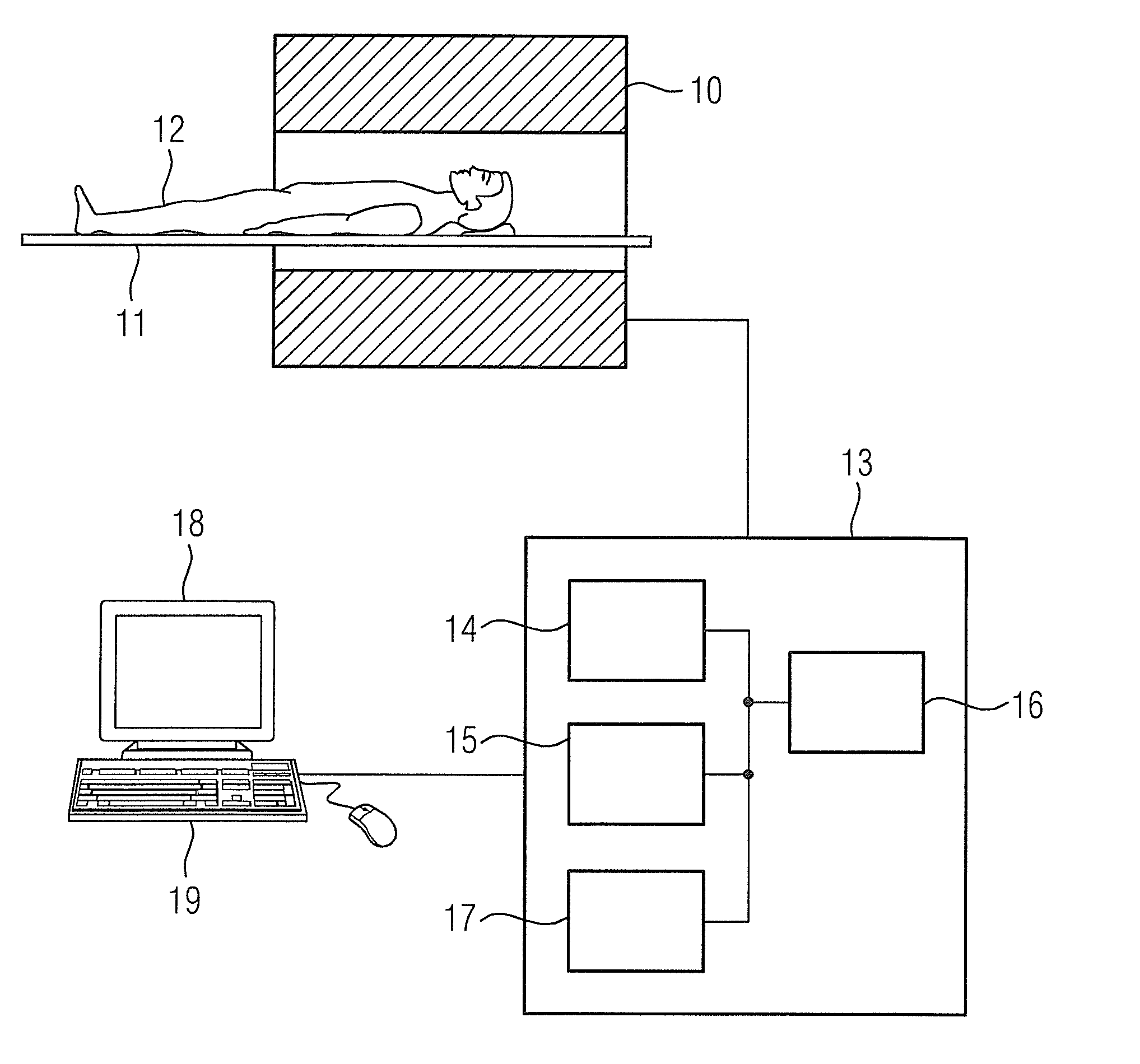
[0030]FIG. 1 shows schematically a diagnostic magnetic resonance apparatus, with which magnetic resonance relaxation parameter maps can be generated. Magnetic resonance relaxation parameter maps show, spatially correctly, the regional distribution of the values of magnetic relaxation constants an area of examination. With the so-called late-gadolinium enhancement examinations (LGE examinations) of the heart is of particular interest the regional distribution of the longitudinal relaxation constant T1.

[0031]The magnetic resonance apparatus has a basic field magnet 10 for the generation of a polarizing field B0, wherein a person 12 to be examined is supported on a table 11 and is moved into the center of the magnet 10 so that spatially encoded magnetic resonance signals can be received from the area of examination. Radiation of radio-frequency pulse sequences and switching of magnetic field gradients allow the magnetization of the nuclear spins to be tilted (“flipped”) out of the alig...


PUM


Abstract

In a method for a rapid determination of spatially resolved magnetic resonance relaxation parameters in an area of examination, a preparation pulse is radiated into the area of examination. During the relaxation of the longitudinal magnetization, spatially encoded magnetic resonance signals are acquired at a minimum of two different points in time using a fast magnetic resonance sequence. At each inversion time, an image data record is reconstructed from the magnetic resonance signals, which are elastically registered to each other. From the recorded image data records, values of magnetic resonance relaxation parameters are spatially accurately determined.

Description

BACKGROUND OF THE INVENTION[0001]1. Field of the Invention[0002]The invention concerns a method for a rapid determination of a spatially resolved magnetic resonance relaxation parameter in one area of examination, such as the longitudinal relaxation constants T1 and the transverse relaxation constants T2.[0003]2. Description of the Prior Art[0004]Medical magnetic resonance imaging is useful in the diagnosis of soft tissue due to the high contrast especially for imaging of different tissues or for the visual representation of the state of the tissue. The spatially resolved determination of the value of magnetic resonance relaxation parameters, such as the longitudinal relaxation constant T1 or the transverse relaxation constant T2, has also an added importance in medical magnetic resonance technique, because it allows to diagnose different tissues or to identify abnormal tissue changes. The spatially resolved and quantified relaxation parameters are represented in the form of paramet...

Claims


Application Information

Patent Timeline
Patent Type & Authority Applications(United States)
IPC IPC(8): G01R33/50
CPCG01R33/50G01R33/56G01R33/56509
Inventor GREISER, ANDREASSCHMITT, PETERXUE, HUI
Owner SIEMENS HEALTHCARE GMBH
Why Eureka
Try Eureka