Reporter genes for magentic resonance imaging and methods of use thereof

a technology of magentic resonance and reporter genes, which is applied in the field of recombinant nucleic acids encoding ferritin fusion proteins, can solve the problems of low resolution imaging, false signal generation, and signal dependency on availability of iron, and achieve the effect of increasing contras

Active Publication Date: 2013-10-24
YEDA RES & DEV CO LTD +1
View PDF4 Cites 22 Cited by
  • Summary
  • Abstract
  • Description
  • Claims
  • Application Information

AI Technical Summary

Benefits of technology

[0019]In one embodiment, this invention provides a Magnetic Resonance Imaging (MRI) contrast agent useful for providing increased contrast in an image of a biological sample comprising: (a) an isolated, recombinant fusion protein comprising a ferritin heavy chain polypeptide fused to a peptide comprising SEQ ID NO: 3, wherein said peptide is fused to the C-terminal end of the ferritin heavy chain polypeptide; and (b) at least one ferromagnetic magnetite iron oxide.

Problems solved by technology

The disadvantages of these current reporters include low resolution imaging, signal dependency on availability of iron, false signal generation, difficulties with accessibility and nonspecific uptake of substrate nanoparticles, delay of change in signal that is dependent on iron availability and ferritin loading factor, and low sensitivity.
However, compared to superparamagnetic iron oxides (8000-63000 μB), native ferritin has a relatively low R2 relaxivity and thus provides relatively low sensitivity as MRI contrast agent.

Method used


Image

  • Reporter genes for magentic resonance imaging and methods of use thereof
  • Reporter genes for magentic resonance imaging and methods of use thereof
  • Reporter genes for magentic resonance imaging and methods of use thereof

Examples


example 1

Recombinant Ferritin:M6A Fusion protein

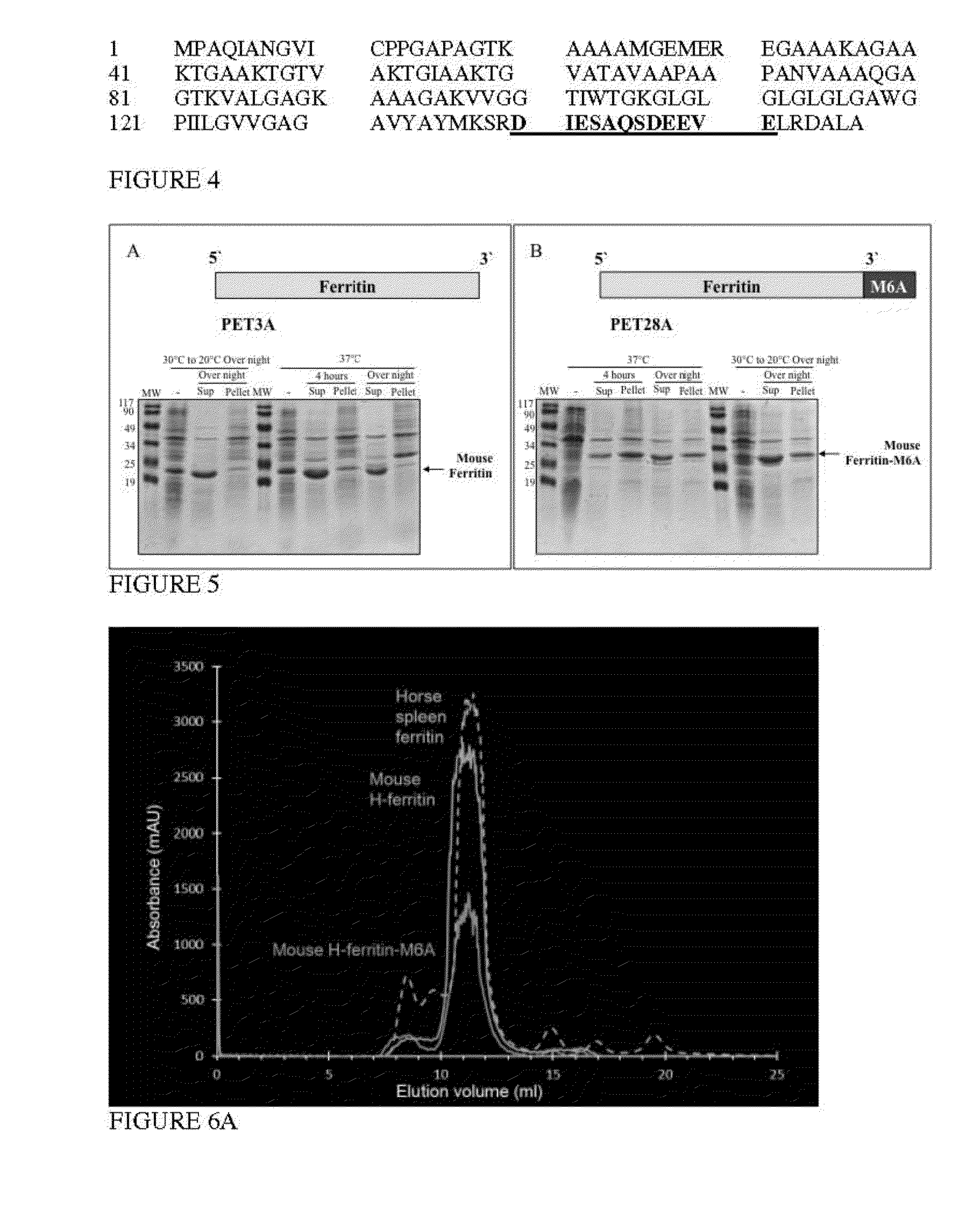
[0180]Recombinant ferritin:M6A fusion protein was designed and produced from a recombinant fusion nucleic acid encoding an M6A peptide, SEQ ID NO: 3 (12 amino acids from Mms6 protein, FIG. 4, with an N-terminal Glycine added) fused to the C-terminal of a mouse heavy chain ferritin polypeptide (SEQ ID NO: 4). The C-terminus of heavy chain ferritin is located in the inner space of a ferritin particle complex. The resulting ferritin:M6A fusion protein (Ferr-M6a; SEQ ID NO: 6) was designed to facilitate conversion of ferrihydrite into magnetite and by this induce contrast for non-invasive MRI measurements.

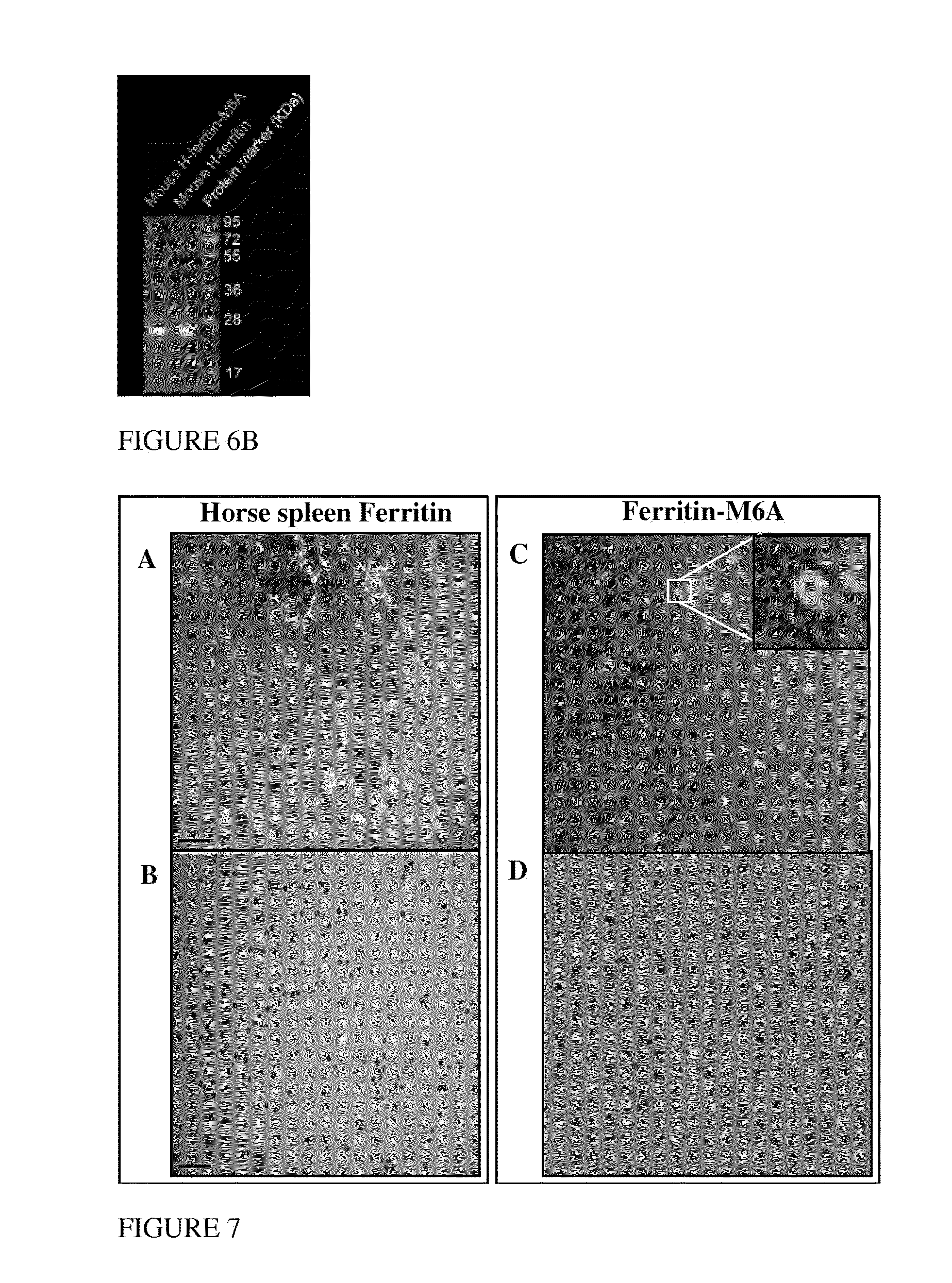
[0181]Recombinant fusion protein, ferritin-M6A, was overexpressed in E. coli Rosetta R3 cell and purified as shown in FIGS. 5A and 5B, and 6A and 6B. FIGS. 5A and 5B show SDS-PAGE results of the different purification steps identifying the ferritin-M6A at each step. FIG. 6A compares size exclusion chromatography results of the isolated ferritin-M6...

example 2

Recombinant iron loaded Ferritin M6A particles

[0182]Purified ferritin-M6A fusion protein was reconstituted with Fe(II) solution under slow oxidative condition at 60° C. and pH 8.5. Thus, a magnetic mineral was synthesized within the inner cavity of purified apo-ferritin-N6A.

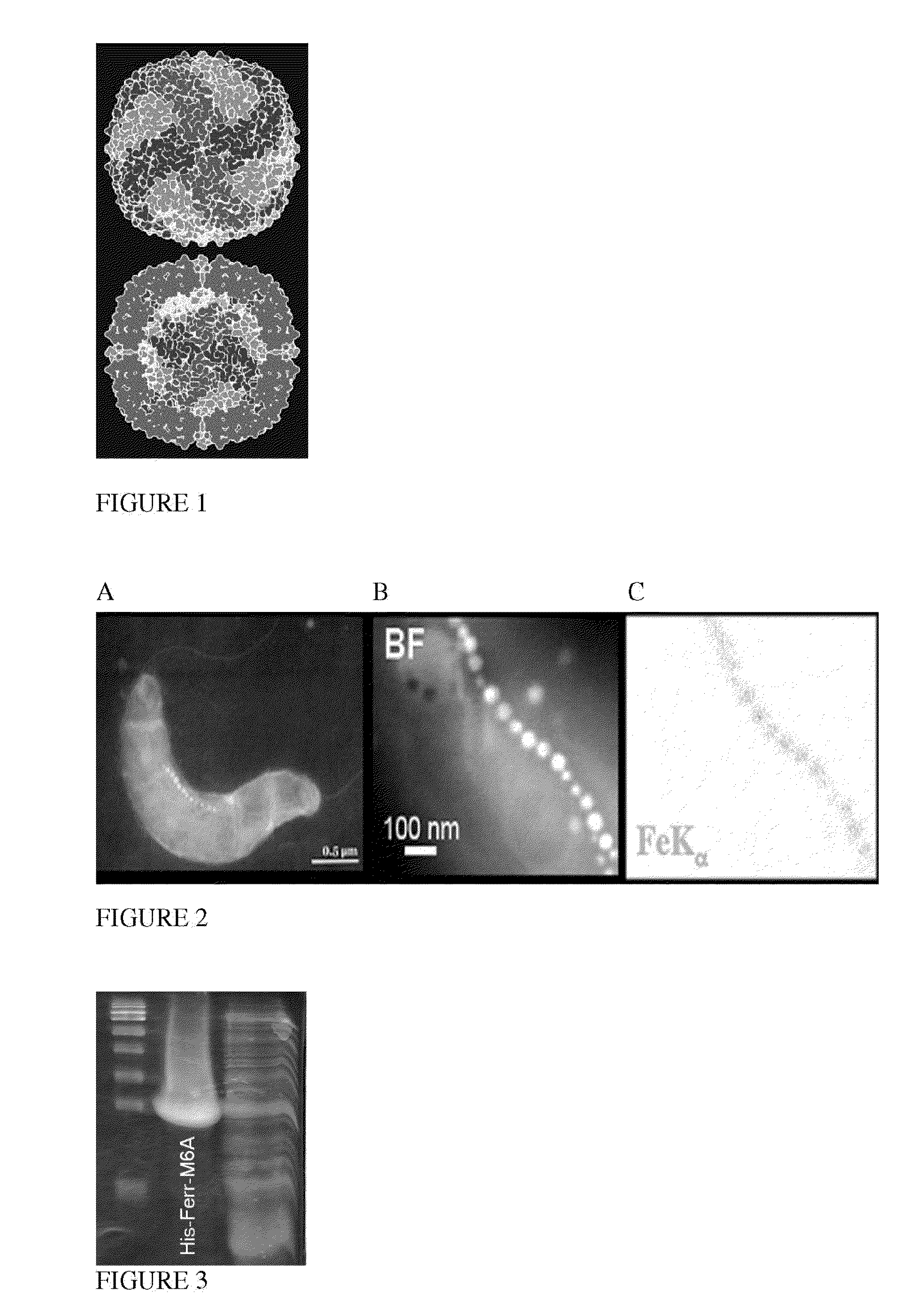
[0183]Ferritin particle consists of a protein shell of 12 nm in diameter and an iron core of about 6 nm in diameter. In the (transmission electron microscopy) TEM mode, unstained ferritin particles can be discerned as approximately 6 nm black particles, as shown in FIG. 7B, and also negatively stained particles can be discerned as approximately 12 nm white rings encircling a black core, as shown in FIG. 7A. TEM images of recombinant fusion protein, Ferritin-M6A, indicated a well formed spherical hollow shell (FIG. 7C) and entrapped within the shell 6 nm spherical mineral particles (FIG. 7D).

example 3

In vitro MRI of Purified Ferritin-M6A

[0184]MRI was applied for examination of R2 relaxation of solutions of reconstituted Ferritin-M6A fusion protein and horse spleen ferritin at different concentrations. The R2 relaxation maps, presented in FIG. 8, show significantly higher relaxation rates of Ferritin-M6A fusion protein samples (spots 1 and 2 of FIG. 8) compared with ferritin samples (spots 5-8 of FIG. 8). Table 1 below, provides the R2, s−1 numerical values of each sample.

TABLE 1SampleR2, S−11Ferritin-M6A 1:127.69902Ferritin-M6A 1:216.01863buffer2.83694buffer2.86715Ferritin 4 mg / ml6.52456Ferritin 2 mg / ml4.70557Ferritin 1 mg / ml3.75488Ferritin 0.5 mg / ml3.5603


PUM

PropertyMeasurementUnit
diameteraaaaaaaaaa
diameteraaaaaaaaaa
diameteraaaaaaaaaa

Abstract

The invention provides a fusion ferritin protein wherein a ferritin heavy chain polypeptide is fused to a peptide, wherein the peptide is fused to the C-terminal end of the ferritin heavy chain; and the peptide includes at least a portion of a Mms6 protein sequence and at least one heterologous amino acid at its N-terminal end. The invention further provides methods of use of the ferritin fusion protein for Magnetic Resonance Imaging.

Description

CROSS REFERENCE TO RELATED APPLICATIONS[0001]This application claims priority from U.S. Provisional Application Ser. No. 61 / 620,462, filed Apr. 5, 2012, which is incorporated in its entirety herein by reference.FIELD OF THE INVENTION[0002]This invention is directed to recombinant nucleic acids encoding ferritin fusion proteins and the use of these ferritin fusion proteins as reporter agents for Magnetic Resonance Imaging (MRI).BACKGROUND OF THE INVENTION[0003]Noninvasive molecular imaging methods are used to provide noninvasive dynamic information on gene expression, transcriptional regulation of gene expression, and tumor microenvironment and the consequences of chemo- and / or radiation therapy.[0004]For example oxygen deficiency, or hypoxia, is a central microenvironmental tumor stress that arises as a consequence of the expansion of solid tumors by cancer cell proliferation, which is unmatched by the expansion and maintenance of the vasculature supply. Tumor cells surrounding func...

Claims


Application Information

Patent Timeline
Patent Type & Authority Applications(United States)
IPC IPC(8): A61K49/14
CPCA61K49/14C07K14/195C07K14/47C07K2319/00C07K2319/42
Inventor NEEMAN, MICHALZARIVACH, RAZRADOUL, MARINACOHEN, BATYAVANDSBURGER, MORIELLEWIN, LIMOR
Owner YEDA RES & DEV CO LTD
Why Eureka
Try Eureka