
Eureka
For R&D, Eureka makes reading and utilizing patents & technical documents easy.
Eureka AIR
Designed for self-driven R&D workflows. Generate viable solutions, solve complex R&D challenges, empower your innovation with AI.

Eureka Materials
Designed for material experts only. Revolutionize your material R&D, from search, analyze, to developing new materials.

TechResearch
Generate reliable direction feasibility study reports for your R&D in just a few steps.

TechSeek
Discover and master advanced knowledge NOW. Basics, ideas, possibilities, all at once.

TechMind
As an expert in R&D Theories, TechMind can generates customized viable solutions instantly.

TechRisk
Analyze your overall solution with one click, know your potential R&D risks in advance.

TechMonitor
Get weekly tech updates, stay abreast of the latest tech innovations and key insights.
3D localisation microscopy and 4d localisation microscopy and tracking methods and systems
- Summary
- Abstract
- Description
- Claims
- Application Information
AI Technical Summary
Benefits of technology
Problems solved by technology
Method used
Image

Examples
example
Materials and Methods
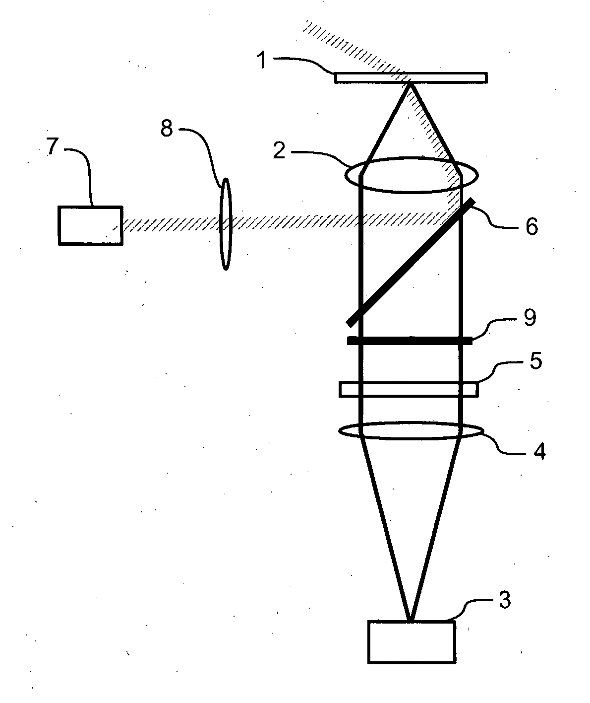
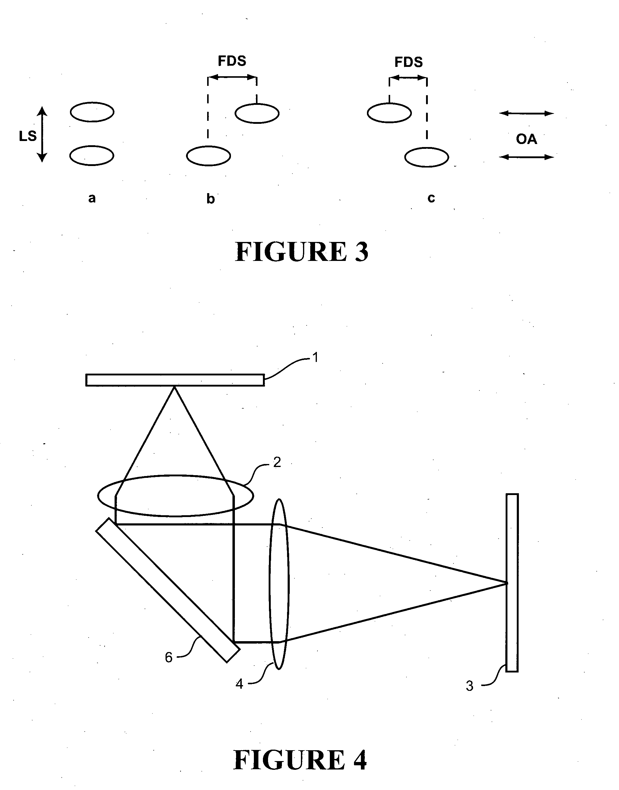
[0057]Cell Isolation and Labelling: Ventricular myocytes were enzymatically isolated from an adult rat and fixed in 2% paraformaldehyde for 10 minutes. They were then labelled for β-tubulin according to a standard immunofluorescence protocol using a primary antibody raised in mouse (T4026, Sigma-Aldrich, NZ), and an Alexa 680 labelled secondary (A21058, Molecular Probes / Invitrogen, NZ). The labelled cells were mounted in a ‘switching buffer’ consisting of 0.5 mg mL−1 glucose oxidase, 40 μg mL−1 catalase, 10% w / v glucose and 50 mM β-mercaptoethylamine in an 80:20 mixture of glycerol and PBS (all obtained from Sigma-Aldrich, NZ).
Imaging: Imaging was performed using a modified Nikon TE2000 microscope with a 60×1.47 NA TIRF objective and using an Andor IXon DV887DCS-BV EMCCD camera for fluorescence detection. Highly inclined plane illumination was used with a 671 nm laser source, leading to a focal plane intensity of approximately 109 W / m2. Approximately 20000 frame...
example 2
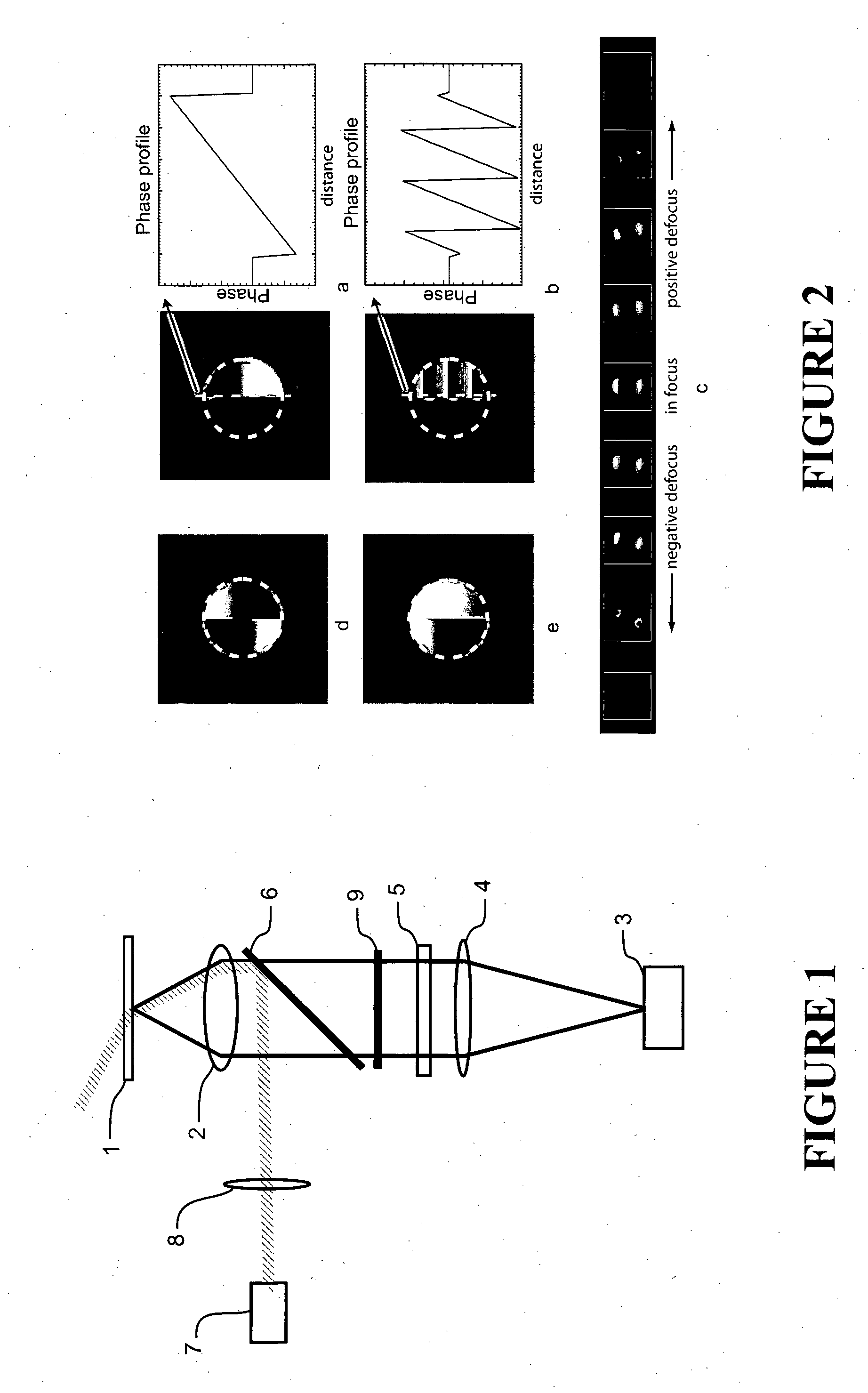
[0063]FIG. 13 shows a theoretical comparison of the axial localisation accuracy of the method (PRI) with two existing 3D localisation methods, namely astigmatic detection and biplane detection. To compare the accuracies point-spread functions for the three methods were simulated and used to calculate the Cramer-Rao lower bound. This is based on the information contained in the point-spread function shape and represents the minimum possible localisation error (that which would be obtained with a perfect analysis algorithm) under given noise conditions. For noise calculations it was assumed that the noise followed poisson statistics with a total of 2000 photons being detected from each fluorescent molecule. It can be seen that the method of the invention is indicated as having superior axial and overall 3D accuracy compared with the other methods, and that the lateral accuracy is maintained over a wider axial range.
example 3
[0064]FIG. 14 illustrates that a wavelength dependence on the relative amplitude of the two lobes can be created using an optical filter. In an experimental imaging system otherwise generally as describe in Example 1, a phase ramp was inserted into one half of the pupil plane and a longpass interference filter into the other. This configuration was used to image fluorescent beads in three different colours and an in-focus section through the point-spread functions corresponding to each of these bead types is shown in FIGS. 14a-c. A line profile through the center of each PSF was plotted as FIG. 14d and shows that the different bead types are clearly distinguishable by their relative lobe heights.
PUM
 Abstract
Description
Claims
Application Information
 - R&D Engineer
- R&D Manager
- IP Professional
- Industry Leading Data Capabilities
- Powerful AI technology
- Patent DNA Extraction
Browse by: Latest US Patents, China's latest patents, Technical Efficacy Thesaurus, Application Domain, Technology Topic, Popular Technical Reports.
© 2024 PatSnap. All rights reserved.Legal|Privacy policy|Modern Slavery Act Transparency Statement|Sitemap|About US| Contact US: help@patsnap.com