Multi-Source Medical Display

a multi-source, medical display technology, applied in the direction of instruments, applications, measurement using nmr, etc., can solve the problems of requiring the use of an additional hand, unable to operate a laser pointer or make indications on a touch screen, and often not being able to spare a surgeon's hand

Active Publication Date: 2014-02-06
KARL STORZ IMAGING INC
View PDF12 Cites 19 Cited by
  • Summary
  • Abstract
  • Description
  • Claims
  • Application Information

AI Technical Summary

Benefits of technology

[0012]The novel method and apparatus have the advantage that the image including the overlay pattern is directly available for further processing outside the sterile area. This further proces

Problems solved by technology

First, the surgeon cannot operate a laser pointer or make indications on a touch screen while also safely performing the surgical procedur

Method used


Image

  • Multi-Source Medical Display
  • Multi-Source Medical Display
  • Multi-Source Medical Display

Examples


Embodiment Construction

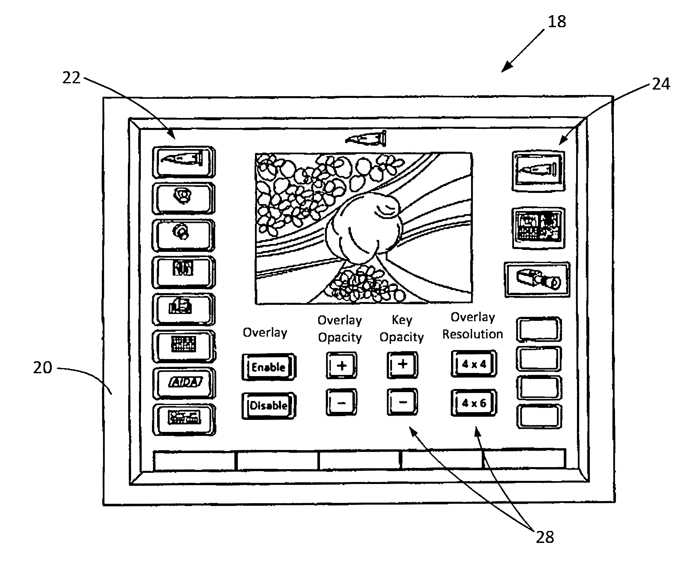
[0047]The present invention provides a system 10 for indicating certain areas of interest in medical or surgical image data by applying an overlay pattern, such as a Cartesian grid, crosshairs, quadrants, etc., on the image. The overlay pattern allows a doctor to then refer or call attention to areas of interest in the image data by referencing the overlay pattern or a portion thereof. As will be discussed in detail below, the overlay may also include an key, which may include alphanumeric labels or coordinates, which may assist the doctor in indicating the area or portion of the overlay to which he / she is referring.

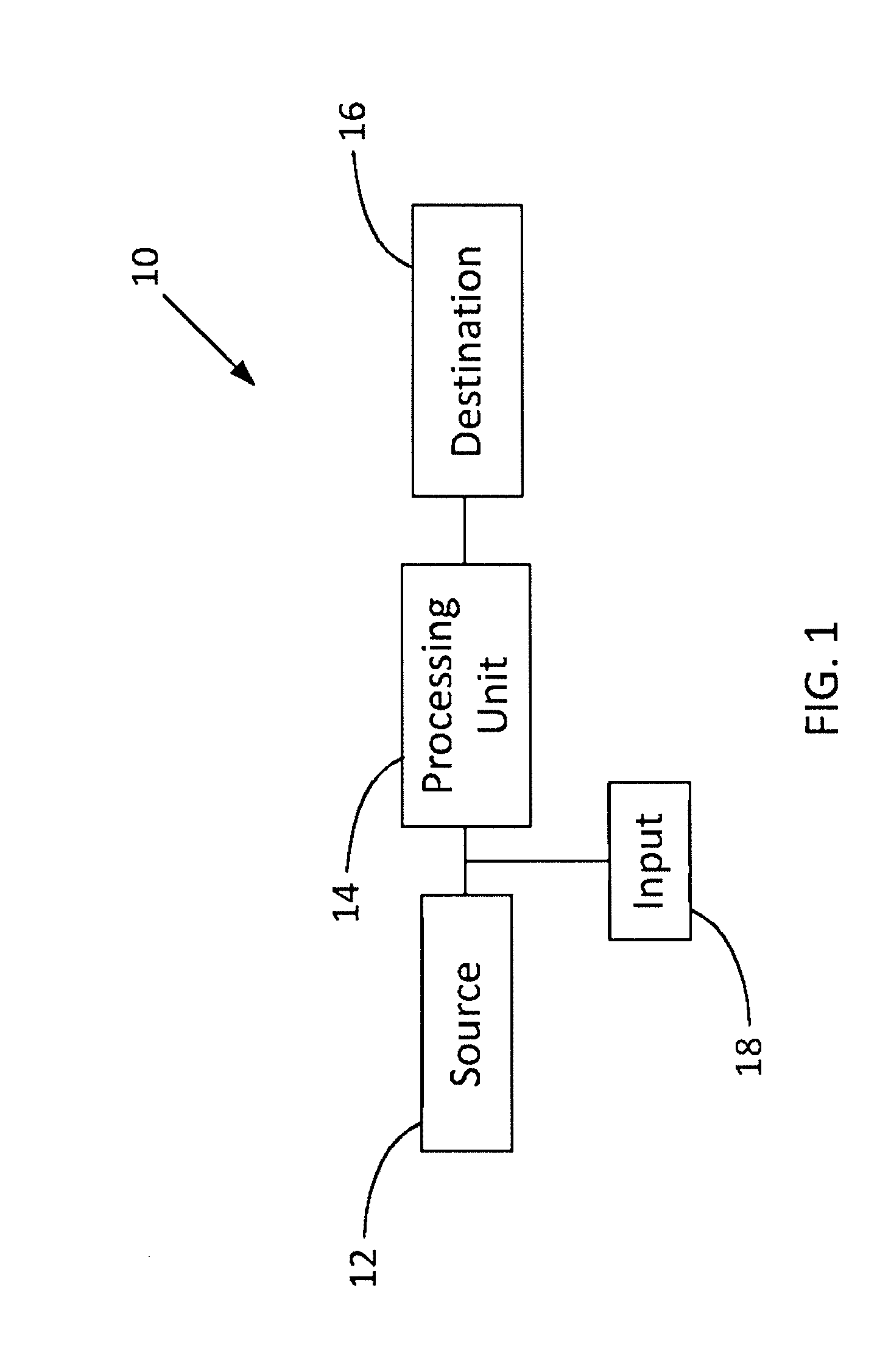
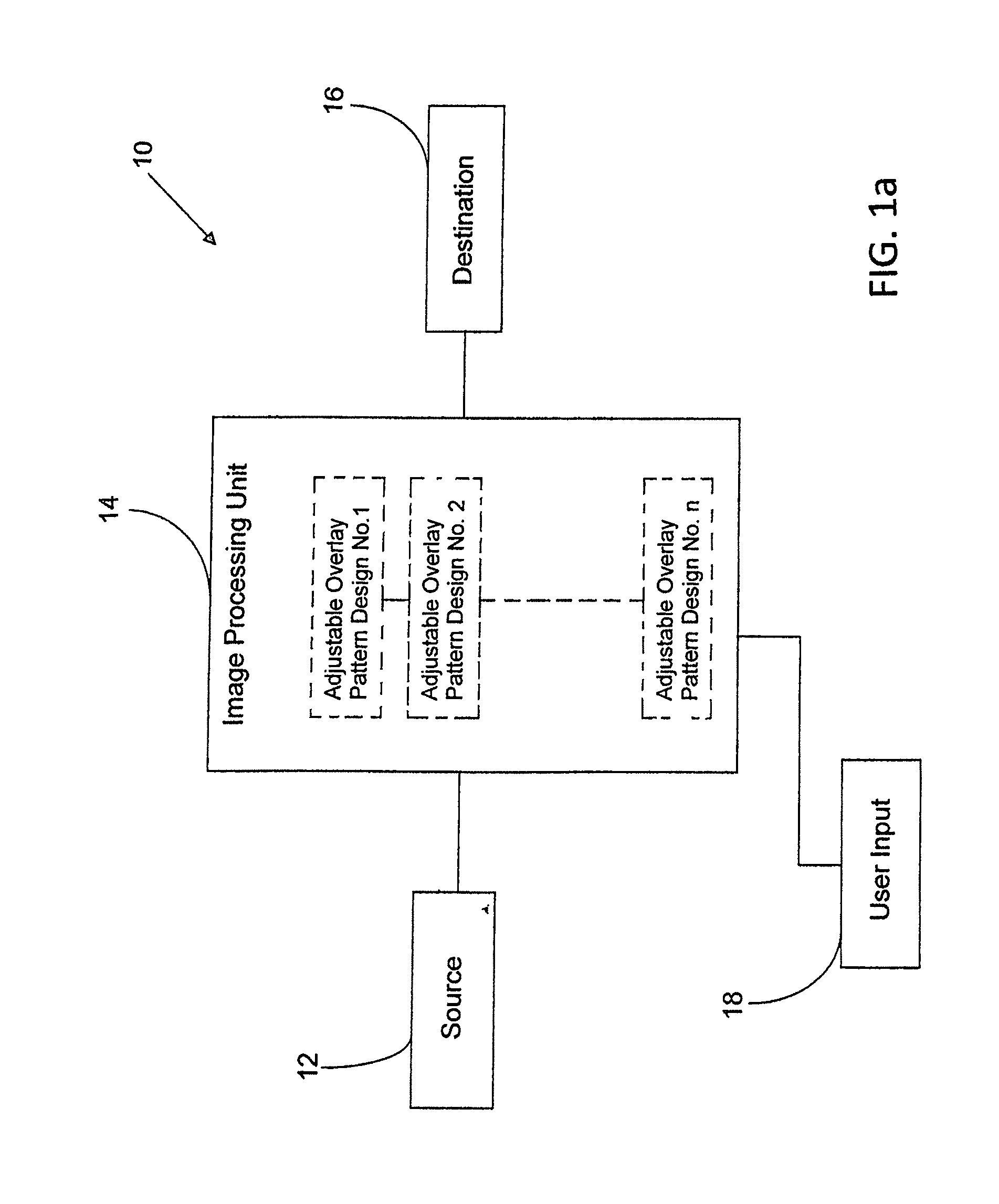
[0048]Referring to FIG. 1, the system 10 includes at least one source 12 of image data in communication with at least one processing unit 14 and at least one destination 16 for the image data. The at least one source 12 of image data connected to the processing unit 14 may include any device, system, or network that generates, acquires, stores, monitors, modifies, or con...


PUM


Abstract

A system for indicating an area of interest on an image, including a source of image data, an image processing unit, a user interface, and a destination, which may be a display. The image data may be ultrasound, X-ray, magnetic resonance imaging, nuclear magnetic resonance imaging, magnetic resonance tomography, computed tomography or surgical image data. The image processing unit may be configured to receive the image data from the source and combine it with a desired overlay pattern selected from a plurality of overlay patterns for indicating an area of interest on the image, which is then displayed on the display. The overlay pattern may include a key with coordinates or labels. Properties of the overlay pattern and the image data may be adjusted independently or automatically.

Description

FIELD OF THE INVENTION[0001]The present invention relates to a system for displaying medical images. Specifically, the present invention relates to a method and apparatus for generating an overlay aid on medical images.BACKGROUND OF THE INVENTION[0002]In modern medicine, treatments are being carried out more and more using technical imaging methods. By way of example, miniaturized cameras are inserted into the body of a patient, and the image taken by the camera is displayed to the physician on a monitor installed in his / her working area. In this way, the physician can, for example, examine an internal organ or a joint for diagnostic purposes and he / she can also carry out surgical operations in a minimally invasive fashion. By arranging a monitor in the working area of the physician, i.e. in the sterile area, the physician may track all the operations that he or she undertakes on the patient live on the monitor, the corresponding monitor image being picked up by the medical imaging ...

Claims


Application Information

Patent Timeline
IPC IPC(8): A61B5/00G06T7/00G01R33/54A61B6/03A61B8/08A61B6/00
CPCA61B5/748A61B8/52G06T7/0012G01R33/54A61B6/032A61B6/52A61B5/0077A61B1/0005A61B5/742
Inventor KING, TIMOTHYPRESCHER, THOMASBARNHILL, KIM
Owner KARL STORZ IMAGING INC
Why Eureka
Try Eureka