Remote Center of Motion Robot for Medical Image Scanning and Image-Guided Targeting

a technology of image scanning and remote center, applied in the field of remote center of motion robot for medical image scanning, can solve the problems of inability to localize or precisely place biopsy needles or implanted brachytherapy seeds, unable to achieve precise placement of brachytherapy seeds, and difficulty in visualization of anatomical structures adjacent to the prostate,

Inactive Publication Date: 2014-02-06
THE JOHN HOPKINS UNIV SCHOOL OF MEDICINE
View PDF5 Cites 60 Cited by
  • Summary
  • Abstract
  • Description
  • Claims
  • Application Information

AI Technical Summary

Benefits of technology

[0012]According to a second aspect of the present invention, a robotic apparatus for positioning at least one imaging probe comprises a support arm, a remote center of motion module operatively connected to the support arm, the remote center of motion module having a parallelogram structure built with belts and having at least two rotational degrees of freedom, and a driver module operatively connected to the remote center of motion module, the driver module for manipulating the imaging probe in at least one degree of freedom, the driver module providing one degree of freedom around a first rotation axis and/or one linear degree of freedom for translation along a second axis.
[0013]According to a third aspect of the present invention, an apparatus for performing ultrasound-guided interventions comprises a remote center of motion module providing at least two rotational degrees of freedom, and a driver module providing at least one degree of freedom for manipulating an imaging probe, the driver module providing one degree of freedom around a first rotation axis and/or one linear degree of freedom for translation along a second axis, aligned with the longitudinal axis of the end-effector, and the driver module further comprising a rotary guide and rail, the rail geometry for allowing an additional medical instr

Problems solved by technology

While 2-D TRUS provides adequate imaging of the soft tissue anatomy, it does not allow for localization or precise placement of biopsy needles or implanted brachytherapy seeds.
During prostatectomy, precise resection of the tumor-containing prostate gland and the preservation of neighboring anatomical structures are critical to preventing tumor recurrence and incontinence, and to preserving sexual potency, However, visualization of anatomical structures adjacent to the prostate can be challenging due to periprostatic connective tissues and intraoperative hemorrhage, even when the site is viewed with surgical loupes during open surgery or under laparoscopic magnification.
However, using TRUS to guide LRP has posed several challenges.
First, TRUS probes are often operated manually; as such, image stability is comprised and the probe position data that is required for 3-D computation is lost.
Also

Method used


Image

  • Remote Center of Motion Robot for Medical Image Scanning and Image-Guided Targeting
  • Remote Center of Motion Robot for Medical Image Scanning and Image-Guided Targeting
  • Remote Center of Motion Robot for Medical Image Scanning and Image-Guided Targeting

Examples


example

[0073]The following Example has been included to provide guidance to one of ordinary skill in the art for practicing representative embodiments of the presently disclosed subject matter. In light of the present disclosure and the general level of skill in the art, those of skill can appreciate that the following Example is intended to be exemplary only and that numerous changes, modifications, and alterations can be employed without departing from the scope of the presently disclosed subject matter. The following Example is offered by way of illustration and not by way of limitation.

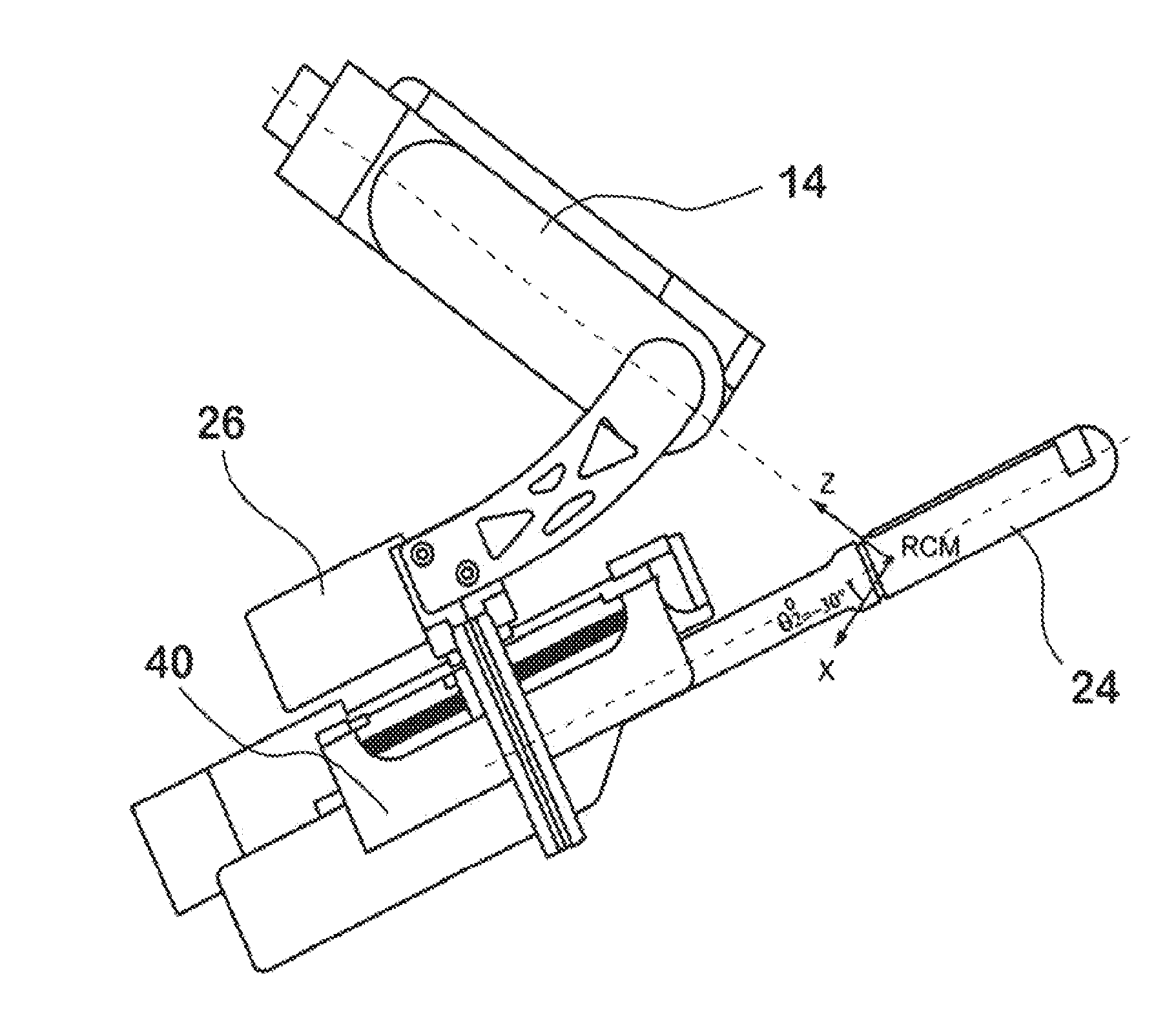
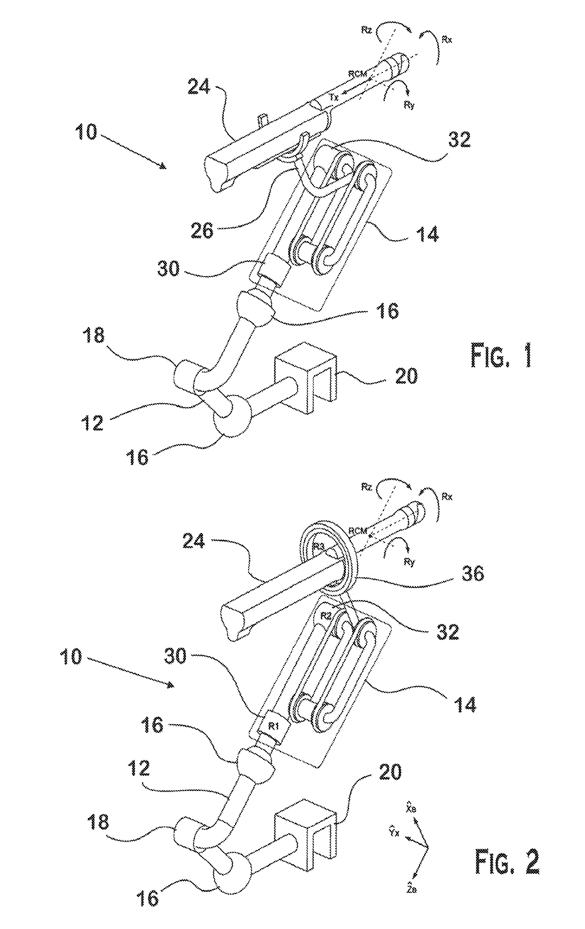
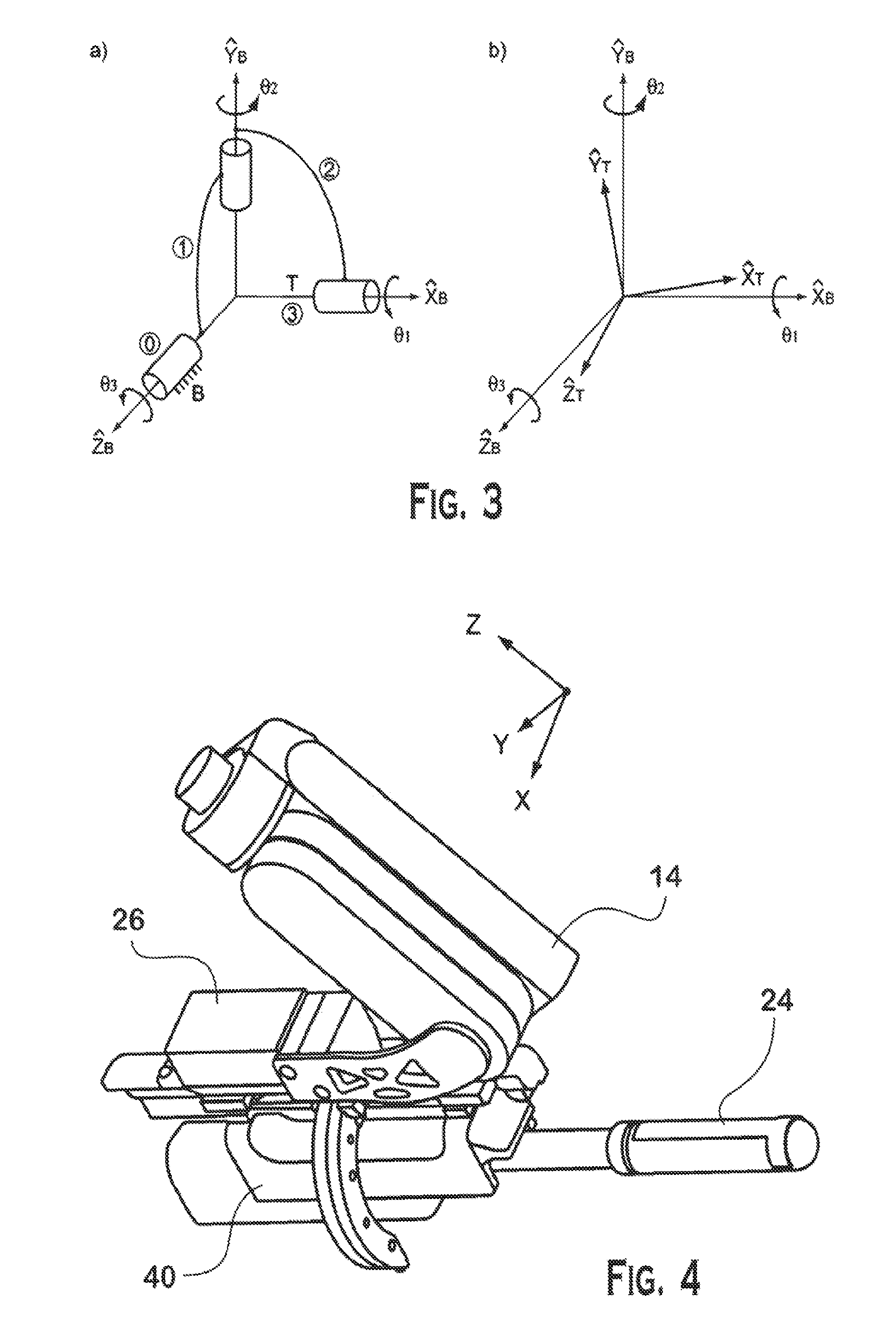
[0074]Two types of prototypes were built in the laboratory—the Euler-R (corresponding to robot with driver module 36) and Euler-RT (corresponding to robot with driver module 26). Both versions presented a compact structure and, due to their RCM kinematic architecture, have a wide range of motion which enables typical positioning and scanning motion (Table 1) that is similar to human maneuverability of a ...


PUM


Abstract

The present invention pertains to a remote center of motion robot for medical image scanning and image-guided targeting, hereinafter referred to as the “Euler” robot. The Euler robot allows for ultrasound scanning for 3-Dimensional (3-D) image reconstruction and enables a variety of robot-assisted image-guided procedures, such as needle biopsy, percutaneous therapy delivery, image-guided navigation, and facilitates image-fusion with other imaging modalities. The Euler robot can also be used with other handheld medical imaging probes, such as gamma cameras for nuclear imaging, or for targeted delivery of therapy such as high-intensity focused ultrasound (HIFU). 3-D ultrasound probes may also be used with the Euler robot to provide automated image-based targeting for biopsy or therapy delivery. In addition, the Euler robot enables the application of special motion-based imaging modalities, such as ultrasound elastography.

Description

REFERENCE TO RELATED APPLICATIONS[0001]This application claims the benefit of U.S. Provisional Patent Application No. 61 / 412,589, filed on Nov. 11, 2010, which is hereby incorporated by reference for all purposes as if fully set forth herein.STATEMENT OF GOVERNMENTAL INTEREST[0002]This invention was made with U.S. government support under grant no. 1R21CA141835-01. The U.S. government has certain rights in the invention.FIELD OF THE INVENTION[0003]The present invention pertains to a remote center of motion robot for medical image scanning. More particularly, the present invention pertains to a remote center of motion robot for medical image scanning and image-guided targeting.BACKGROUND OF THE INVENTION[0004]Prostate cancer is the most common form of cancer in American men. Like other cancers, early diagnosis and treatment is critical to the clinical management of the disease, and to preserving patients' quality of life and increasing life expectancy. In furtherance of these objecti...

Claims


Application Information

Patent Timeline
IPC IPC(8): A61B8/00A61B19/00
CPCA61B19/2203A61B8/4444A61B8/0841A61B8/085A61B8/12A61B8/4218A61B8/483A61B8/485A61B8/5207A61N7/02A61B10/02A61B10/0241A61B8/4254A61B34/30A61B2090/378A61N7/00A61B8/14
Inventor STOIANOVICI, DANPETRISOR, DORUSCHAFER, FELIXHAN, MISOPKIM, CHUNWOO
Owner THE JOHN HOPKINS UNIV SCHOOL OF MEDICINE
Why Eureka
Try Eureka