Method for improved estimation of tracer uptake in physiological image volumes

Inactive Publication Date: 2014-05-01
UNIVERSITY OF OSLO
View PDF17 Cites 14 Cited by
  • Summary
  • Abstract
  • Description
  • Claims
  • Application Information

AI Technical Summary

Benefits of technology

[0041]The method may hence be used to correct errors in the PET signal (PET image) arising from the limited spatial resolution of the PET scanner which means that the true and/or improved physiological tracer uptake can be estimated at the voxel level. The method of a preferred embodiment takes the original PET image (PET volume) together with anatomical structures segmented from e.g. MR-images as input, and provides as an output an estimate of the true physiological tracer uptake in each voxel of the image volume. The partial volume effect problem in positron emission tomography (PET) imaging can thus be solved at a voxel level and this is achieved by using regional information from co-registered magnetic resonance images (MRI). The distribution of the tracer in the corrected physiological image volume is an estimated quantity which takes into account the diffusion effect that would occur in respect of each class of tissue in each voxel.
[0042]T

Problems solved by technology

A number of phenomena introduced by the imaging

Method used


Image

  • Method for improved estimation of tracer uptake in physiological image volumes
  • Method for improved estimation of tracer uptake in physiological image volumes
  • Method for improved estimation of tracer uptake in physiological image volumes

Examples


Embodiment Construction

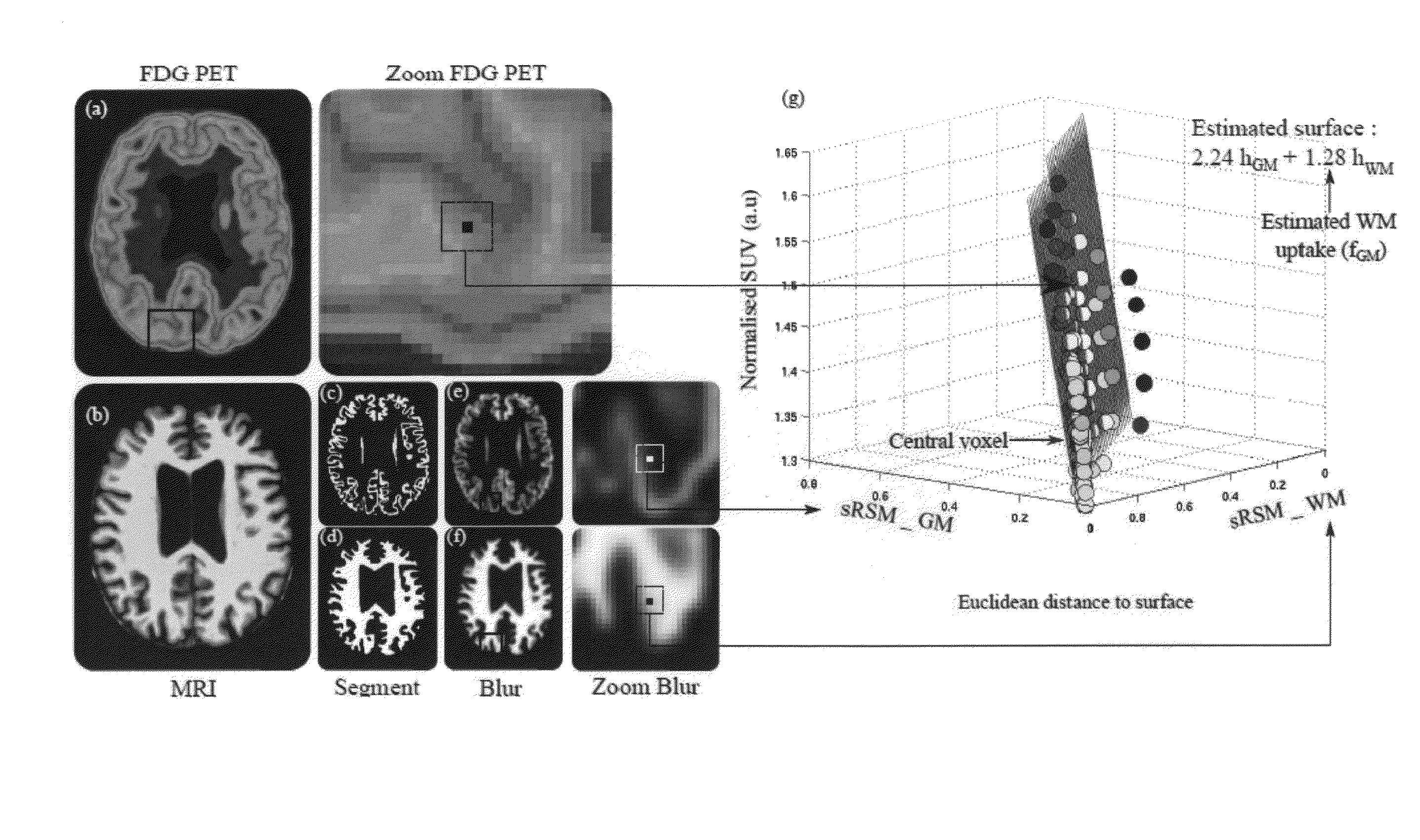
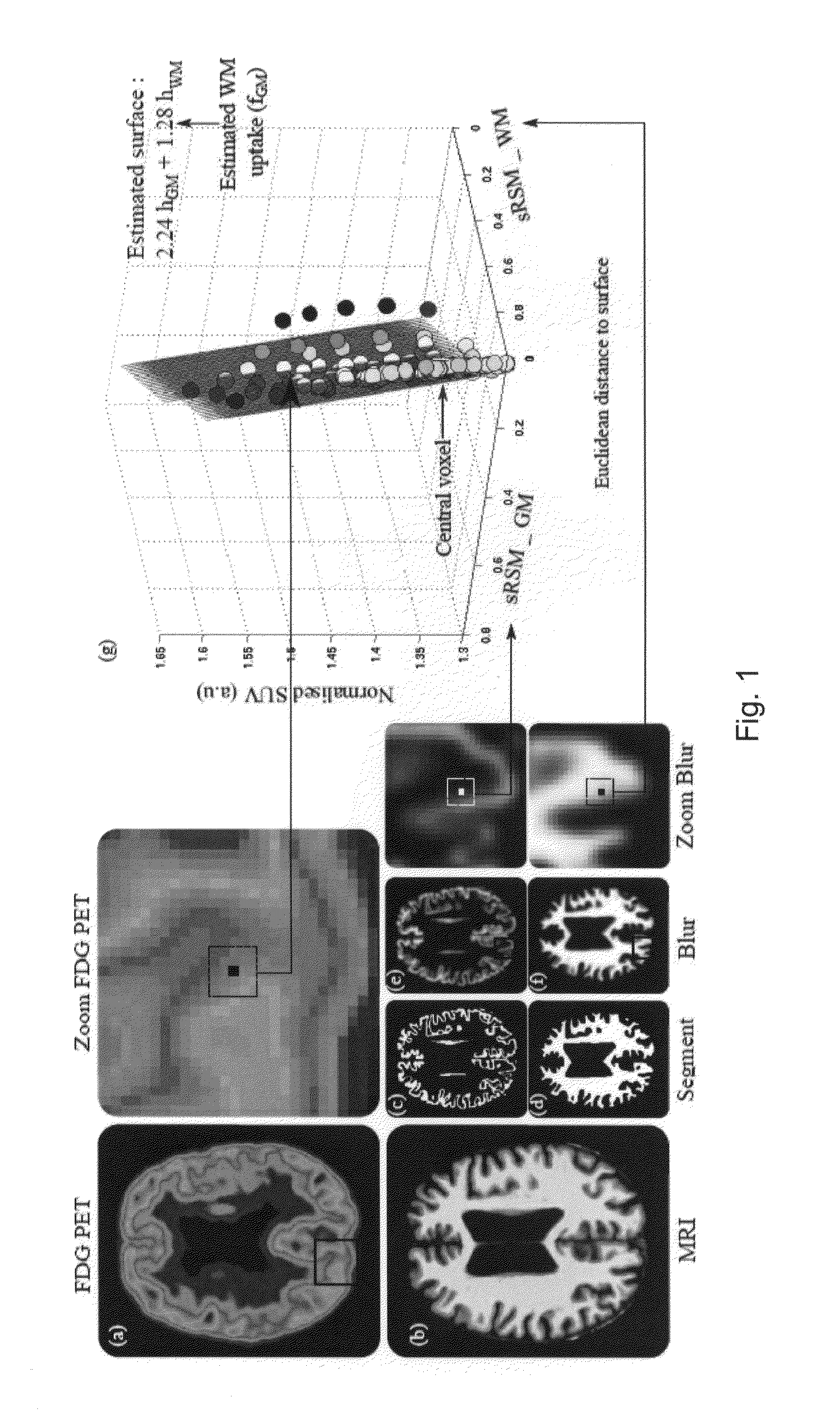
[0087]A preferred embodiment is shown in summary by FIG. 1. For this example the anatomical imaging is MRI and the physiological imaging is PET. FIG. 1 illustrates the case where the brain was segmented based on the MRI images into white and grey matter regions. The left column show the original physiologic (a) and anatomical images (b), which are coregistered. The small images (c) and (d) of the lower row show the segmented grey and white matter regions of the MRI image, followed by the results of blurring in (e) and (f). The blown-up cut-outs (PET on top and blurred white and grey matter in the lower part) identify a cube for which the regression analysis was carried out. The result of the regression analysis of corresponding voxels (within the cube) in the three image sets is illustrated by the 3D plot of the regression surface

g(r)=hGM(r)·fGM(r)+hWM(r)·fWM(r)+η(r)  (8)

[0088]in which g(r) is plotted against the blurred region hGM(r) and hWM(r). The single points on which the regre...


PUM


Abstract

A method of estimating the true uptake of a tracer in a physiological image acquired with a physiological image modality that has limited spatial resolution comprises: co-registering an anatomical image volume and the physiological image volume such that both image volumes have corresponding voxels; segmenting the anatomical image volume into a number of non-overlapping anatomical regions; combining each of the segmented anatomical regions with a function representative of the limited spatial resolution of the physiological image modality to generate a blurred anatomical region volume for each of the segmented regions; assuming the physiological image in each voxel to be a linear sum of the image intensities of the corresponding voxels in the blurred region volumes in that voxel; estimating, for each voxel, a value representative of the contribution to that voxel from each blurred region volume based on the uptake measured by the physiological image for that voxel and for nearby voxels; and using the estimated values for the voxels to estimate a true uptake of the tracer in each voxel and to thereby establish a corrected physiological image volume.

Description

COLOR DRAWING STATEMENT[0001]The patent or application file contains at least one drawing executed in color. Copies of this patent or patent application publication with color drawing(s) will be provided by the Office upon request and payment of the necessary fee.CROSS REFERENCE TO RELATED APPLICATION[0002]This application is related to and claims the benefit of British Patent Application Number 1219403.1 filed on 29 Oct. 2012, the contents of which are herein incorporated by reference in their entirety.TECHNICAL FIELD[0003]The present invention relates to a method of estimating the true uptake of a physiological tracer in a physiological image acquired with a physiological image modality that has limited spatial resolution.BACKGROUND[0004]Physiological imaging techniques such as emission tomography, including single photon emission computed tomography (SPECT) and positron emission tomography (PET), make it possible to estimate parameters characterizing physiological and molecular p...

Claims


Application Information

Patent Timeline
IPC IPC(8): G06T7/00
CPCG06T7/0012G06T7/0016G06T2207/10104G06T2207/30016
Inventor SKRETTING, ARNECOELLO, CHRISTOPHER
Owner UNIVERSITY OF OSLO
Why Eureka
Try Eureka