Evoked response testing system for neurological disorders

a neurological disorder and evoked response technology, applied in the field of electroencephalogram (eeg) signal capture methods and apparatuses, can solve the problems of long signal latencies, abnormally high peak voltages, and difficult to distinguish cognitive and physiological contributors to the observed eeg, and achieves convenient testing and configuration. and use.

Active Publication Date: 2014-05-08
NEURONETRIX SOLUTIONS
View PDF10 Cites 20 Cited by
  • Summary
  • Abstract
  • Description
  • Claims
  • Application Information

AI Technical Summary

Benefits of technology

[0068]The invention overcomes the above-noted and other deficiencies of the prior art by providing an auditory and / or visual ERP testing system which is easy to configure and use; enables standardized protocols and methods; facilitates testing of a broad range of neurological attributes and disorders; and delivers reliable and reproducible results. This system employs an integrated, software controlled headset which presents the stimuli and records the ERPs. The headset is programmed through a web-enabled software control panel. The testing protocols and stimuli are downloaded from an online protocol database. The resultant test data is uploaded from the headset to an online database. Various neurological attributes are classified using an automatic analysis method. And finally, results are available for display via a web-enabled software application. Such analyses are facilitated by associating testing paradigms and patient identification information with test results. Thereby, multiple types of ERP analyses by be performed for a range of neurological conditions.

Problems solved by technology

ERPs from dyslexic children often show abnormally high peak voltages and long signal latencies.
These characteristics may correlate to higher than normal energy requirements to process sounds and slower discrimination and sound-to-symbol mapping, the outward manifestations of which will primarily be difficulty in reading and writing.
This intermixing of signals makes it difficult to separate cognitive and physiological contributors to the observed EEG.
The smaller size of the ERP relative to other physiological events can also make it difficult to discern the relevant signal.
However, information recorded at the scalp cannot capture all of the generated electrical activity.
It may be difficult to detect a signal if the distance from the cortical or subcortical regions generating the signal to the scalp is too great relative to the signal's strength.
Challenges exist for a comprehensive approach to detection and analysis due to generally-known techniques for source localization that rely on different principles which can produce conflicting results.
Thus, findings from intracranial recordings, functional magnetic resonance imaging (fMRI), magnetoencephalography (MEG), brain electromagnetic source analysis (BESA), positron emission tomography (PET), or low-resolution brain electromagnetic tomography (LORETA) may not always agree.
Other ERP components such as the Contingent Negative Variation, the Left Anterior Negativity, the Late Positive Potential, and the Positive Slow Wave are not included in the current review due to a sparcity of information regarding their sources and / or the limited space available to cover a large amount of research.
However, it is difficult to differentiate the peak- specific locations because dipole source analysis is still relatively primitive, making it difficult to disentangle the individual effects of the neighboring structures.
However, this effect was not present for the scrambled pictures.
Given the use of ERP averaging to remove noise from the data, researchers must maintain a balance between presenting enough deviant trials to obtain low-noise average responses, and not allowing the subject to habituate to the deviant, thus diminishing the effect.
The MMN for visual stimuli has been difficult to obtain, although there is some evidence that it can be captured with optical techniques.
The possible sources were investigated and reported that selecting only one region (e.g., hippocampus or thalamus) resulted in poor model fit, but combining the different locations produced a better model.
The amount of effortful semantic processing required is also unclear.
In addition, it is often larger for correctly recognized words than falsely recognized lures and can be affected by depth of processing, and the amount of retrieved episodic information.
In the early years of electrophysiological research, equipment limitations made it very difficult or impossible to record and / or analyze more than a single peak or to record from more than a few electrode sites.
Further, it clear that peak characteristics can be affected by the procedures used to record ERPs.
Differences in number of trials or length of intertrial intervals, variations in stimulus intensity or modality can contribute to inconsistent outcomes.
There are potential problems of interpretation, directly linking the scalp distribution of an ERP component with brain structures located below the specific electrodes.
The tremendous clinical potential of the ERP method has been well recognized although because of certain system and methodological limitations this potential has only rarely been realized.
These problems generally fall into four areas: (1) complicated, difficult to use, and incompatible hardware systems; (2) lack of standardized testing protocols including: stimuli, stimuli sequencing and timing, signal processing, testing environment; (3) analytical methods which do not provide statistically powerful or reproducible results, due to signal processing limitations, non-algorithmic peak detection methods, and disregard for information in the ERPs other than the peaks, and (4) requirement to perform large population studies in order to discriminate small neurological variations.
The equipment expense, level of training required to perform these brainwaves studies, and unreliable results renders them impractical for widespread screening and diagnostic use.

Method used


Image

  • Evoked response testing system for neurological disorders
  • Evoked response testing system for neurological disorders
  • Evoked response testing system for neurological disorders

Examples


Embodiment Construction

[0103]In the drawings where like members are given the same reference numeral, in

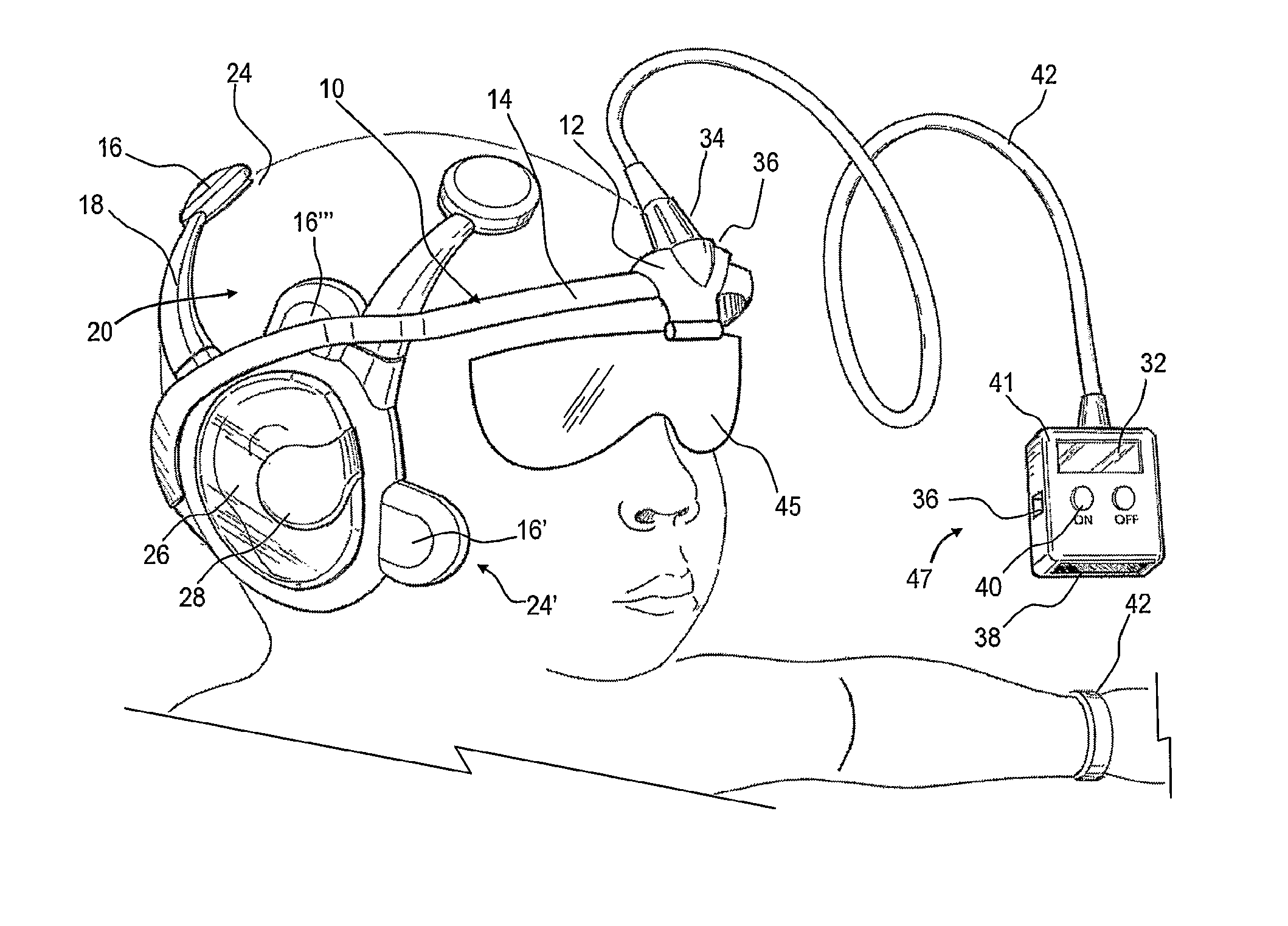
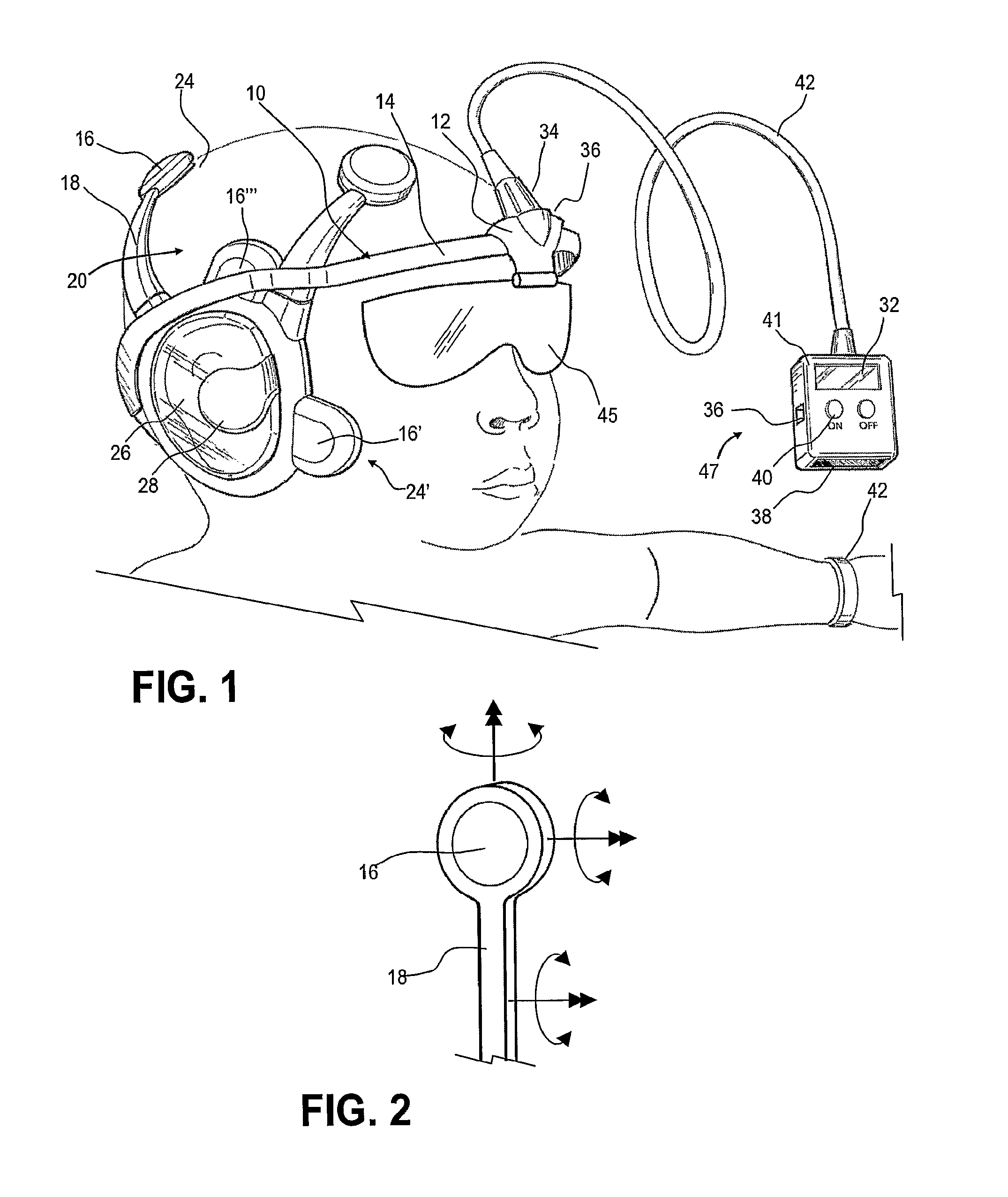
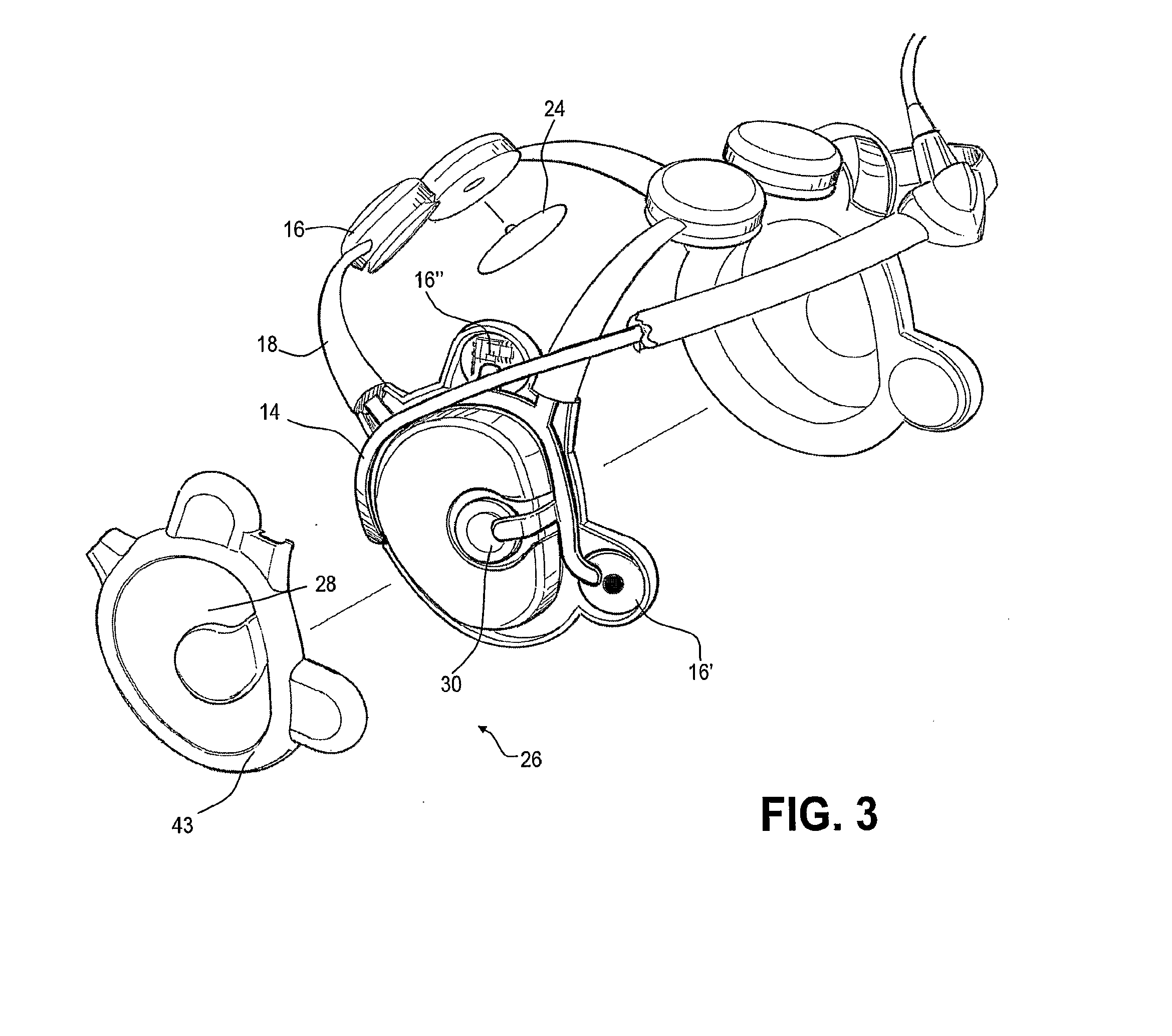
[0104]FIG. 1, an integrated Evoked Response Potential (ERP) headset 10 includes embedded features that enable clinicians to readily perform an ERP test without the necessity of extensive training. Portability of diagnostic data taking allows use whenever and wherever desired. Economy of use is achieved by centralized processing of the diagnostic data so that a great number of headsets 10 may be used without the necessity of expensive waveform processing equipment at each location. Collecting data from many screened individuals enables enhanced and improved diagnostic algorithms to be created and implemented. Furthermore, the headset 10 includes features that speed its use while avoiding human error and the need for extensive training.

[0105]To these ends, the ERP headset 10 incorporates a control module 12 that advantageously allows the headset 10 to be portable and to be used in a clinical setting by in...


PUM


Abstract

A dyslexia screening system suitable for clinical use includes an integrated headset that efficiently and conveniently performs an auditory evoked response (ERP) test by positioning electrodes about the ears of the subject. An integral control module automatically performs the test, providing simplified controls and indications to the clinician. A number of screening tests that are stored in the headset are periodically uploaded for billing, remote analysis and result reporting. A paradigm that characterizes testing performed for a subject along with the patient identification and / or patient demographics are stored in an associated fashion for later fusion and analyses with similar but not necessarily identically constructed ERP tests.

Description

[0001]The present application is a continuation of U.S. patent application Ser. No. 11 / 570,630, entitled “Evoked Response Testing System for Neurological Disorders,” filed May 8, 2007, the disclosure of which is incorporated herein, and which is a National Stage Entry of PCT International Patent App. No. PCT / U.S.05 / 021272, entitled “Evoked Response Testing System for Neurological Disorders,” filed Jun. 16, 2005, the disclosure of which is incorporated by reference herein, which in turn claims the benefit of U.S. patent application Ser. No. 60 / 580,853, entitled “Auditory Evoked Response Mapping System for Auditory Neurome,” filed Jun. 18, 2004, the disclosure of which is incorporated by reference herein.[0002]Subject matter disclosed in the present application is also related to subject matter disclosed in PCT International Patent App. No. PCT / U.S.05 / 021257, entitled “Wireless Electrode for Biopotential Measurement,” filed Jun. 16, 2005, the disclosure of which is incorporated by ref...

Claims


Application Information

Patent Timeline
Patent Type & Authority Applications(United States)
IPC IPC(8): A61B5/16A61B5/0484A61B5/00
CPCA61B5/16A61B5/04845A61B5/04842A61B5/0002A61B5/6814A61B2560/045A61B2562/08A61B5/378A61B5/38A61B5/291A61B5/165
Inventor FADEM, KALFORD C.
Owner NEURONETRIX SOLUTIONS
Why Eureka
Try Eureka